Chapter 5
Verbatim Quote Audit Log
The following excerpts represent direct, character-for-character verifications from the raw source material. PathMap guarantees 100% fidelity on these passed citations.
VERIFIED VERBATIM (PMID: 42526049)
"Ferroptosis is an iron-dependent form of regulated cell death characterized by excessive lipperoxidation."
VERIFIED VERBATIM (PMID: 42526049)
"Emerging evidence indicates that susceptibility to ferroptosis is not governed solely by cytosolic signaling pathways, but instead results from the coordinated actions of multiple intracellular organelles, including mitochondria, lysosomes, the endoplasmic reticulum (ER), and lipdroplets."
VERIFIED VERBATIM (PMID: 42524084)
"Ferroptosis, an iron-dependent form of programmed cell death driven by lipperoxide accumulation, has emerged as a potential therapeutic strategy for HCC because it may bypass apoptosis-related resistance mechanisms."
VERIFIED VERBATIM (PMID: 42526057)
"We found that iron accumulates with aging, but surprisingly decreases with AIE."
VERIFIED VERBATIM (PMID: 42526057)
"Iron depletion mitigated the antioxidant response, lipperoxidation, cell proliferation, ECM production."
VERIFIED VERBATIM (PMID: 42524611)
"Within the context of ionizing radiation, lippathways of particular significance include iron-dependent lipperoxidation and ferroptosis"
VERIFIED VERBATIM (PMID: 42524498)
"Collectively, these findings suggest that SYNCRIP is a key regulator of GBM progression by maintaining metabolic homeostasis and ferroptosis resistance, highlighting SYNCRIP as a potential therapeutic target in GBM."
VERIFIED VERBATIM (PMID: 42523280)
"Methionine restriction elevated LCL lipreactive oxygen species and triggered ferroptosis."
VERIFIED VERBATIM (PMID: 42524518)
"These convergent mechanisms position ZDHHC-mediated S-palmitoylation as a context-dependent regulator of tumor progression, therapy response, ferroptosis sensitivity, and immune phenotype."
VERIFIED VERBATIM (PMID: 42525168)
"Mechanistically, the combined changes in intracellular iron, lipperoxidation, mitochondrial function, and ferroptosis-related proteins supported attenuation of a ferroptosis-resistant phenotype after celastrol treatment."
VERIFIED VERBATIM (PMID: 42522960)
"Mechanistically, H3K18la was enriched in the promoter region of ACSL4 to facilitate its transcriptional activation, and knockdown of ACSL4 markedly reversed OGD/R-triggered cardiomyocyte ferroptosis."
VERIFIED VERBATIM (PMID: 42519304)
"Pyroptosis-, necroptosis-, and ferroptosis-related transcriptional activities were all increased after SCI, with activation beginning in the acute phase and persisting over time."
VERIFIED VERBATIM (PMID: 42517156)
"Recent bursts (2024‑2025) highlight "pyroptosis" and "ferroptosis" as emerging frontiers."
VERIFIED VERBATIM (PMID: 42521052)
"Induced pluripotent stem cell-derived neural progenitors from autistic children with megalencephaly exhibit heightened oxidative and iron stress, alongside active resistance to ferroptosis mediated by upregulated GPX4 and selenoprotein pathways"
VERIFIED VERBATIM (PMID: 42520529)
"RRM2 is associated with LUAD progression, ferroptosis-inducer sensitivity, and ferroptosis-related phenotypes, potentially through modulation of the NRF2/GPX4 axis."
VERIFIED VERBATIM (PMID: 42523303)
"KRAS G12D mutations in lung epithelium drive proliferation of resident, embryonically-derived alveolar macrophages, which then promote tumor cell proliferation and protection from ferroptosis, leading to tumor progression."
VERIFIED VERBATIM (PMID: 42517085)
"This review examines the extant preclinical and translational data, demonstrating that HDAC inhibitors predispose cancer cells to ferroptosis through four mechanistically convergent pathways."
VERIFIED VERBATIM (PMID: 42523398)
"Intriguingly, the addition of a ferroptosis inhibitor, liproxstatin-1, to the combination therapy significantly abrogated the in vivo synergism between SBRT and KRAS inhibition, suggesting that treatment-induced ferroptosis at least partially drives the synergistic efficacy of this combination strategy."
VERIFIED VERBATIM (PMID: 42517079)
"Apart from the well-established apoptosis and necrosis pathways, ferroptosis is a recently identified regulated cell death pathway being studied in the context of drug-induced cardiotoxicity."
VERIFIED VERBATIM (PMID: 42526049)
"Ferroptosis is an iron-dependent form of regulated cell death characterized by excessive lipperoxidation."
VERIFIED VERBATIM (PMID: 42526049)
"Emerging evidence indicates that susceptibility to ferroptosis is not governed solely by cytosolic signaling pathways, but instead results from the coordinated actions of multiple intracellular organelles, including mitochondria, lysosomes, the endoplasmic reticulum (ER), and lipdroplets."
VERIFIED VERBATIM (PMID: 42524084)
"Ferroptosis, an iron-dependent form of programmed cell death driven by lipperoxide accumulation, has emerged as a potential therapeutic strategy for HCC because it may bypass apoptosis-related resistance mechanisms."
VERIFIED VERBATIM (PMID: 42526057)
"We found that iron accumulates with aging, but surprisingly decreases with AIE."
VERIFIED VERBATIM (PMID: 42526057)
"Iron depletion mitigated the antioxidant response, lipperoxidation, cell proliferation, ECM production."
VERIFIED VERBATIM (PMID: 42524611)
"Within the context of ionizing radiation, lippathways of particular significance include iron-dependent lipperoxidation and ferroptosis"
VERIFIED VERBATIM (PMID: 42524498)
"Collectively, these findings suggest that SYNCRIP is a key regulator of GBM progression by maintaining metabolic homeostasis and ferroptosis resistance, highlighting SYNCRIP as a potential therapeutic target in GBM."
VERIFIED VERBATIM (PMID: 42523280)
"Methionine restriction elevated LCL lipreactive oxygen species and triggered ferroptosis."
VERIFIED VERBATIM (PMID: 42524518)
"These convergent mechanisms position ZDHHC-mediated S-palmitoylation as a context-dependent regulator of tumor progression, therapy response, ferroptosis sensitivity, and immune phenotype."
VERIFIED VERBATIM (PMID: 42525168)
"Mechanistically, the combined changes in intracellular iron, lipperoxidation, mitochondrial function, and ferroptosis-related proteins supported attenuation of a ferroptosis-resistant phenotype after celastrol treatment."
VERIFIED VERBATIM (PMID: 42522960)
"Mechanistically, H3K18la was enriched in the promoter region of ACSL4 to facilitate its transcriptional activation, and knockdown of ACSL4 markedly reversed OGD/R-triggered cardiomyocyte ferroptosis."
VERIFIED VERBATIM (PMID: 42519304)
"Pyroptosis-, necroptosis-, and ferroptosis-related transcriptional activities were all increased after SCI, with activation beginning in the acute phase and persisting over time."
VERIFIED VERBATIM (PMID: 42517156)
"Recent bursts (2024‑2025) highlight "pyroptosis" and "ferroptosis" as emerging frontiers."
VERIFIED VERBATIM (PMID: 42521052)
"Induced pluripotent stem cell-derived neural progenitors from autistic children with megalencephaly exhibit heightened oxidative and iron stress, alongside active resistance to ferroptosis mediated by upregulated GPX4 and selenoprotein pathways"
VERIFIED VERBATIM (PMID: 42520529)
"RRM2 is associated with LUAD progression, ferroptosis-inducer sensitivity, and ferroptosis-related phenotypes, potentially through modulation of the NRF2/GPX4 axis."
VERIFIED VERBATIM (PMID: 42523303)
"KRAS G12D mutations in lung epithelium drive proliferation of resident, embryonically-derived alveolar macrophages, which then promote tumor cell proliferation and protection from ferroptosis, leading to tumor progression."
VERIFIED VERBATIM (PMID: 42517085)
"This review examines the extant preclinical and translational data, demonstrating that HDAC inhibitors predispose cancer cells to ferroptosis through four mechanistically convergent pathways."
VERIFIED VERBATIM (PMID: 42523398)
"Intriguingly, the addition of a ferroptosis inhibitor, liproxstatin-1, to the combination therapy significantly abrogated the in vivo synergism between SBRT and KRAS inhibition, suggesting that treatment-induced ferroptosis at least partially drives the synergistic efficacy of this combination strategy."
VERIFIED VERBATIM (PMID: 42517079)
"Apart from the well-established apoptosis and necrosis pathways, ferroptosis is a recently identified regulated cell death pathway being studied in the context of drug-induced cardiotoxicity."
VERIFIED VERBATIM (PMID: 42524582)
"Our results demonstrate that LCD triggered a substantial accumulation of reactive oxygen species and induced depolarization of the mitochondrial membrane, leading to profound mitochondrial dysfunction."
VERIFIED VERBATIM (PMID: 42519304)
"Pyroptosis-, necroptosis-, and ferroptosis-related transcriptional activities were all increased after SCI, with activation beginning in the acute phase and persisting over time."
VERIFIED VERBATIM (PMID: 42519304)
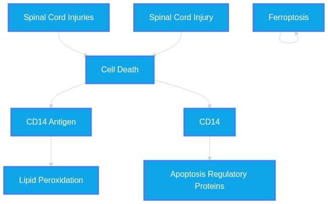
"Integrated multi-model analysis identified CD14 as the most robust candidate hub gene associated with the lytic cell death index."
VERIFIED VERBATIM (PMID: 42519304)
"Enrichment analyses showed that lytic cell death-associated genes were mainly involved in inflammatory responses, immune regulation, myeloactivation, and related signaling pathways."
VERIFIED VERBATIM (PMID: 42519304)
"However, the current findings are primarily associative, and further functional studies are required to determine whether CD14 directly modulates lytic cell death-related pathways and contributes to secondary injury progression after SCI."
VERIFIED VERBATIM (PMID: 42341849)
"Rather than being viewed only as an endpoint of cell death, ferroptosis in microglia is increasingly recognized as a process that links iron dyshomeostasis, lipperoxidation, oxidative stress, and immune-inflammatory activation, thereby contributing to the sustained amplification of neuroinflammation."
VERIFIED VERBATIM (PMID: 42341847)
"These findings provide the first evidence that ACSL4-mediated ferroptosis is a key driver of acute spinal motor neuron injury following CA."
VERIFIED VERBATIM (PMID: 42317798)
"We demonstrated that SCD1 deficiency is the driving force behind aberrant ERMCS expansion, leading to increased lipperoxidation and neuronal ferroptosis."
VERIFIED VERBATIM (PMID: 42292377)
"Ninjurin1 (NINJ1) is a cell-surface molecule that has gained considerable attention for its role in mediating plasma membrane rupture (PMR). Originally identified as an adhesion molecule induced after nerve injury, NINJ1 is now recognized as a common terminal executor of PMR across multiple forms of lytic cell death, including pyroptosis, necroptosis, and ferroptosis."
VERIFIED VERBATIM (PMID: 42289170)
"PANoptosis is a newly identified cell death mode that synergistically initiates pyroptosis, apoptosis and necroptosis via activation of PANoptosome. It is closely associated with oxidative stress, neuroinflammation, and secondary injury following TBI and SCI"
VERIFIED VERBATIM (PMID: 42337999)
"Administering anti-HMGB1 antibody to D-SCI rats could significantly augment their activity distance, movement speed and sucrose preference rate, while also suppressing the ferroptosis level and the expression of ferroptosis-related proteins in the hippocampus."
VERIFIED VERBATIM (PMID: 42448629)
"Contusive SCI is characterised by neuronal loss due to S1P2 receptor activation in spinal neurones. S1P2 receptor activation increases lipperoxidation, which induces neuronal ferroptosis."
VERIFIED VERBATIM (PMID: 42464547)
"SCI activates the Piezo1 channel in microglia, triggering mitochondrial dysfunction and mediating cellular ferroptosis, thereby aggravating secondary neuroinflammation."
VERIFIED VERBATIM (PMID: 42486345)
"Collectively, these findings suggest that GADD45A promotes neuronal ferroptosis and apoptosis after SCI by regulating the NF-κB pathway, and that GADD45A may be a potential therapeutic target for SCI."
VERIFIED VERBATIM (PMID: 42327731)
"Because BMECs form the vascular interface between the circulation and the brain parenchyma, ferroptotic injury in this cell population may represent an immunovascular mechanism through which endothelial redox stress is translated into barrier dysfunction and neuroinflammatory amplification."
VERIFIED VERBATIM (PMID: 42313207)
"Recent studies indicate that STAT3 exhibits pronounced spatiotemporal and cell-type-specific activation after SCI. Depending on the upstream trigger and cellular compartment involved, STAT3 can amplify or restrain neuroinflammation, shape astrocyte and microglial reactivity, influence mitochondrial bioenergetics and oxidative stress, modulate ferroptosis and apoptosis"
VERIFIED VERBATIM (PMID: 42313317)
"The results indicate that the mechanisms by which TMP exerts its effects in SCI treatment include promoting nerve regeneration, improving vascular dysfunction, exerting anti-inflammatory effects, inhibiting neuronal apoptosis, reducing oxidative stress, regulating iron metabolism, maintaining ion homeostasis, alleviating pyroptosis, and modulating autophagy."
VERIFIED VERBATIM (PMID: 42526057)
"Iron depletion mitigated the antioxidant response, lipperoxidation, cell proliferation, ECM production."
VERIFIED VERBATIM (PMID: 42517904)
"The present study aimed to develop a bioactive therapeutic platform based on an adipose-derived extracellular matrix (ECM) hydrogel incorporating cytokine-releasing PLGA microspheres to promote spinal cord repair."
VERIFIED VERBATIM (PMID: 42519304)
"Pyroptosis-, necroptosis-, and ferroptosis-related transcriptional activities were all increased after SCI, with activation beginning in the acute phase and persisting over time."
VERIFIED VERBATIM (PMID: 42519304)
"Integrated multi-model analysis identified CD14 as the most robust candidate hub gene associated with the lytic cell death index."
VERIFIED VERBATIM (PMID: 42519304)
"Enrichment analyses showed that lytic cell death-associated genes were mainly involved in inflammatory responses, immune regulation, myeloactivation, and related signaling pathways."
VERIFIED VERBATIM (PMID: 42519304)
"However, the current findings are primarily associative, and further functional studies are required to determine whether CD14 directly modulates lytic cell death-related pathways and contributes to secondary injury progression after SCI."
VERIFIED VERBATIM (PMID: 42341849)
"Rather than being viewed only as an endpoint of cell death, ferroptosis in microglia is increasingly recognized as a process that links iron dyshomeostasis, lipperoxidation, oxidative stress, and immune-inflammatory activation, thereby contributing to the sustained amplification of neuroinflammation."
VERIFIED VERBATIM (PMID: 42341847)
"These findings provide the first evidence that ACSL4-mediated ferroptosis is a key driver of acute spinal motor neuron injury following CA."
VERIFIED VERBATIM (PMID: 42317798)
"We demonstrated that SCD1 deficiency is the driving force behind aberrant ERMCS expansion, leading to increased lipperoxidation and neuronal ferroptosis."
VERIFIED VERBATIM (PMID: 42292377)
"Ninjurin1 (NINJ1) is a cell-surface molecule that has gained considerable attention for its role in mediating plasma membrane rupture (PMR). Originally identified as an adhesion molecule induced after nerve injury, NINJ1 is now recognized as a common terminal executor of PMR across multiple forms of lytic cell death, including pyroptosis, necroptosis, and ferroptosis."
VERIFIED VERBATIM (PMID: 42289170)
"PANoptosis is a newly identified cell death mode that synergistically initiates pyroptosis, apoptosis and necroptosis via activation of PANoptosome. It is closely associated with oxidative stress, neuroinflammation, and secondary injury following TBI and SCI"
VERIFIED VERBATIM (PMID: 42337999)
"Administering anti-HMGB1 antibody to D-SCI rats could significantly augment their activity distance, movement speed and sucrose preference rate, while also suppressing the ferroptosis level and the expression of ferroptosis-related proteins in the hippocampus."
VERIFIED VERBATIM (PMID: 42448629)
"Contusive SCI is characterised by neuronal loss due to S1P2 receptor activation in spinal neurones. S1P2 receptor activation increases lipperoxidation, which induces neuronal ferroptosis."
VERIFIED VERBATIM (PMID: 42464547)
"SCI activates the Piezo1 channel in microglia, triggering mitochondrial dysfunction and mediating cellular ferroptosis, thereby aggravating secondary neuroinflammation."
VERIFIED VERBATIM (PMID: 42486345)
"Collectively, these findings suggest that GADD45A promotes neuronal ferroptosis and apoptosis after SCI by regulating the NF-κB pathway, and that GADD45A may be a potential therapeutic target for SCI."
VERIFIED VERBATIM (PMID: 42327731)
"Because BMECs form the vascular interface between the circulation and the brain parenchyma, ferroptotic injury in this cell population may represent an immunovascular mechanism through which endothelial redox stress is translated into barrier dysfunction and neuroinflammatory amplification."
VERIFIED VERBATIM (PMID: 42313207)
"Recent studies indicate that STAT3 exhibits pronounced spatiotemporal and cell-type-specific activation after SCI. Depending on the upstream trigger and cellular compartment involved, STAT3 can amplify or restrain neuroinflammation, shape astrocyte and microglial reactivity, influence mitochondrial bioenergetics and oxidative stress, modulate ferroptosis and apoptosis"
VERIFIED VERBATIM (PMID: 42313317)
"The results indicate that the mechanisms by which TMP exerts its effects in SCI treatment include promoting nerve regeneration, improving vascular dysfunction, exerting anti-inflammatory effects, inhibiting neuronal apoptosis, reducing oxidative stress, regulating iron metabolism, maintaining ion homeostasis, alleviating pyroptosis, and modulating autophagy."
VERIFIED VERBATIM (PMID: 42526057)
"Iron depletion mitigated the antioxidant response, lipperoxidation, cell proliferation, ECM production."
VERIFIED VERBATIM (PMID: 42517904)
"The present study aimed to develop a bioactive therapeutic platform based on an adipose-derived extracellular matrix (ECM) hydrogel incorporating cytokine-releasing PLGA microspheres to promote spinal cord repair."
VERIFIED VERBATIM (PMID: 42499235)
"Single-cell RNA sequencing (scRNA-seq) and spatial transcriptomics (ST) analysis revealed that Tlr4 was upregulated in myelocells (microglia and macrophages) and played a pivotal role in triggering downstream necroptosis, which was confirmed by protein levels."
VERIFIED VERBATIM (PMID: 42517042)
"Lp(a) activates p38 by increasing intracellular ROS levels and promotes ferroptosis in cardiomyocytes via SLC7A11 inhibition, which depends on p53 activation."
VERIFIED VERBATIM (PMID: 42519304)
"Lytic cell death-related programs are dynamically and persistently activated after SCI and are closely associated with immune-inflammatory responses during secondary injury."
VERIFIED VERBATIM (PMID: 42498720)
"PLIN2 interacts with the mitochondrial outer membrane protein PGAM5 to regulate lipdroplet-mitochondria contacts."
VERIFIED VERBATIM (PMID: 42456380)
"PANoptosis, with multi-target characteristics, provides a new idea to overcome the bottleneck of single-target strategy for CaOx-induced kidney injury."
VERIFIED VERBATIM (PMID: 42453609)
"This concept may be especially useful in neurological diseases. It helps explain why neuronal death, sustained inflammatory activation, and tissue injury often develop together and reinforce one another."
VERIFIED VERBATIM (PMID: 42403480)
"Upon secondary acute trauma, the sudden influx of heme and labile iron ignites an uncontrollable "ferroptotic storm," synergizing with neuroinflammation to drive irreversible neural loss."
VERIFIED VERBATIM (PMID: 42378634)
"under chemotherapeutic stress, the Lysine acetyltransferase 8 (KAT8) catalyzes the specific lactylation of the inner mitochondrial membrane ADP/ATP translocator 2 (ANT2) at K92."
VERIFIED VERBATIM (PMID: 42517186)
"a growing body of evidence suggests that mitochondrial dysfunction is a central rheostat that enables necroptotic execution."
VERIFIED VERBATIM (PMID: 42519304)
"Lytic cell death-related programs are dynamically and persistently activated after SCI and are closely associated with immune-inflammatory responses during secondary injury."
VERIFIED VERBATIM (PMID: 42519304)
"CD14 was identified as a candidate hub gene associated with myeloinflammatory activation and lytic cell death-related signatures."
VERIFIED VERBATIM (PMID: 42498720)
"PLIN2 interacts with the mitochondrial outer membrane protein PGAM5 to regulate lipdroplet-mitochondria contacts."
VERIFIED VERBATIM (PMID: 42498720)
"Genetic deletion of Plin2 markedly reduced lipdroplet burden, attenuated neuroinflammation, and promoted neuronal survival and functional recovery in vivo."
VERIFIED VERBATIM (PMID: 42403480)
"Upon secondary acute trauma, the sudden influx of heme and labile iron ignites an uncontrollable "ferroptotic storm," synergizing with neuroinflammation to drive irreversible neural loss."
VERIFIED VERBATIM (PMID: 42517186)
"a growing body of evidence suggests that mitochondrial dysfunction is a central rheostat that enables necroptotic execution."
VERIFIED VERBATIM (PMID: 42456380)
"PANoptosis, with multi-target characteristics, provides a new idea to overcome the bottleneck of single-target strategy for CaOx-induced kidney injury."
VERIFIED VERBATIM (PMID: 42453609)
"This concept may be especially useful in neurological diseases. It helps explain why neuronal death, sustained inflammatory activation, and tissue injury often develop together and reinforce one another."
VERIFIED VERBATIM (PMID: 42378634)
"under chemotherapeutic stress, the Lysine acetyltransferase 8 (KAT8) catalyzes the specific lactylation of the inner mitochondrial membrane ADP/ATP translocator 2 (ANT2) at K92."
VERIFIED VERBATIM (PMID: 42468674)
"Allicin can reduce myocardial PANoptosis and ameliorate MI/R injury by inhibiting TLR4 activation."
VERIFIED VERBATIM (PMID: 42501927)
"We summarize how apoptosis, necroptosis, pyroptosis, and ferroptosis operate as an interlocking network in autoimmune microenvironments, frequently co-existing as context-dependent mixed-death states driven by shared triggers such as cytokines, PRR ligands, and oxidative stress, and governed by decision hubs including RIPK1 and caspase-8."
VERIFIED VERBATIM (PMID: 42506907)
"Here, we found that intracellular levels of the metabolic cofactor nicotinamide adenine dinucleotide (NAD+) were depleted in response to cell death triggers that drive pyroptosis, necroptosis, PANoptosis, and ferroptosis."
VERIFIED VERBATIM (PMID: 42317798)
"SCD1 deficiency is the driving force behind aberrant ERMCS expansion, leading to increased lipperoxidation and neuronal ferroptosis."
VERIFIED VERBATIM (PMID: 42484540)
"STED-microscopy quantification showed that WBV increased the linear density of VGAT + and VGLUT1 + perisomatic terminals, as well as the number of SER + fibers."
VERIFIED VERBATIM (PMID: 42476817)
"Our data underscore the importance of isoform-paralog interplay in studying regional vulnerability in neurodegenerative diseases."
VERIFIED VERBATIM (PMID: 42426407)
"Mechanistically, MANF attenuated mitochondrial dysfunction and ER stress of NP-MSCs under acidic conditions by maintaining MAM integrity, as demonstrated by the complete abolition of this protection upon treatment with the MAM uncoupler FATE1."
VERIFIED VERBATIM (PMID: 42388246)
"BoNT/A exerts neuroprotective effects in chronic SCI by reducing neuroinflammation and supporting neuronal and oligodendroglial preservation."
VERIFIED VERBATIM (PMID: 42505382)
"Dysregulated LD remodeling mediates metabolic flexibility, immune escape and drug resistance in obesity, metabolic dysfunction-associated steatotic liver disease (MASLD), neurodegeneration, viral infection and cancer."
VERIFIED VERBATIM (PMID: 42490372)
"These studies reveal that 65% of structures preferred conformers with a conserved adenine-nicotinamide separation while allowing limited flexibility in the pyrophosphate."
VERIFIED VERBATIM (PMID: 42519304)
"Lytic cell death-related programs are dynamically and persistently activated after SCI and are closely associated with immune-inflammatory responses during secondary injury."
VERIFIED VERBATIM (PMID: 42519304)
"CD14 was identified as a candidate hub gene associated with myeloinflammatory activation and lytic cell death-related signatures."
VERIFIED VERBATIM (PMID: 42498720)
"PLIN2 interacts with the mitochondrial outer membrane protein PGAM5 to regulate lipdroplet-mitochondria contacts."
VERIFIED VERBATIM (PMID: 42498720)
"Genetic deletion of Plin2 markedly reduced lipdroplet burden, attenuated neuroinflammation, and promoted neuronal survival and functional recovery in vivo."
VERIFIED VERBATIM (PMID: 42403480)
"Upon secondary acute trauma, the sudden influx of heme and labile iron ignites an uncontrollable "ferroptotic storm," synergizing with neuroinflammation to drive irreversible neural loss."
VERIFIED VERBATIM (PMID: 42517186)
"a growing body of evidence suggests that mitochondrial dysfunction is a central rheostat that enables necroptotic execution."
VERIFIED VERBATIM (PMID: 42456380)
"PANoptosis, with multi-target characteristics, provides a new idea to overcome the bottleneck of single-target strategy for CaOx-induced kidney injury."
VERIFIED VERBATIM (PMID: 42453609)
"This concept may be especially useful in neurological diseases. It helps explain why neuronal death, sustained inflammatory activation, and tissue injury often develop together and reinforce one another."
VERIFIED VERBATIM (PMID: 42378634)
"under chemotherapeutic stress, the Lysine acetyltransferase 8 (KAT8) catalyzes the specific lactylation of the inner mitochondrial membrane ADP/ATP translocator 2 (ANT2) at K92."
VERIFIED VERBATIM (PMID: 42468674)
"Allicin can reduce myocardial PANoptosis and ameliorate MI/R injury by inhibiting TLR4 activation."
VERIFIED VERBATIM (PMID: 42501927)
"We summarize how apoptosis, necroptosis, pyroptosis, and ferroptosis operate as an interlocking network in autoimmune microenvironments, frequently co-existing as context-dependent mixed-death states driven by shared triggers such as cytokines, PRR ligands, and oxidative stress, and governed by decision hubs including RIPK1 and caspase-8."
VERIFIED VERBATIM (PMID: 42506907)
"Here, we found that intracellular levels of the metabolic cofactor nicotinamide adenine dinucleotide (NAD+) were depleted in response to cell death triggers that drive pyroptosis, necroptosis, PANoptosis, and ferroptosis."
VERIFIED VERBATIM (PMID: 42317798)
"SCD1 deficiency is the driving force behind aberrant ERMCS expansion, leading to increased lipperoxidation and neuronal ferroptosis."
VERIFIED VERBATIM (PMID: 42484540)
"STED-microscopy quantification showed that WBV increased the linear density of VGAT + and VGLUT1 + perisomatic terminals, as well as the number of SER + fibers."
VERIFIED VERBATIM (PMID: 42476817)
"Our data underscore the importance of isoform-paralog interplay in studying regional vulnerability in neurodegenerative diseases."
VERIFIED VERBATIM (PMID: 42426407)
"Mechanistically, MANF attenuated mitochondrial dysfunction and ER stress of NP-MSCs under acidic conditions by maintaining MAM integrity, as demonstrated by the complete abolition of this protection upon treatment with the MAM uncoupler FATE1."
VERIFIED VERBATIM (PMID: 42388246)
"BoNT/A exerts neuroprotective effects in chronic SCI by reducing neuroinflammation and supporting neuronal and oligodendroglial preservation."
VERIFIED VERBATIM (PMID: 42505382)
"Dysregulated LD remodeling mediates metabolic flexibility, immune escape and drug resistance in obesity, metabolic dysfunction-associated steatotic liver disease (MASLD), neurodegeneration, viral infection and cancer."
VERIFIED VERBATIM (PMID: 42490372)
"These studies reveal that 65% of structures preferred conformers with a conserved adenine-nicotinamide separation while allowing limited flexibility in the pyrophosphate."
VERIFIED VERBATIM (PMID: 42468674)
"Allicin can reduce myocardial PANoptosis and ameliorate MI/R injury by inhibiting TLR4 activation."
Chapter 8
Abstract Repository
Raw text abstracts programmatically cached during the evaluation phase. Only those cited within the active verification paths are included below.
PMID: 42289170
Mapped to Reference [23]
ID: 42289170
Title: Mitochondrial homeostasis imbalance-triggered PANoptosis in traumatic brain and spinal cord injury: from mechanism to therapeutic strategies.
Abstract: Traumatic injury to the central nervous system (CNS), also known as traumatic brain injury (TBI) and spinal cord injury (SCI), is characterized by high disability and mortality worldwide. PANoptosis is a newly identified cell death mode that synergistically initiates pyroptosis, apoptosis and necroptosis via activation of PANoptosome. It is closely associated with oxidative stress, neuroinflammation, and secondary injury following TBI and SCI, yet the key pathogenic factors and mechanisms underlying PANoptosis remain incompletely elucidated. Mitochondria, as a central organelle for energy synthesis and oxidative stress, its health and homeostasis are the cornerstone of cell survival and biological function. Emerging evidence suggests that the loss of mitochondrial homeostasis plays a fundamental role in the activation and execution of PANoptosis across various cell types. Here, we review the detailed manifestations of mitochondrial homeostasis imbalance in TBI and SCI, such as impaired biogenesis, abnormal dynamics, mitophagy dysfunction, and mitochondria-derived vesicles. Meanwhile, we systematically analyze the characteristics and pathological effects of PANoptosis cascade following TBI and SCI, with a focus on the regulatory patterns, mechanisms, and potential targets of injured mitochondria driving PANoptosis. In addition, we discuss the advancements and future perspectives of mitochondria-based strategies for modulating PANoptosis in TBI and SCI. Taken together, despite considerable challenges in governing post-traumatic mitochondria homeostasis, its multiple targeting of the upstream PANoptosome and downstream cell death signaling offers a promising approach to improve the outcome of CNS trauma.
PMID: 42292377
Mapped to Reference [22]
ID: 42292377
Title: Digging deeper into NINJ1: its multifaceted role in central nervous system diseases.
Abstract: Ninjurin1 (NINJ1) is a cell-surface molecule that has gained considerable attention for its role in mediating plasma membrane rupture (PMR). Originally identified as an adhesion molecule induced after nerve injury, NINJ1 is now recognized as a common terminal executor of PMR across multiple forms of lytic cell death, including pyroptosis, necroptosis, and ferroptosis. This function positions NINJ1 as a key link between cell death and inflammatory activation. However, the precise role of NINJ1 in the central nervous system (CNS) remains unclear. This review systematically outlines the molecular structure, expression, activation, and regulation of NINJ1, with a focus on its multifaceted roles in CNS disorders, including multiple sclerosis, ischemic stroke, traumatic brain injury, spinal cord injury, neuropsychiatric disorders and neurodegenerative diseases. We also highlight critical knowledge gaps, particularly regarding cell type-specific functions in the CNS. Finally, we evaluate therapeutic strategies targeting NINJ1 (including monoclonal antibodies, functional peptides, and small-molecule inhibitors) and their potential applications in neurological diseases. By integrating current evidence and identifying unresolved questions, this review aims to provide a foundation for future mechanistic and translational studies of NINJ1 in the CNS.
PMID: 42313207
Mapped to Reference [29]
ID: 42313207
Title: STAT3 Signaling in Spinal Cord Injury: Neurochemical Mechanisms Linking Neuroinflammation, Mitochondrial Stress, and Glial Remodeling.
Abstract: Spinal cord injury (SCI) is a devastating neurological disorder marked by profound disturbances in cytokine signaling, redox balance, mitochondrial homeostasis, and glial-neuronal communication. Although many therapeutic strategies have been explored to attenuate secondary injury, effective molecularly targeted interventions remain limited. Increasing evidence identifies signal transducer and activator of transcription 3 (STAT3) as a central signaling node in the neurochemical response to SCI. Recent studies indicate that STAT3 exhibits pronounced spatiotemporal and cell-type-specific activation after SCI. Depending on the upstream trigger and cellular compartment involved, STAT3 can amplify or restrain neuroinflammation, shape astrocyte and microglial reactivity, influence mitochondrial bioenergetics and oxidative stress, modulate ferroptosis and apoptosis, and alter the regenerative state of the injured spinal cord. In this review, we frame STAT3 not simply as a downstream effector of the JAK/STAT cascade, but as an integrative regulator of SCI neurochemistry that links cytokine-driven signaling to metabolic stress, glial remodeling, and axonal repair. We emphasize how injury phase, cell type, and subcellular localization influence STAT3-dependent outcomes, discuss emerging therapeutic strategies that converge on STAT3-centered pathways, and outline the key challenges that must be addressed for precise translational targeting.
PMID: 42313317
Mapped to Reference [30]
ID: 42313317
Title: Mechanisms ofra Tetmethylpyrazine in spinal cord injury: a narrative review.
Abstract: Spinal cord injury (SCI) is characterized by irreversible loss of motor and sensory function, imposing a substantial burden on patients and their families. Tetramethylpyrazine (TMP), a bioactive compound derived from traditional Chinese medicine, possesses a wide range of pharmacological activities and has demonstrated potential therapeutic effects in the treatment of SCI. Therefore, this article provides a comprehensive review of the mechanisms by which TMP promotes spinal cord repair. This review compiles a large body of in vitro, in vivo, and clinical studies, including a total of 86 publications documenting the effects of TMP on SCI. The results indicate that the mechanisms by which TMP exerts its effects in SCI treatment include promoting nerve regeneration, improving vascular dysfunction, exerting anti-inflammatory effects, inhibiting neuronal apoptosis, reducing oxidative stress, regulating iron metabolism, maintaining ion homeostasis, alleviating pyroptosis, and modulating autophagy. Through these mechanisms, TMP contributes to the restoration of spinal cord morphology, motor function, and electrophysiological parameters in experimental animal models. Clinical reports on the use of TMP injection for SCI are relatively limited, and its clinical efficacy requires further investigation. The combined application of nanotechnology or hydrogels provides an efficient targeted delivery and sustained-release system for TMP in the spinal cord, thereby significantly enhancing its bioavailability. Overall, TMP shows promising potential in SCI treatment and may serve as a valuable adjunctive therapeutic strategy.
PMID: 42317798
Mapped to Reference [21]
ID: 42317798
Title: LXRα/SCD1-Mediated Endoplasmic Reticulum-Mitochondria Crosstalk in Inhibiting Neuronal Ferroptosis after Spinal Cord Injury.
Abstract: Spinal cord injury (SCI) causes extensive neuronal loss, in which ferroptosis is critically involved. Although lipid transport at endoplasmic reticulum-mitochondria contact sites (ERMCSs) has been implicated in facilitating ferroptosis, the neuron-specific regulatory mechanisms remain elusive. Here, we show that neuronal ferroptosis is characterized by excessive ERMCS formation. Mechanistically, a systematic screening revealed the down-regulation of stearoyl-CoA desaturase 1 (SCD1), a critical enzyme in the synthesis of monounsaturated fatty acids (MUFAs) in neurons, following SCI (in vivo) or erastin treatment (in vitro). We demonstrated that SCD1 deficiency is the driving force behind aberrant ERMCS expansion, leading to increased lipid peroxidation and neuronal ferroptosis. Conversely, SCD1 overexpression could reverse these effects. Furthermore, we identified liver X receptor alpha (LXRα) as a direct transcriptional activator of SCD1. Pharmacological activation of LXRα with T0901317 upregulated SCD1 expression, which in turn restrained ERMCS formation, elevated MUFA levels, and ultimately inhibited neuronal ferroptosis. In murine SCI models, both adeno-associated virus-mediated neuronal SCD1 overexpression and LXRα agonist treatment effectively mitigated excessive ERMCS, reduced lesion size, preserved neuronal architecture, and improved functional recovery. Collectively, our study establishes the LXRα-SCD1 axis as a novel and druggable pathway for reducing neuronal loss and improving functional recovery by modulating ERMCS-dependent lipid exchange dynamics, revealing promising therapeutic targets after central nervous system trauma.
PMID: 42327731
Mapped to Reference [28]
ID: 42327731
Title: Endothelial ferroptosis in blood-brain barrier dysfunction and neuroinflammation: mechanisms and immune-vascular crosstalk.
Abstract: Ferroptosis is an iron-dependent form of regulated cell death driven by phospholipid peroxidation. In the central nervous system (CNS), most ferroptosis research has focused on neurons and glial cells, whereas the vulnerability of brain microvascular endothelial cells (BMECs) and its consequences for blood-brain barrier (BBB) integrity remain less clearly defined. Because BMECs form the vascular interface between the circulation and the brain parenchyma, ferroptotic injury in this cell population may represent an immunovascular mechanism through which endothelial redox stress is translated into barrier dysfunction and neuroinflammatory amplification. In this review, we summarize molecular pathways that may promote or restrain BMEC ferroptosis, including iron handling, antioxidant defense mediated by the solute carrier family 7 member 11 (SLC7A11)-glutathione peroxidase 4 (GPX4) axis and nuclear factor erythroid 2-related factor 2 (Nrf2) signaling, lipid peroxidation, and junctional remodeling. We then discuss how ferroptosis-associated endothelial injury may contribute to BBB leakage, damage-associated molecular pattern release, innate immune sensing, leukocyte recruitment, glial activation, and self-amplifying inflammatory feedback at the neurovascular interface. We organize the available literature according to the strength and cellular specificity of evidence, separating BMEC-specific findings, BBB-focused in vivo studies, indirect CNS evidence, and mechanistic analogies from non-CNS endothelial systems. Finally, we evaluate disease-specific evidence in ischemic stroke and selected neurodegenerative or inflammatory conditions, together with therapeutic strategies, BMEC-targeting considerations, candidate clinical biomarkers, and translational barriers for modulating endothelial ferroptosis. This review frames endothelial ferroptosis as a promising but incompletely established immunovascular link between BBB dysfunction and neuroinflammation, and highlights the need for BMEC-specific models, human BBB systems, endothelial ferroptosis biomarkers, biomarker-guided monitoring, BMEC-targeted delivery approaches, and careful evaluation of the physiological risks of systemic or prolonged ferroptosis blockade.
PMID: 42337999
Mapped to Reference [24]
ID: 42337999
Title: Anti-HMGB1 Antibody Therapy Ameliorates Depression Following Spinal Cord Injury in Rats by Inhibiting Ferroptosis.
Abstract: Depression following spinal cord injury (D-SCI) refers to a depressive state that occurs in an individual after a major spinal cord injury (SCI), characterized mainly by low mood and reduced interest. This study aims to investigate the regulatory role of anti-HMGB1 antibody in the depressive-like behaviour of D-SCI rats and to explore its underlying mechanisms. A depression model was established in rats 5 weeks after SCI. The expression of HMGB1 and ferroptosis markers (MDA, GSH and iron ion deposition) in the hippocampus were examined in both the sham group and the D-SCI group. Subsequently, D-SCI rats were treated with an anti-HMGB1 antibody, and the depression-like behaviours of each group were assessed using open field and sucrose preference tests. Ferroptosis levels in the hippocampus, as well as the expression of ferroptosis-related proteins (ACSL4, SLC7A11 and GPX4), were also investigated. The co-localization of HMGB1 and NeuN in the rat hippocampus was detected by immunofluorescence double staining. Furthermore, at the cellular level, the effect of the anti-HMGB1 antibody on Erastin-induced ferroptosis in rat hippocampal neurons was analysed. The results indicated that compared to the sham group, the levels of HMGB1 and ferroptosis in the hippocampus of rats in the D-SCI group were significantly elevated. Administering anti-HMGB1 antibody to D-SCI rats could significantly augment their activity distance, movement speed and sucrose preference rate, while also suppressing the ferroptosis level and the expression of ferroptosis-related proteins in the hippocampus. Moreover, HMGB1 and NeuN were co-expressed in the rat hippocampus. The results from primary rat hippocampal neurons indicated that anti-HMGB1 antibody could inhibit erastin-induced ferroptosis in rat hippocampal neurons. Taken together, anti-HMGB1 antibody therapy can ameliorate depressive behaviour in D-SCI rats; the possible mechanism may involve the inhibition of ferroptosis in hippocampal neurons.
PMID: 42341847
Mapped to Reference [20]
ID: 42341847
Title: Edaravone attenuates ACSL4-dependent ferroptosis in spinal motor neurons following cardiac arrest in rats.
Abstract: The contribution of acute spinal motor neuron injury following cardiac arrest (CA) remains poorly understood. This study aimed to investigate the role of ferroptosis in CA-induced spinal cord injury and to evaluate the neuroprotective effects of edaravone. Asphyxial CA was induced in rats for 5 min, followed by resuscitation. Edaravone was administered immediately after the return of spontaneous circulation (ROSC). At 24 h post-ROSC, The CA group exhibited significant hindlimb motor deficits and reduced survival rates. Histological analysis revealed selective injury of choline acetyltransferase (ChAT)-positive motor neurons in the lumbar spinal cord, accompanied by mitochondrial shrinkage and membrane rupture, which are characteristic of ferroptosis. Immunofluorescence demonstrated a selective upregulation of the pro-ferroptotic enzyme acyl-CoA synthetase long-chain family member 4 (ACSL4) specifically in ChAT-positive motor neurons, whereas glutathione peroxidase 4 (GPX4) expression remained relatively preserved. Edaravone treatment significantly improved neurological outcomes and survival, attenuated lipid peroxidation (evidenced by decreased malondialdehyde and preserved glutathione levels), and effectively suppressed ACSL4 upregulation in the motor neurons. Furthermore, edaravone mitigated neuroinflammation by reducing astrogliosis and microglial activation. These findings provide the first evidence that ACSL4-mediated ferroptosis is a key driver of acute spinal motor neuron injury following CA. Edaravone exerts potent neuroprotection by targeting this pathway, suggesting its therapeutic potential for ameliorating spinal cord injury in patients with CA.
PMID: 42341849
Mapped to Reference [19]
ID: 42341849
Title: Microglial ferroptosis mediated neuroinflammation in central nervous system diseases.
Abstract: Microglial ferroptosis has become an important pathological mechanism in studies of central nervous system (CNS) diseases. Rather than being viewed only as an endpoint of cell death, ferroptosis in microglia is increasingly recognized as a process that links iron dyshomeostasis, lipid peroxidation, oxidative stress, and immune-inflammatory activation, thereby contributing to the sustained amplification of neuroinflammation. In this review, we summarize the molecular mechanisms by which microglial ferroptosis mediates neuroinflammatory responses, with a focus on iron homeostasis disruption, lipid peroxidation and ROS amplification, collapse of the GPX4-dependent antioxidant defense, mitochondrial ROS generation, and inflammasome activation. We further classify related CNS diseases into three categories according to disease course and pathological features: chronic neurodegenerative and demyelinating diseases, acute CNS injuries, and neuropsychiatric or systemic inflammation-related brain dysfunction. Within this framework, we compare the pathological significance of microglial ferroptosis across different disease contexts. We also discuss potential therapeutic strategies targeting iron homeostasis, lipid peroxidation, antioxidant defenses, inflammatory amplification networks, and microglia-specific delivery systems. Finally, we address current challenges in the field, including insufficient cell-type specificity, inconsistent detection criteria, disease-stage heterogeneity, and barriers to clinical translation. This review provides an integrated perspective on the mechanisms by which microglial ferroptosis drives neuroinflammation and highlights its potential relevance for precision intervention in CNS diseases.
PMID: 42378634
Mapped to Reference [39]
ID: 42378634
Title: Lactate-Driven Restriction of Mitochondrial Permeability Transition Promotes Resistance to Chemo-Immunotherapy by Suppressing Tumor PANoptosis.
Abstract: Intrinsic resistance limits chemo-immunotherapy efficacy in triple-negative breast cancer (TNBC). While metabolic reprogramming is linked to immune evasion, the precise mechanistic orchestration remains unclear. Here, utilizing single-cell transcriptomics and quantitative lactylome profiling, we show that elevated tumor lactate drives resistance by broadly suppressing PANoptosis. Mechanistically, under chemotherapeutic stress, the Lysine acetyltransferase 8 (KAT8) catalyzes the specific lactylation of the inner mitochondrial membrane ADP/ATP translocator 2 (ANT2) at K92. Lactylated ANT2 recruits the phosphoglycerate mutase 5 (PGAM5) to dephosphorylate Cyclophilin D (CypD). This cascade restricts mitochondrial permeability transition pore (mPTP) opening, preserving mitochondrial homeostasis and averting immunogenic cell death. Crucially, a cell-penetrating competitive peptide targeting the KAT8-ANT2 interface effectively uncouples this metabolic lock, re-sensitizing TNBC tumors to cytotoxic stress and restoring chemo-immunotherapy efficacy in vivo. Our findings unveil a profound mechanistic link between the Warburg effect and mitochondrial homeostasis, establishing KAT8-mediated ANT2 lactylation as a targetable vulnerability to improve chemo-immunotherapy efficacy.
PMID: 42388246
Mapped to Reference [46]
ID: 42388246
Title: A translational preclinical strategy for chronic spinal cord injury: neuroprotective and regenerative potential of botulinum neurotoxin type A combined with muscle atrophy prevention via electrostimulation.
Abstract: Spinal cord injury (SCI) triggers persistent neuroinflammation, gliosis, neuronal loss, and demyelination, leading to motor deficits and neuropathic pain (NeP). Botulinum neurotoxin type A (BoNT/A) has shown anti-inflammatory and neuroprotective effects in acute SCI, but its potential in the chronic phase remains unclear. This study investigates whether combining BoNT/A with electrical muscle stimulation (EMS) enhances recovery in chronic SCI. Adult mice with severe thoracic SCI (paraplegic) underwent EMS (30 min/d for 10 non-consecutive days starting 3 d post-injury) or no stimulation. Fifteen days after SCI, animals received a single intrathecal injection of BoNT/A (15 pg/5 μl) or saline. Functional recovery was assessed up to 60 d as well as in moderate and mild SCI mice. NeP onset and maintenance were evaluated. Spinal cord tissue was analysed for astrocytic and microglial morphology, neuronal and oligodendroglial survival, myelin protein expression, and in vitro effects on oligodendrocyte precursor cells (OPCs). The phenotype of hindlimb muscles was evaluated through morphological and gene expression analyses. EMS was able to counteract muscle atrophy and fibrosis, and when combined with BoNT/A, also denervation. Moreover, the combination restored hindlimb motor function in chronic SCI, whereas BoNT/A or EMS alone were ineffective. NeP, a common comorbidity associated with SCI, was mitigated by BoNT/A treatment even when administered in the chronic phase. BoNT/A reduced astrocytic hypertrophy and excitatory synapse association and was associated with a morphology-based redistribution of microglial profiles toward a resting-like classification, decreased apoptosis, and increased neuronal and oligodendroglial survival. Myelin basic protein (MBP) expression was significantly elevated in vivo. In vitro, BoNT/A promoted OPC differentiation into myelinating oligodendrocytes, increased process complexity, and upregulated MBP, galactocerebroside C, proteolipid protein, and myelin oligodendrocyte glycoprotein under both proliferative and differentiating conditions. Cleaved synaptosomal-associated protein 25 colocalization with OPC confirmed direct BoNT/A internalization and activity. BoNT/A exerts neuroprotective effects in chronic SCI by reducing neuroinflammation and supporting neuronal and oligodendroglial preservation. When combined with EMS, it also promotes remyelination and improves muscle homeostasis, suggesting that early stimulation creates a permissive environment for recovery. These findings support the clinical evaluation of BoNT/A as a therapeutic strategy for chronic SCI.
PMID: 42403480
Mapped to Reference [35]
ID: 42403480
Title: The ferroptosis-mediated domino effect: metabolic crosstalk from intervertebral disc degeneration to spinal deformity and cord injury: a mini review.
Abstract: Spinal degeneration, spinal deformity, and spinal cord injury (SCI) are classically managed as discrete biomechanical or neurological entities. However, emerging evidence reveals them as an interconnected pathological continuum. This mini-review introduces the "ferroptosis-mediated domino effect" as the core metabolic driver linking these conditions. The cascade initiates within the avascular intervertebral disc, where aberrant mechanotransduction (e.g., via Piezo1) provokes severe oxidative stress and subsequent ferroptosis, leading to extracellular matrix degradation and structural collapse. The ensuing spinal deformity chronically compresses the spinal microvasculature, disrupting the blood-spinal cord barrier (BSCB) and facilitating localized iron deposition. This chronic ischemic insult generates a metabolically "primed" spinal cord characterized by extreme vulnerability. Upon secondary acute trauma, the sudden influx of heme and labile iron ignites an uncontrollable "ferroptotic storm," synergizing with neuroinflammation to drive irreversible neural loss. Finally, we evaluate cutting-edge translational interventions-including reactive oxygen species (ROS)-responsive nanoparticles and nanozyme-loaded hydrogels-that offer spatiotemporal precision to halt this pathological crosstalk. By dismantling disciplinary silos, this framework advocates for next-generation, dual-action therapeutic strategies that simultaneously restore biomechanical stability and mitigate metabolic collapse.
PMID: 42426407
Mapped to Reference [45]
ID: 42426407
Title: MANF safeguards mitochondria-associated endoplasmic reticulum membrane integrity in nucleus pulposus-derived mesenchymal stem cells to maintain homeostasis of the intervertebral disc.
Abstract: Intervertebral disc (IVD) degeneration (IDD) is a leading cause of low back pain, with limited treatment options. The degenerative disc's harsh microenvironment promotes nucleus pulposus-derived mesenchymal stem cells (NP-MSCs) death and hinders self repair. Mesencephalic astrocyte-derived neurotrophic factor (MANF), an atypical neurotrophic factor, has protective effects in degenerative diseases. However, its role in IDD is unclear. Assessment of MANF expression was conducted in both human nucleus pulposus tissues and a rat IVD puncture model. An in vitro model of degeneration was established by acid treatment of NP-MSCs, and the functional role of MANF was explored through its knockdown and overexpression. RNA sequencing was employed to identify downstream targets. The therapeutic potential of MANF-overexpressing NP-MSCs was evaluated in a rat puncture model. MANF expression was markedly downregulated in degenerated IVD tissues from both human patients and rat models. Correspondingly, in vitro experiments demonstrated that MANF knockdown exacerbated, while its overexpression mitigated, acid-induced apoptosis of NP-MSCs. Mechanistically, MANF attenuated mitochondrial dysfunction and ER stress of NP-MSCs under acidic conditions by maintaining MAM integrity, as demonstrated by the complete abolition of this protection upon treatment with the MAM uncoupler FATE1. Transcriptomic analysis and subsequent validation identified receptor expression-enhancing protein 1 (REEP1) as a critical downstream effector through which MANF safeguards MAM integrity. We further elucidated that MANF upregulates REEP1 expression by directly inhibiting miR-33b-5p. In vivo, transplantation of MANF-overexpressing NP-MSCs effectively attenuated IDD in a rat model. MANF protected NP-MSCs from acidosis by sustaining MAM integrity via the MiR-33b-5p/REEP1 axis. These findings reveal MANF's mechanism and therapeutic potential for IDD.
PMID: 42448629
Mapped to Reference [25]
ID: 42448629
Title: Targeting sphingosine-1-phosphate receptor-2 attenuates spinal cord injury by preventing neuronal ferroptosis.
Abstract: Spinal cord injury (SCI) imposes severe physiological and psychological burdens on patients. We investigated the role of sphingosine-1-phosphate receptor 2 (S1P2 receptor) in contusive spinal cord injury and evaluated the therapeutic effects of an S1P2 receptor antagonist S118 (C₁₉H₁₄Cl2FN₇O) in a rat model of SCI. The SCI model was established using a 10 g weight dropped onto the T10 vertebrae in female rats. After functional testing, spinal cords were harvested for biochemical and histopathological assays at different time points. Nissl and Prussian blue staining were used to analyse neuronal death. Neuronal ferroptosis in spinal cords was examined using transmission electron microscopy, and lipid peroxidation in the cultured neurones was analysed. After SCI, S1P (Sphingosine 1-phosphate) was released from crushed spinal cords and subsequently activated the neuronal S1P2 receptor to increase lipid peroxidation, which injured neurones via inducing neuronal ferroptosis through the P-ERK/ERK/ACSL4 pathway, resulting in limb paralysis. S1P2 receptor inhibition significantly blocked S1P2 receptor activation and attenuated neuronal ferroptosis. Thus, S1P2 receptor was a therapeutic target for the treatment of SCI. Systemic administration of the S1P2 receptor antagonist S118 (C₁₉H₁₄Cl2FN₇O) effectively promoted locomotor function recovery by attenuating neuronal ferroptosis in rat spinal cords. S118 impeded neuronal ferroptosis by inhibiting lipid peroxidation. Contusive SCI is characterised by neuronal loss due to S1P2 receptor activation in spinal neurones. S1P2 receptor activation increases lipid peroxidation, which induces neuronal ferroptosis. S118 (C₁₉H₁₄Cl2FN₇O) improves locomotor functional recovery by preserving the spinal cord structure after SCI.
PMID: 42453609
Mapped to Reference [38]
ID: 42453609
Title: PANoptosis in neurological disorders: from inflammatory cell death mechanisms to neuroprotective strategies.
Abstract: PANoptosis is now regarded as an inflammatory form of programmed cell death (PCD). It reflects the coordinated involvement of apoptosis, pyroptosis, and necroptosis, usually through the PANoptosome in a shared pathological environment. This concept may be especially useful in neurological diseases. It helps explain why neuronal death, sustained inflammatory activation, and tissue injury often develop together and reinforce one another. Neural tissue is particularly sensitive to oxidative stress, mitochondrial dysfunction, immune-mediated inflammation, and blood-brain barrier disruption. These pathological changes are common in many forms of neural injury. Therefore, abnormal PANoptosis activation may provide a common mechanism linking different types of nervous system damage. This review summarizes the historical evolution, molecular mechanisms, disease-related roles, and intervention strategies of PANoptosis in neurological disorders. It focuses on PANoptosome assembly and key mechanistic nodes, including NOD-like receptor family pyrin domain-containing 3 (NLRP3), caspase-8, the receptor-interacting serine/threonine protein kinase 1 (RIPK1)/receptor-interacting serine/threonine protein kinase 3 (RIPK3)/mixed lineage kinase domain-like protein (MLKL) axis, gasdermin D (GSDMD), and Ninjurin 1 (NINJ1). It also highlights current translational limitations, such as disease heterogeneity, incomplete cell-specific validation, and insufficient clinical evidence.
PMID: 42456380
Mapped to Reference [37]
ID: 42456380
Title: Therapeutic potential of PANoptosis in calcium oxalate crystal-induced kidney injury: An integrated view of cell death pathways.
Abstract: Calcium oxalate (CaOx) stones account for more than 80% of kidney stones and are one of the most common diseases in the urinary system. The core pathological event of CaOx crystals is the damage of renal tubular epithelial cells (RTECs). Recent studies have shown that CaOx crystals can induce a variety of programmed cell death (PCD) pathways, such as apoptosis, pyroptosis, necroptosis, and ferroptosis, in RTECs at the same time, and there are complex compensations and crosstalk between these death pathways, resulting in the limited efficacy of a single targeting strategy. Therefore, exploring the mechanisms that can integrate the regulation of multiple cell death pathways has become an important direction in this field. PANoptosis is an inflammatory PCD mode driven by the PANoptosome complex, which synchronously triggers the characteristic events of three death pathways in the same cell through the cooperative integration of the core molecular components of pyroptosis, apoptosis, and necroptosis. In this process, cysteinyl aspartate-specific proteinase-8 (Caspase-8) and receptor-interacting serine/threonine kinase 3 (RIPK3), as the core components of the PANoptosome, jointly determine whether the cell goes to a single programmed death or an integrated PANoptosis. The limited studies' evidence supports that CaOx crystals induce concurrent activation of apoptosis, pyroptosis, and necroptosis, suggesting the possibility of PANoptosis in CaOx‑induced kidney injury. At the same time, the rupture of the cell membrane caused by PANoptosis, similar to other forms of PCD, releases a large number of damage-associated molecular patterns (DAMPs), which activate innate immunity to form an inflammatory cascade and further aggravates tissue damage. PANoptosis, with multi-target characteristics, provides a new idea to overcome the bottleneck of single-target strategy for CaOx-induced kidney injury. In the future, new biomarkers and multi-target intervention strategies should be developed based on PANoptosis, which is expected to open up a new path for the prevention and treatment of CaOx-induced kidney injury.
PMID: 42464547
Mapped to Reference [26]
ID: 42464547
Title: [Mechanisms of Piezo1-mediated microglial ferroptosis in inhibiting spinal cord injury repair].
Abstract: To investigate the mechanism of the mechanosensitive ion channel Piezo1 in microglial ferroptosis following spinal cord injury (SCI), and to assess the effects of Piezo1 inhibition on ameliorating the injury microenvironment and promoting neurological functional recovery. Primary microglia cells were extracted from neonatal 1-2 days C57BL/6 mice and divided into control group, Yoda1 (Piezo1 agonist) group, and Yoda1+GsMTx4 (Piezo1 inhibitor) group. Live/dead cell staining, reactive oxygen species (ROS) fluorescence staining, 5, 5', 6, 6'-tetrachloro-1, 1', 3, 3'-tetraethylbenzimidazolylcarbocyanine iodide (JC-1) mitochondrial membrane potential detection, and transmission electron microscopy were utilized to assess microglial ferroptosis and mitochondrial functional characteristics. SPF female C57BL/6 mice aged 6 to 8 weeks were used to detect the expression of Piezo1 at different time points after SCI by Western blot, and the two time points with no significant change and the most significant change in Piezo1 expression after SCI were selected for subsequent experiments. T 8, T 9 SCI models were established by modified Allen's method, and were divided into sham operation group, injury group, and injury+shPiezo1 group (Piezo1-targeted interfering virus AAV-shPiezo1 was injected in situ to knock down the expression of Piezo1 14 days before modeling). Colocalization of Piezo1 with microglial markers purinergic receptor P2Y12 (P2ry12), and the expressions of glutathione peroxidase 4 (GPX4) and acyl coenzyme A synthetase long chain member 4 (ACSL4) were observed by immunofluorescence staining. Basso Mouse Scale (BMS) score was used to assess hindlimb motor function in mice. The level of ROS was detected by dihydroethidium (DHE) staining; the content of malondialdehyde (MDA) was detected by MDA kit; the levels of tumor necrosis factor α (TNF-α) and interleukin 10 (IL-10) were detected by ELISA assay; the pathological morphology of spinal cord was observed by HE staining. In vitro experiments showed that compared with the control group, the Yoda1 group had typical ultrastructural changes of ferroptosis, such as increased microglial cell death, enhanced ROS fluorescence, mitochondrial membrane potential depolarization, mitochondrial shrinkage and mitochondrial cristae breakage (all P<0.05), while the GsMTx4 group could partially reverse the above effects ( P<0.05). In vivo experiments demonstrated that the expression of Piezo1 in spinal cord tissue was up-regulated sequentially after SCI, and reached the peak on the 7th day after SCI ( P<0.05), and it was mainly localized in P2ry12-positive microglia. Compared with the injury group, in the injury+shPiezo1 group, the expression of ferroptosis core protein GPX4 in microglia was increased, the expression of ACSL4 was decreased, the levels of ROS and MDA in spinal cord tissue were decreased ( P<0.05), the level of pro-inflammatory factor TNF-α was decreased, and the level of anti-inflammatory factor IL-10 was increased ( P<0.05). In addition, the BMS score was significantly higher than that of the injury group ( P<0.05) from the 14th day after operation, and the spinal cord tissue structure was relatively well preserved, and the cavity area was reduced. SCI activates the Piezo1 channel in microglia, triggering mitochondrial dysfunction and mediating cellular ferroptosis, thereby aggravating secondary neuroinflammation. Targeted inhibition of Piezo1 effectively blocks the ferroptosis process, ameliorates the immune microenvironment, and promotes tissue repair and locomotor functional recovery after SCI. 探究机械敏感性离子通道Piezo1在脊髓损伤(spinal cord injury,SCI)后小胶质细胞铁死亡中的作用机制,并评估抑制Piezo1对改善损伤微环境及促进神经功能恢复的影响。. 提取新生1~2 d C57BL/6小鼠原代小胶质细胞,分为对照组、Yoda1(Piezo1激动剂)组及Yoda1+GsMTx4(Piezo1抑制剂)组。利用活死细胞染色、活性氧(reactive oxygen species,ROS)荧光染色、5,5’,6,6’-四氯-1,1’,3,3’-四乙基苯并咪唑碳花青碘化物(5,5’,6,6’-tetrachloro-1,1’,3,3’-tetraethylbenzimidazolylcarbocyanine iodide,JC-1)线粒体膜电位检测及透射电镜观察小胶质细胞铁死亡及线粒体功能特征。取6~8周龄SPF级雌性C57BL/6小鼠,采用Western blot检测Piezo1在SCI后不同时间点的表达规律,选取损伤后Piezo1表达未见明显改变及变化最显著的2个时间点进行后续实验。采用改良Allen法制备T 8、T 9 SCI模型;实验分为假手术组、损伤组和损伤+shPiezo1组(造模前14 d原位注射靶向Piezo1的干扰病毒AAV-shPiezo1以敲低Piezo1表达)。免疫荧光染色观察Piezo1与小胶质细胞标志物嘌呤能受体P2Y12(purinergic receptor P2Y12,P2ry12)的共定位及谷胱甘肽过氧化物酶4(glutathione peroxidase 4,GPX4)、酰基辅酶A合成酶长链家族成员4(acyl coenzyme A synthetase long chain member 4,ACSL4)的表达;Basso Mouse Scale(BMS)评分评估小鼠后肢运动功能;二氢乙锭(dihydroethidium,DHE)染色检测组织ROS水平;丙二醛(malondialdehyde,MDA)试剂盒检测MDA含量;ELISA检测炎症因子TNF-α、IL-10水平;HE染色观察脊髓组织病理形态。. 体外实验示,与对照组相比,Yoda1组小胶质细胞死亡增多、ROS荧光增强、线粒体膜电位去极化、线粒体出现皱缩及嵴断裂等铁死亡典型超微结构改变(均 P<0.05);而GsMTx4组可部分逆转上述效应( P<0.05)。体内实验示,SCI后脊髓组织中Piezo1表达呈时序性上调,术后7 d达峰值( P<0.05),且主要定位于P2ry12阳性小胶质细胞。与损伤组比较,损伤+shPiezo1组小胶质细胞内铁死亡核心蛋白GPX4表达回升、ACSL4表达下降,脊髓组织内ROS及MDA水平降低( P<0.05),同时促炎因子TNF-α水平下降、抗炎因子IL-10水平升高( P<0.05);此外,自术后14 d起BMS评分显著高于损伤组( P<0.05),且脊髓组织结构保存相对完好,空洞面积减小。. SCI通过激活小胶质细胞Piezo1通道,引发线粒体功能障碍并介导细胞铁死亡,进而加重继发性神经炎症;靶向抑制Piezo1可有效阻断铁死亡进程,改善免疫微环境,促进SCI后组织修复与运动功能恢复。.
PMID: 42468674
Mapped to Reference [40]
ID: 42468674
Title: Allicin alleviates myocardial PANoptosis during ischemia-reperfusion by inhibiting TLR4 activation.
Abstract: PANoptosis is a newly identified form of programmed cell death characterized by necroptosis, pyroptosis, and apoptosis. However, the mechanism of myocardial PANoptosis in myocardial ischemia-reperfusion (MI/R) remains unclear. Allicin is a promising drug for MI/R treatment, and the targets for myocardial PANoptosis remain to be explored. This study aims to clarify the mechanism of myocardial PANoptosis during MI/R and therapeutic targets of allicin. Sprague-Dawley rats were used to establish MI/R models. Allicin (3.6 mg/kg) was injected via the tail vein 5 min before reperfusion. Myocardial damage (cardiac function, structure, cTnT, CK-MB and apoptosis), PANoptosome components (RIPK1/3, caspase-8, ASC and NLRP3), PANoptosis indicators (MLKL, GSDMD, IL-1β/18 and caspase-3) were assessed to evaluate the cardioprotective effects of allicin. Subsequently, the potential signaling pathway related to PANoptosis and therapeutic targets of allicin were screened through transcriptomic analysis, and TLR4 signaling was selected for verification. Then, H9C2 cells were used to establish an oxygen-glucose deprivation/reperfusion (OGD/R) model. The TLR4 inhibitor TAK-242, agonist RS09, and allicin were used to clarify the pathological role of TLR4 in myocardial PANoptosis and the therapeutic target of allicin by measuring the indicators of myocardial damage, PANoptosis and TLR4 expression. In vivo experiments revealed that allicin alleviated MI/R injury and reduced both myocardial PANoptosome components and PANoptosis. Based on transcriptomic analysis and published studies, the TLR4 signaling pathway was selected to verify the pathological role in PANoptosis and the therapeutic effects of allicin. In vitro experiments demonstrated that TLR4 activation further aggravated OGD/R-induced PANoptosis and increased TLR4 expression. Conversely, both allicin and the TLR4 inhibitor suppressed myocardial PANoptosis and TLR4 expression. Allicin can reduce myocardial PANoptosis and ameliorate MI/R injury by inhibiting TLR4 activation. These findings provide a new target and strategy for the treatment of MI/R injury.
PMID: 42476817
Mapped to Reference [44]
ID: 42476817
Title: Functional divergence of Capicua isoforms explains differential tissue vulnerability in neurological disease.
Abstract: Many neurological diseases impact specific brain regions despite widespread expression of the disease-related protein. Spinocerebellar ataxia type 1 (SCA1) primarily affects the cerebellum, though Ataxin-1 (ATXN1) is widely expressed. We previously showed that intensified interaction between mutant ATXN1 and Capicua (CIC) drives SCA1 pathogenesis in the cerebellum, whereas ATXN1 loss augments amyloid β production in the hippocampus and cortex. CIC, however, forms a complex with ATXN1 and its paralog, Ataxin-1-like (ATXN1L), yet knockout of either yields completely different phenotypes. To determine whether this could be due to CIC having two isoforms, we generated mice bearing either the long (CIC-L) or short (CIC-S) isoform. Loss of CIC-L led to cognitive deficits, whereas loss of CIC-S caused early postnatal lethality, phenocopying ATXN1 and ATXN1L knockout mice, respectively. Furthermore, CIC-L preferentially interacts with ATXN1, and CIC-S with ATXN1L. Our data underscore the importance of isoform-paralog interplay in studying regional vulnerability in neurodegenerative diseases.
PMID: 42484540
Mapped to Reference [43]
ID: 42484540
Title: Whole-Body Vibration After Compressive Spinal Cord-Injury in Rats Restores Dorsal Horn Synaptic Relations and Alleviates Pain-Associated Behavior.
Abstract: Spinal cord injury (SCI) induces neuronal loss and demyelination, leading to maladaptive neuronal circuits that drive persistent central neuropathic pain (PCNP). While pharmacological, psychological, and physiotherapeutic approaches have been applied, including whole-body vibration (WBV), synaptic-level mechanisms of WBV remain largely unexplored. Here, we assessed the post-SCI pain-associated behavior index (PAB, based on established behavioral criteria) and compared synapse counts (SYN+, VGLUT1+, ChAT+, VGAT+), CGRP+- and SER+-structures, as well as astrocytic and microglial populations in the lumbar dorsal horn following thoracic SCI in WBV-treated and untreated rats. Animals received WBV from postoperative week 3 to 12, and outcomes were compared with non-treated controls. PAB was consistently reduced in WBV-treated animals. STED-microscopy quantification showed that WBV increased the linear density of VGAT + and VGLUT1 + perisomatic terminals, as well as the number of SER + fibers. Conversely, WBV reduced CGRP + structures in the dorsal horn, decreased the density of CGRP + perisomatic and axo-axonic synapses, and lowered astrocytic and microglial populations. Our data indicate that the WBV-induced frequent (15-30 Hz) muscle contractions and proprioceptive impulses contribute to spasticity modulation (via VGAT-related mechanisms) and attenuation of post-SCI hyperalgesia (CGRP-associated). Together with the reduced astro- and microglia amounts, the described synaptic alterations are considered essential prerequisites for better motor recovery. These findings provide preclinical evidence for the functional benefits of WBV in an animal SCI model and warrant further investigations to determine mechanisms underpinning this non-invasive, low-cost and easily applicable rehabilitation approach.
PMID: 42486345
Mapped to Reference [27]
ID: 42486345
Title: Novel role of GADD45A in synergistic regulation of neuronal ferroptosis and apoptosis after spinal cord injury via NF-κB signaling.
Abstract: Ferroptosis and apoptosis are major mechanisms of neuronal injury after spinal cord injury (SCI), but regulators that coordinate both processes remain poorly defined. In this study, we analyzed 188 ferroptosis-related differentially expressed genes (FRDEGs) at 7 day (7d) after SCI and identified GADD45A as a central gene in the post-SCI ferroptosis network, with a functional profile closely linked to apoptosis. GADD45A was markedly upregulated in injured spinal cord tissue. In vivo, GADD45A knockdown improved neurological recovery and promoted tissue repair by modulating markers of ferroptosis and apoptosis. In H2O2-treated PC12 cells, GADD45A knockdown reduced the expression of Cleaved Caspase-3, BAX, Cleaved Caspase-9, 4-HNE, and ACSL4, while increasing the expression of BCL-2, GPX4, FTH1, and FPN. It also attenuated H2O2-induced cellular injury. Mechanistically, GADD45A knockdown inhibited NF-κB signaling and reduced nuclear translocation of NF-κB-p65. These protective effects were reversed by the NF-κB activator CU-T12-9. Collectively, these findings suggest that GADD45A promotes neuronal ferroptosis and apoptosis after SCI by regulating the NF-κB pathway, and that GADD45A may be a potential therapeutic target for SCI.
PMID: 42490372
Mapped to Reference [48]
ID: 42490372
Title: Conformational diversity and interaction signatures of NADH across protein families.
Abstract: Nicotinamide adenine dinucleotide (NADH) is a ubiquitous redox cofactor that participates in a wide range of enzymatic and regulatory processes. These include metabolism, signalling, and diseases such as cancer and neurodegeneration. Despite the abundance of NADH-protein complex structures, the general principles governing how proteins shape NADH conformation and interaction modes remain unclear, limiting our ability to rationally interpret cofactor specificity, catalytic efficiency, and off-target effects of inhibitors. Here, we present a comprehensive structural analysis of NADH recognition across protein families using 345 NADH-bound crystal structures from the Protein Data Bank. We adopted a descriptor-driven strategy that quantitatively captures the internal geometry of NADH using angles, dihedrals, and interatomic distances, enabling direct comparison of cofactor shapes independent of protein fold. These studies reveal that 65% of structures preferred conformers with a conserved adenine-nicotinamide separation while allowing limited flexibility in the pyrophosphate. The interaction profiles demonstrate that NADH recognition is dominated by hydrogen bonding and electrostatic interactions involving nearly all heteroatoms, while most carbon positions remain non-interacting. Residue- and moiety-level analyses further show that the nicotinamide region serves as the primary interaction hotspot across enzyme classes, while only a handful of structures exhibit adenine-centric recognition. Together, this study establishes a unified biophysical framework that links NADH shape, interaction signatures, and protein context, providing rational insights for cofactor engineering and the design of NADH-targeted inhibitors.
PMID: 42498720
Mapped to Reference [34]
ID: 42498720
Title: PLIN2-PGAM5-regulated lipid droplet-mitochondria contacts drive microglial neuroinflammation after spinal cord injury via fatty acid metabolic reprogramming.
Abstract: Persistent neuroinflammation is a hallmark of central nervous system (CNS) injury, driving neuronal loss and functional deficits. While microglial lipid metabolic reprogramming, particularly lipid droplet accumulation, has been implicated in chronic inflammation, the underlying mechanisms remain poorly understood. Using single-cell transcriptomics, we identified a previously defined lipid droplet-accumulating microglia (LDAM) subpopulation in spinal cord injury (SCI), characterized by elevated PLIN2 expression. Genetic deletion of Plin2 markedly reduced lipid droplet burden, attenuated neuroinflammation, and promoted neuronal survival and functional recovery in vivo. Mechanistically, PLIN2 interacts with the mitochondrial outer membrane protein PGAM5 to regulate lipid droplet-mitochondria contacts. This interaction inverts canonical metabolic flux by redirecting fatty acids from mitochondria back to lipid droplets, thereby suppressing β-oxidation and inducing mitochondrial dysfunction, which subsequently triggers STING-dependent inflammation. Notably, the PLIN2 220-392 domain is essential for PGAM5 binding and contact formation; disrupting this interaction attenuates STING signaling and enhances neuroprotection and functional recovery in vivo. Our findings identify the PLIN2-PGAM5 as a critical metabolic-immune pathway and suggest that targeting organelle-contact-mediated metabolic reprogramming represents a potential therapeutic strategy for CNS injury.
PMID: 42499235
Mapped to Reference [32]
ID: 42499235
Title: Multiomics Profiling Identifies Tlr4 as a Therapeutic Target of Necroptosis in Spinal Cord Injury.
Abstract: Spinal cord injury (SCI) leads to a complex cascade of cellular events, among which necroptosis plays a critical role in exacerbating neuronal injury and inflammation. In this study, we aimed to identify and validate key genes associated with necroptosis in SCI using bulk RNA-seq data, followed by differential analysis and weighted gene coexpression network analysis (WGCNA). We identified several candidate necroptosis-related genes, and further least absolute shrinkage and selection operator (LASSO) regression highlighted five SCI-necroptosis differentially expressed genes (DEGs): toll-like receptor 4 (Tlr4), Nlrp3, Il1b, Tnfaip3, and Stat4. These genes were validated using RT-qPCR and western blot experiments. Our analysis revealed that necroptosis scores were significantly elevated following SCI. Single-cell RNA sequencing (scRNA-seq) and spatial transcriptomics (ST) analysis revealed that Tlr4 was upregulated in myeloid cells (microglia and macrophages) and played a pivotal role in triggering downstream necroptosis, which was confirmed by protein levels. In vitro and in vivo experiments confirmed that Tlr4 inhibition attenuated necroptosis and inflammation. This study is the first to establish Tlr4 as a direct upstream regulator of the pRIPK1/pRIPK3/pMLKL necroptotic axis in SCI, distinct from its role as a general inflammatory mediator, suggesting Tlr4 as a promising therapeutic target for functional recovery.
PMID: 42501927
Mapped to Reference [41]
ID: 42501927
Title: Programmed cell death in autoimmune diseases.
Abstract: Autoimmune diseases (AIDs) are chronic inflammatory disorders in which loss of self-tolerance intersects with tissue stress and damage. Increasing evidence indicates that regulated cell death (RCD) can act as an upstream amplifier in selected autoimmune settings, while in other settings it may mainly report downstream collateral injury caused by cytotoxic lymphocytes, immune complexes, complement activation, or tissue hypoxia. Accordingly, this review distinguishes causal death execution from associative pathway signatures and highlights the types of longitudinal, cell-type-resolved, and perturbational evidence needed to make that distinction. We summarize how apoptosis, necroptosis, pyroptosis, and ferroptosis operate as an interlocking network in autoimmune microenvironments, frequently co-existing as context-dependent mixed-death states driven by shared triggers such as cytokines, PRR ligands, and oxidative stress, and governed by decision hubs including RIPK1 and caspase-8. We highlight integrated concepts such as PANoptosis to explain pathway convergence and compensatory switching into parallel lytic branches when a single node is constrained. The review further connects mechanistic insights to translational priorities, emphasizing biomarker strategies that report pathway engagement, targeted modulation of executors or upstream sensing and cytokine circuits, and lesion-localized delivery approaches to improve the therapeutic window. Finally, we outline key gaps that must be addressed to enable precision interventions, including spatial and cell-type resolved validation of death programs, longitudinal profiling across flare-remission trajectories, and harmonized composite panels capable of capturing mixed-death dynamics in heterogeneous AIDs.
PMID: 42505382
Mapped to Reference [47]
ID: 42505382
Title: Lipid Droplets as Metabolic-Epigenetic Signaling Hubs: Interplay Between Phase Separation, Cellular Adaptation, and Disease.
Abstract: Lipid droplets (LDs) were long thought to be passive organelles merely for neutral lipid storage. Mounting evidence redefines LDs as dynamic metabolic signaling hubs orchestrating cellular stress adaptation, with multifaceted roles in organelle crosstalk, metabolic reprogramming, redox balance and immune signaling. LD function is tightly intertwined with liquid-liquid phase separation (LLPS) and epigenetic remodeling, bridging cellular metabolism to gene expression and cell fate control. LD biogenesis relies on ER lipid structures, phase-separated protein assemblies and lipid regulatory proteins. Via contacts with multiple organelles, LDs regulate lipid catabolism, ferroptosis, inflammation and chromatin accessibility, while their metabolites directly reshape epigenetic modifications and transcription. LLPS-driven biomolecular condensates further coordinate LD-linked metabolic and stress signaling. Dysregulated LD remodeling mediates metabolic flexibility, immune escape and drug resistance in obesity, metabolic dysfunction-associated steatotic liver disease (MASLD), neurodegeneration, viral infection and cancer. This review summarizes progress in LD biogenesis and metabolism, dissects mechanistic crosstalk between LDs, LLPS and epigenetic control, and outlines LD-driven pathogenic reprogramming across human disorders. We also discuss therapeutic approaches targeting LD and LLPS pathways. Despite promising translational prospects, unresolved mechanistic and clinical hurdles persist. Further research on LD biology will reshape our framework linking metabolism, chromatin regulation and stress adaptation.
PMID: 42506907
Mapped to Reference [42]
ID: 42506907
Title: NAD+ depletion links metabolic stress to drive innate immune priming and selectively control PANoptosis.
Abstract: The innate immune system can detect infection, tissue damage, and other homeostatic disruptions to initiate an immune response, drive inflammation, and promote programmed cell death. While these responses can be beneficial in host defense, aberrant activation of inflammatory, lytic cell death pathways can be pathogenic. Emerging evidence suggests that cellular metabolic disruption can promote inflammatory cell death, but the mechanistic connections between these processes are not well understood, limiting our ability to identify regulatory nodes that can be therapeutically targeted. Here, we found that intracellular levels of the metabolic cofactor nicotinamide adenine dinucleotide (NAD+) were depleted in response to cell death triggers that drive pyroptosis, necroptosis, PANoptosis, and ferroptosis. However, restoring NAD+ inhibited PANoptosis but not the other forms of cell death. Mechanistically, NAD+ restoration reduced the expression of PANoptotic sensors or regulators, including the transcription factor IRF1, a critical factor for innate immune sensor priming in PANoptosis. Our findings thereby suggest that NAD+ depletion is an early cell death signaling event and that restoring NAD+ levels specifically blocks PANoptosis by suppressing priming. Hence, targeting NAD+ metabolism represents a potential therapeutic strategy for infectious and inflammatory diseases associated with dysregulated PANoptosis.
PMID: 42517042
Mapped to Reference [33]
ID: 42517042
Title: Mechanistic insights into lipoprotein(a)-induced cardiomyocyte ferroptosis via ROS/p38/p53 signaling.
Abstract: Lipoprotein(a) [Lp(a)], a low-density lipoprotein-like molecule covalently linked to apolipoprotein (a), is a residual cardiovascular risk factor with established atherogenic and antifibrinolytic properties. However, its direct involvement in cardiomyocyte injury mechanisms remains unclear. This study aimed to investigate the effects of Lp(a) on cardiomyocytes. A combination of in vitro cell culture and in vivo small animal models were used for investigations. Lp(a) induced ferroptosis through a redox-sensitive pathway via sequential p38 MAPK activation and p53-mediated transcriptional regulation. Exposure of AC16 human cardiomyocytes to Lp(a) triggered hallmark ferroptotic events, including intracellular Fe2+ accumulation, an increase in malondialdehyde (MDA) levels, and concurrent increases in p38 MAPK (p-p38) phosphorylation. Pharmacological blockade of p38 using SB203580 or siRNA-mediated p38 silencing significantly attenuated these ferroptotic markers, confirming the central role of p38 in sensitizing cardiomyocytes to ferroptosis. p38 activation drove the nuclear translocation of p53, with both pharmacological p53 inhibition (pifithrin-α) and genetic p53 knockdown effectively mitigating Lp(a)-induced lipid peroxidation and cell death. Furthermore, Lp(a) promoted an increase in intracellular reactive oxygen species (ROS) levels and initiated p38 phosphorylation, subsequently activating p53 to suppress SLC7A11 expression. These cellular findings were validated in vivo using Lp(a)-treated C57BL/6J mice, which recapitulated cardiac dysfunction, as indicated by characteristic ferroptotic markers: myocardial Fe2+/MDA elevation, glutathione/cysteine depletion, and p38-p53 axis activation. Lp(a) activates p38 by increasing intracellular ROS levels and promotes ferroptosis in cardiomyocytes via SLC7A11 inhibition, which depends on p53 activation.
PMID: 42517079
Mapped to Reference [17]
ID: 42517079
Title: Doxorubicin-induced cardiotoxicity: Is ferroptosis the primary driver or a downstream amplifier?
Abstract: Doxorubicin (Dox) is one of the most effective anticancer agents used to treat a wide range of solid tumors as well as hematological malignancies. However, its associated cardiotoxicity poses a major challenge for its therapeutic use. There are numerous studies exploring the underlying cellular mechanisms behind Dox-induced cardiotoxicity. Apart from the well-established apoptosis and necrosis pathways, ferroptosis is a recently identified regulated cell death pathway being studied in the context of drug-induced cardiotoxicity. Under normal physiology, cardiomyocytes maintain a highly regulated iron homeostasis, while the polyunsaturated fatty acid-rich membrane also renders it susceptible to peroxidation, a hallmark of ferroptosis. Dox-induced cardiotoxicity disrupts the coordinated control of iron metabolism, generating reactive oxygen species, propagating lipid peroxidation, and impairing mitochondrial function. Progressive structural damage and functional loss of cardiomyocytes culminate in permanent cardiac cell death. Therefore, targeting regulatory nodes of ferroptosis may be beneficial for ameliorating Dox-induced cytotoxicity. However, it is still not clear whether the ferroptotic process merely acts as an initiator or can further act as an amplifier to upregulate the downstream signaling molecules in this cell death cascade. This review offers an overview of perspectives on the ferroptotic pathway and introduces readers to a novel driver-amplifier concept. See also the graphical abstract(Fig. 1).
PMID: 42517085
Mapped to Reference [15]
ID: 42517085
Title: HDAC inhibitors as ferroptosis sensitizers in cancer: Epigenetic regulation of redox balance and iron metabolism.
Abstract: The evasion of programmed cell death significantly contributes to therapeutic failure in cancer, with resistance to apoptosis being the most prevalent form of resistance in multidrug-refractory diseases. Ferroptosis, an iron-dependent, non-apoptotic form of regulated cell death characterized by the lethal accumulation of lipid peroxides, represents a pharmacologically significant vulnerability in cancers that are resistant to apoptosis and tolerant to drugs. The resistance to ferroptosis, induced by the aberrant overexpression of the epigenetic enzyme histone deacetylases (HDACs) and the sustained transcriptional activity of key antiferroptotic targets, particularly GPX4 and SLC7A11, is enforced through epigenetic mechanisms. This review examines the extant preclinical and translational data, demonstrating that HDAC inhibitors predispose cancer cells to ferroptosis through four mechanistically convergent pathways. These pathways include the transcriptional silencing of SLC7A11 and subsequent glutathione depletion, disruption of intracellular iron homeostasis via ferroportin downregulation, enhancement of mitochondrial ROS-induced lipid peroxidation, and suppression of the HDAC3-NRF2-GPX4 antiferroptotic axis. The specific roles of HDAC1, HDAC3, and HDAC10 in colorectal, lung, gastric, and hematological cancers are elucidated. Additionally, the review discusses hybrid molecules of HDAC-ferroptosis, combination strategies with GPX4 inhibitors, and immunochemotherapy. Considerations such as isoform selectivity, biomarker development, and clinical translation are addressed, highlighting HDAC inhibitor-mediated ferroptosis sensitization as a promising strategy to overcome drug resistance in cancer. See also the graphical abstract(Fig. 1).
PMID: 42517156
Mapped to Reference [11]
ID: 42517156
Title: Bibliometric Trends in Inflammasome‑Driven Pyroptosis and Cardiovascular Disease.
Abstract: This bibliometric study provides the first comprehensive synthesis of inflammasome‑driven pyroptosis research in cardiovascular disease (CVD), systematically mapping its evolution. Pyroptosis, an inflammatory form of programmed cell death triggered by inflammasome activation, plays a critical role in various CVDs, including hypertension, ischemia‑reperfusion injury (I/R injury), atherosclerosis, and heart failure (HF). Despite rapid growth of the literature, no bibliometric analysis has specifically focused on this area. Data were retrieved from the Web of Science Core Collection (1998-April 27, 2025). Bibliometric and visual analyses were performed using CiteSpace and VOSviewer to examine publication trends, country/region, funding agency, institution, author, journal, subject category, co‑cited reference, keyword co‑occurrence, and emerging hotspots. A total of 4,511 documents (2,918 original articles and 1,593 reviews) were included. China contributed 2,259 publications (50.1% of total) with 56,326 citations; the United States contributed 1,022 publications (22.7%) with 79,057 citations and the highest country‑level h‑index (147); and Italy ranked third with 290 publications (6.4%). Harvard University and its affiliated institutions led in both publication quantity and impact (h‑index, citations per article). Keyword co‑occurrence identified four clusters: pyroptosis mechanisms, NLRP3 inflammasome, signaling pathways, and CVDs. Recent bursts (2024‑2025) highlight "pyroptosis" and "ferroptosis" as emerging frontiers. This bibliometric study identifies NLRP3 as the central research focus in inflammasome‑driven pyroptosis research, with the strongest citation burst. The findings reveal a shift from basic mechanistic studies toward translational research, highlighting emerging priorities such as the crosstalk between pyroptosis and ferroptosis and the need for patient stratification in future clinical trials.
PMID: 42517186
Mapped to Reference [36]
ID: 42517186
Title: Mitochondrial Dysfunction at the Crossroads of Necroptosis: Mechanisms, Molecular Mediators, and Therapeutic Opportunities.
Abstract: The conceptual landscape of cell death has evolved beyond the traditional dichotomy of apoptosis and necrosis to encompass diverse regulated pathways including necroptosis, autophagy, ferroptosis, and pyroptosis. Necroptosis, a caspase-independent inflammatory form of programmed cell death, has emerged as a critical driver of the pathogenesis of cardiovascular disorders, neurodegenerative diseases, and cancer. Concurrently, our understanding of mitochondrial biology has undergone a paradigm shift: mitochondria are no longer viewed merely as bioenergetic powerhouses, but as dynamic signalling hubs that orchestrate metabolic reprogramming, cellular homeostasis, and ultimate cell fate decisions. In this regard, a growing body of evidence suggests that mitochondrial dysfunction is a central rheostat that enables necroptotic execution. This review delineates the mechanistic interplay between necroptosis and mitochondrial dysfunction and systematically analyzes the key molecular mediators and pathological pathways through which mitochondrial dysregulation drives necroptotic activation. Furthermore, this review identifies actionable therapeutic targets and translational strategies for modulating necroptosis in related diseases.
PMID: 42517904
Mapped to Reference [31]
ID: 42517904
Title: Bioactive adipose-derived ECM hydrogel loaded with cytokine-releasing microspheres promotes spinal cord repair by suppressing inflammation, apoptosis, oxidative stress, and ferroptosis.
Abstract: Spinal cord injury (SCI) is a devastating neurological condition characterized by severe neuronal loss, inflammation, oxidative stress, and various forms of regulated cell death that collectively impair functional recovery. The present study aimed to develop a bioactive therapeutic platform based on an adipose-derived extracellular matrix (ECM) hydrogel incorporating cytokine-releasing PLGA microspheres to promote spinal cord repair. The hydrogel was fabricated from decellularized adipose tissue and combined with microspheres encapsulating interleukin-10 (IL-10), vascular endothelial growth factor (VEGF), and glial cell line-derived neurotrophic factor (GDNF) to achieve sustained cytokine delivery. Seventy-five male Sprague-Dawley rats were randomly allocated into five experimental groups, including control, SCI, hydrogel, microsphere, and Hydrogel + Mic groups. Tissue specimens were subsequently harvested from the lesion site for further analyses. In a rat model of SCI, treatment with the cytokine-releasing microsphere-loaded hydrogel significantly improved electrophysiological conduction and locomotor recovery compared with untreated SCI animals and groups receiving individual treatments. Molecular analyses demonstrated that the combined treatment markedly suppressed the expression of pro-inflammatory cytokines TNF-α and IL-1β. Additionally, apoptosis-related markers showed substantial modulation, characterized by decreased Caspase-3 and Bax expression and increased Bcl-2 levels. The therapy also improved the oxidative balance by increasing antioxidant markers including GSH, SOD, and CAT while reducing the lipid peroxidation marker MDA. Furthermore, ferroptosis-associated biomarkers were significantly regulated, with elevated levels of GSH, GPX4, and SLC7A11 and reduced ACSL4 expression. Histological analyses revealed significant preservation of spinal cord architecture, reduced cavity formation, enhanced neuronal survival, and decreased glial activation in animals treated with the composite hydrogel system. Collectively, these findings demonstrate that adipose-derived ECM hydrogel loaded with cytokine-releasing microspheres provides a multifunctional therapeutic strategy that attenuates inflammation, apoptosis, oxidative stress, and ferroptosis, ultimately promoting structural and functional recovery following spinal cord injury.
PMID: 42519304
Mapped to Reference [10]
ID: 42519304
Title: Dynamic activation of lytic cell death-related programs identifies CD14 as a candidate hub gene associated with secondary injury after spinal cord injury.
Abstract: Secondary spinal cord injury (SCI) involves persistent inflammation, oxidative stress, and multiple forms of programmed cell death. However, the dynamic activation of lytic cell death-related programs and their key regulatory nodes during SCI progression remain unclear. Public transcriptomic datasets were analyzed using single-sample gene set enrichment analysis (ssGSEA) to assess pyroptosis-, necroptosis-, and ferroptosis-related transcriptional activities, and integrated lytic cell death-related indices were constructed. Differential expression analysis, weighted gene co-expression network analysis, functional enrichment, and multiple machine learning models were combined to identify candidate hub genes associated with lytic cell death-related signatures. Key findings were further evaluated using an external human SCI-related cohort, RT-qPCR and immunofluorescence validation in a rat SCI model, and published single-cell RNA-seq data. Pyroptosis-, necroptosis-, and ferroptosis-related transcriptional activities were all increased after SCI, with activation beginning in the acute phase and persisting over time. Enrichment analyses showed that lytic cell death-associated genes were mainly involved in inflammatory responses, immune regulation, myeloid activation, and related signaling pathways. Integrated multi-model analysis identified CD14 as the most robust candidate hub gene associated with the lytic cell death index. External validation in a human SCI-related peripheral blood cohort, RT-qPCR and immunofluorescence validation in a rat SCI model, and single-cell reanalysis further supported the upregulation of CD14 and its association with myeloid inflammatory activation and pyroptosis-, necroptosis-, and ferroptosis-related signatures. Lytic cell death-related programs are dynamically and persistently activated after SCI and are closely associated with immune-inflammatory responses during secondary injury. CD14 was identified as a candidate hub gene associated with myeloid inflammatory activation and lytic cell death-related signatures. However, the current findings are primarily associative, and further functional studies are required to determine whether CD14 directly modulates lytic cell death-related pathways and contributes to secondary injury progression after SCI.
PMID: 42520529
Mapped to Reference [13]
ID: 42520529
Title: RRM2 promotes lung adenocarcinoma progression and is associated with ferroptosis-inducer sensitivity through the NRF2/GPX4 signaling axis.
Abstract: Lung adenocarcinoma (LUAD) is a leading cause of cancer-related mortality, characterized by aggressive progression and therapy resistance. Ferroptosis, an iron-dependent form of regulated cell death, has emerged as a promising therapeutic avenue. However, the role of Ribonucleotide Reductase M2 (RRM2) in ferroptosis regulation and its relevance to LUAD progression remain incompletely understood. We integrated bulk transcriptomic, proteomic, WGCNA, and single-cell datasets to evaluate the clinical and biological relevance of RRM2 in LUAD. Functional validation was performed using RRM2 knockdown, ferroptosis-inducer sensitivity assays, ferroptosis-related biochemical assays, NRF2/GPX4 pathway analysis, rescue experiments, and xenograft models. RRM2 was significantly upregulated in LUAD tissues and was associated with poor overall survival. Single-cell analysis localized high RRM2 expression to a proliferative tumor cell subpopulation enriched in cell cycle- and immune-related pathways. Functionally, RRM2 knockdown suppressed LUAD cell proliferation and tumor growth and was accompanied by increased ROS, lipid ROS, Fe²⁺, and MDA levels and decreased GSH levels. RRM2 depletion also increased ferroptosis-inducer sensitivity, with enhanced erastin and RSL3 sensitivity in A549 cells and clear RSL3 sensitization in PC9 cells. In parallel, RRM2 silencing was associated with reduced NRF2 and GPX4 expression, decreased NRF2 nuclear-to-cytosolic signal intensity, and increased ACSL4 expression. NRF2 overexpression partially restored GPX4 immunofluorescence intensity in RRM2-knockdown cells. Moreover, NRF2 overexpression or Ferr-1 treatment partially reversed the growth-suppressive effects induced by RRM2 deficiency in vitro and in vivo. RRM2 is associated with LUAD progression, ferroptosis-inducer sensitivity, and ferroptosis-related phenotypes, potentially through modulation of the NRF2/GPX4 axis. These findings support RRM2 as a candidate prognostic biomarker and a potential therapeutic target in LUAD, while the precise molecular relationship between RRM2 and the NRF2/GPX4 axis warrants further investigation.
PMID: 42521052
Mapped to Reference [12]
ID: 42521052
Title: Exploring Ferroptosis: Unraveling Its Potential Role in Autistic Spectrum Disorder.
Abstract: Autism spectrum disorder (ASD) is a diverse neurodevelopmental disorder characterized by ambiguous etiological mechanisms and the absence of recognized disease-modifying pharmacotherapies. Ferroptosis, an iron-dependent and lipid peroxidation-driven mechanism of regulated cell death, has been associated with neurodevelopment and neurodegeneration, prompting interest in its potential role in ASD. This narrative review consolidates from molecular and clinical studies, animal models, and in vitro systems to assess ferroptosis as a candidate mechanistic pathway, biomarker source, and therapeutic target in ASD. Peripheral transcriptomic analyses reveal differentially expressed ferroptosis-related genes, ferroptosis-based molecular clusters, and immune-activated subtype in children with ASD, facilitating the development of ferroptosis-derived diagnostic and scoring models with modest yet reproducible discrimination. Clinical data associate maladaptive polyunsaturated fatty acid profiles, increased lipid peroxidation products, and adverse docosahexaenoic acid/arachidonic acid ratio with autistic social impairments, aligning with ferroptosis-prone conditions. In rodent models, genetic or pharmacological modulation of DDIT4-PI3K/Akt signaling, Nrf2/GPX4/xCT antioxidant systems, and ferritinophagy mitigates ASD-like social deficits, repetitive behaviors, anxiety-like phenotypes, and liver pathology. Induced pluripotent stem cell-derived neural progenitors from autistic children with megalencephaly exhibit heightened oxidative and iron stress, alongside active resistance to ferroptosis mediated by upregulated GPX4 and selenoprotein pathways, indicating subtype-specific ferroptosis resistance. These findings suggest a complex, context-dependent role of ferroptosis and ferroptosis resistance in ASD, interacting with immune dysregulation, redox imbalance, and peripheral organ involvement. Nevertheless, longitudinal and interventional studies integrating brain, peripheral, and cellular data are required to establish causality, define meaningful ferroptosis-related signatures, and evaluate the safety and efficacy of ferroptosis-modulating interventions.
PMID: 42522960
Mapped to Reference [9]
ID: 42522960
Title: Lactate/AARS1-mediated H3K18la in the modulation of ACSL4 transcription to trigger ferroptosis in myocardial ischemia reperfusion.
Abstract: Hypertension serves as a pivotal risk factor for myocardial ischemia reperfusion injury (MIRI). Reciprocally, MIRI exacerbates hypertension by inducing oxidative stress, inflammatory responses, cardiomyocyte death, fibrosis-associated myocardial remodeling, and RAAS system disruption, forming a vicious feedback cycle. This study aimed to investigate the regulatory role and underlying molecular mechanism of the lactate-related signaling axis in cardiomyocyte ferroptosis during MIRI, and to identify novel potential therapeutic targets for interrupting this detrimental feedback loop. In vivo mouse MIRI models, in vitro cardiomyocyte oxygen‒glucose deprivation/reoxygenation (OGD/R) models, and spontaneously hypertensive rat (SHR) models were successfully established. Oxaloacetate and β-alanine were administered to inhibit lactate production and protein lactylation, respectively. Hematoxylin‒eosin (HE) and Masson staining were performed to evaluate myocardial histopathological damage and fibrosis. Immunohistochemistry (IHC) and Western blotting were used to detect the protein expression levels of lysine lactylation (Kla), H3K18la, alanyl-tRNA synthetase 1 (AARS1), and acyl-CoA synthetase long-chain family member 4 (ACSL4). An enzyme-linked immunosorbent assay (ELISA) was adopted to quantify the lactate content and ferroptosis-related marker levels. Transmission electron microscopy (TEM), immunofluorescence staining, and chromatin immunoprecipitation (ChIP) assays were separately utilized to observe the mitochondrial ultrastructure, assess cellular lipid peroxidation, and verify gene promoter enrichment. Lactate, Kla, and H3K18la levels were markedly elevated in the MIRI and OGD/R models, accompanied by severe myocardial injury, fibrosis, and excessive cardiomyocyte ferroptosis. Inhibition of lactate production effectively reduced lactylation levels and mitigated ferroptosis as well as myocardial structural damage. Mechanistically, H3K18la was enriched in the promoter region of ACSL4 to facilitate its transcriptional activation, and knockdown of ACSL4 markedly reversed OGD/R-triggered cardiomyocyte ferroptosis. AARS1 overexpression strengthened lactylation and ACSL4 expression to promote ferroptosis, while its mutant did not. Notably, hypertension aggravated MIRI, promotes further increases in the level of histone lactylation mediated by AARS1, and exacerbates ferroptosis. Pharmacological intervention with β-alanine blocked the lactate/AARS1/H3K18la/ACSL4 axis and attenuated MIRI-induced myocardial damage. Abnormal lactate accumulation facilitates H3K18la modification via AARS1-dependent regulation, which transcriptionally activates ACSL4 and modulates cardiomyocyte ferroptosis, ultimately contributing to the pathological progression of MIRI. Targeting the lactate/AARS1/H3K18la/ACSL4 regulatory axis is a promising and viable therapeutic strategy for MIRI intervention.
PMID: 42523280
Mapped to Reference [6]
ID: 42523280
Title: Epstein-Barr virus transformation creates a methionine-dependent ferroptosis vulnerability in B cells.
Abstract: Epstein-Barr virus (EBV) causes over 200,000 cancers annually, including immunoblastic lymphomas in immunosuppressed hosts. Most transformed cells arrest, yet survive when deprived of the essential amino acid methionine. We instead find that EBV transformed lymphoblastoid cell lines (LCLs), which model the EBV latency III program-driven B-cell lymphoproliferative diseases of immunosuppressed hosts, rapidly die upon methionine restriction. Methionine restriction elevated LCL lipid reactive oxygen species and triggered ferroptosis. Whereas methionine restriction hypomethylates the EBV genome and triggers viral reactivation in latency I Burkitt cells by lowering the cellular methylation potential, the LCL latency III program instead redirected methionine toward redox defense, without altering the SAM/SAH ratio. Stable-isotope tracing revealed that latency III strongly induces transsulfuration, synthesizing cysteine de novo to support glutathione pools. The EBV oncoprotein LMP2A, which mimics B-cell receptor signaling, supported newly infected human B cell cystathionine-β-synthase and cystathionine-γ-lyase expression and methionine dependence, phenocopied by immunoglobulin crosslinking. In vivo , dietary methionine restriction impaired LCL xenograft outgrowth and depleted tumor cystine. Combined methioninase and cyst(e)inase administration blocked both cysteine sources, collapsed tumor glutathione levels, and triggered ferroptosis. Our results define methionine metabolism as a targetable ferroptosis vulnerability of EBV-transformed B cells. Methionine restriction triggers EBV-transformed lymphoblastoid B cell ferroptosisEBV latency III induces transsulfuration to sustain LCL cysteine and glutathione Methioninase or dietary methionine restriction strongly impair LCL growth in vivo Methioninase plus cyst(e)inase collapses xenograft GSH levels and drives ferroptosis.
PMID: 42523303
Mapped to Reference [14]
ID: 42523303
Title: An IL-34-IGF-1 inflammatory axis fuels KRAS-mutant lung cancer progression.
Abstract: Macrophages are innate immune cells of embryonic or adult origin with tissue specific roles in homeostasis, disease surveillance, and wound repair that can be co-opted to promote tumor growth and spread 1-11 . An understanding of the specific roles of macrophage subsets in lung tumor initiation and progression could promote new therapeutic approaches for this deadly disease. Here, we show that KRAS G12D mutations in lung epithelium drive proliferation of resident, embryonically-derived alveolar macrophages, which then promote tumor cell proliferation and protection from ferroptosis, leading to tumor progression. Using genetically engineered mouse models of mutant KRAS G12D non-small cell lung cancer 12,13 , we found that alveolar macrophages accumulate by proliferation in response to tumor cell-secreted IL-34, recapitulating events observed in late embryonic lung development. Tumor alveolar macrophages in turn drive IGF-1-dependent tumor cell proliferation. Neutralization or deletion of IL-34 suppresses IGF-1 expression, reduces macrophage and tumor cell proliferation and inhibits tumor progression. High IL34 and IGF1 correlate with poor survival in KRAS G12D/V lung adenocarcinomas and in other solid tumors, indicating that bi-directional proliferative signaling between resident macrophages and tumor cells can drive human lung tumor progression. These studies identify resident macrophage-tumor cell interactions as key interception points for lung cancer therapy.
PMID: 42523398
Mapped to Reference [16]
ID: 42523398
Title: Concurrent Stereotactic Body Radiation Therapy and KRAS Inhibition Synergistically Improve Pre-clinical Pancreatic Cancer Treatment.
Abstract: Pancreatic ductal adenocarcinoma (PDAC) is one of the most challenging cancers to treat due to the dismal survival rate, poor post-treatment outcome and profound resistance to a wide range of therapies. With mutant KRAS being a key driver, small molecule inhibitors targeting KRAS or pan-RAS (KRASi) have demonstrated exciting preclinical and early clinical anti-tumor efficacy, and the pan-RAS(ON) inhibitor daraxonrasib (RMC-6236) recently achieved Phase 3 clinically meaningful improvements in patient survival compared to chemotherapy. But resistance to RAS/KRAS inhibitor inevitably develops, which limits and compromises the treatment outcome. In this study, we investigated the combination of stereotactic body radiation therapy (SBRT) and KRAS inhibition (MRTX1133 and daraxonrasib) in the treatment of preclinical PDAC models. We found that this combination strategy synergistically suppresses PDAC cell growth in vitro and enhances tumor control while minimizing local recurrence in orthotopically implanted KPC ( LSL-Kras G12D/+ ;Trp53 R172H/+ ;Pdx1-Cre ) murine PDAC tumors in vivo . As radiation therapy (RT) induces ferroptosis in multiple cancer types and mutant KRAS promotes various anti-ferroptotic mechanisms, we tested the role of ferroptosis in promoting tumor-control efficacy. Intriguingly, the addition of a ferroptosis inhibitor, liproxstatin-1, to the combination therapy significantly abrogated the in vivo synergism between SBRT and KRAS inhibition, suggesting that treatment-induced ferroptosis at least partially drives the synergistic efficacy of this combination strategy. Our study indicates that this SBRT-KRASi combination has the potential to overcome treatment resistance and improve outcomes in PDAC patients. These data directly support the design of a planned multi-center Phase 2 clinical trial with this combination strategy in locally advanced PDAC.
PMID: 42524084
Mapped to Reference [2]
ID: 42524084
Title: Ferroptosis regulatory networks as therapeutic sensitizers in combination therapy for hepatocellular carcinoma (Review).
Abstract: Hepatocellular carcinoma (HCC) is a leading cause of cancer-related mortality worldwide, and multidrug resistance remains a major barrier to effective treatment. Ferroptosis, an iron-dependent form of programmed cell death driven by lipid peroxide accumulation, has emerged as a potential therapeutic strategy for HCC because it may bypass apoptosis-related resistance mechanisms. The present narrative review summarizes current evidence on ferroptosis-mediated sensitization mechanisms in combination therapy for HCC, focusing on core regulatory networks, including glutathione peroxidase 4, System Xc- and iron metabolism pathways, and their interactions with key signaling pathways, such as activating transcription factor 4/signal transducer and activator of transcription 3, p53 and Wnt/β-catenin. The current review also discusses the synergistic effects and molecular mechanisms of ferroptosis inducers combined with targeted therapy, chemotherapy and immunotherapy. Furthermore, the potential value of ferroptosis-related biomarkers for predicting treatment response and prognosis is evaluated, and unresolved mechanistic questions and barriers to clinical translation are highlighted. Finally, the present review outlines future research directions, including the development of targeted nanodelivery systems and biomarker-based clinical trials, to support more precise ferroptosis-based combination strategies for HCC.
PMID: 42524498
Mapped to Reference [5]
ID: 42524498
Title: SYNCRIP drives ferroptosis resistance and metabolic activation via SIRT1 and HK2 in glioblastoma.
Abstract: Synaptotagmin-binding cytoplasmic RNA-interacting protein (SYNCRIP) is an RNA-binding protein (RBP) implicated in the pathogenesis of various cancers through involvement in regulating multiple cellular processes. Notably, this study identified that SYNCRIP expression is significantly elevated in glioblastoma (GBM) and is associated with poor prognosis and tumor progression. Mechanistically, SYNCRIP upregulates SIRT1 expression at both the transcriptional and post-transcriptional levels by stabilizing SIRT1 mRNA. Meanwhile, loss of SYNCRIP leads to reduced SIRT1 expression, accumulation of reactive oxygen species (ROS), and induction of ferroptosis. Notably, restoration of SIRT1 rescues cells from ferroptotic cell death, supporting the critical role of SIRT1 in SYNCRIP-mediated ferroptosis resistance. SYNCRIP also enhances hexokinase 2 (HK2) expression through transcriptional activation and internal ribosome entry site (IRES)-mediated translation, thereby promoting glycolytic activity in GBM. Furthermore, depletion of SYNCRIP results in mitochondrial dysfunction and impairs GBM cell migration and invasion by downregulating epithelial-mesenchymal transition (EMT)-associated factors. Collectively, these findings suggest that SYNCRIP is a key regulator of GBM progression by maintaining metabolic homeostasis and ferroptosis resistance, highlighting SYNCRIP as a potential therapeutic target in GBM.
PMID: 42524518
Mapped to Reference [7]
ID: 42524518
Title: ZDHHC-Mediated Protein S-Palmitoylation in Cancer: Epigenetic Interfaces, Structural Logic and Therapeutic Targeting.
Abstract: Protein S-palmitoylation, the reversible thioesterification of cysteine side chains, is emerging as a druggable post-translational modification that couples membrane topology to oncogenic, metabolic, immune, and epigenetic networks in cancer. ZDHHC palmitoyltransferases and depalmitoylating enzymes, including acyl-protein thioesterases and palmitoyl-protein thioesterase 1, constitute a dynamic circuitry that governs the localization, stability, and signaling competence of key regulators of tumor growth, metabolic adaptation, and immune phenotype. Here, we synthesize recent structural and chemical biology advances that clarify how human ZDHHC enzymes achieve acyl-chain recognition and substrate engagement. Structural studies show that these enzymes adopt a four-transmembrane, "tent-like" fold, in which the helices create a membrane-embedded cavity for acyl-chain accommodation. We also discuss how ankyrin-repeat domains and accessory partners shape substrate recruitment and subcellular localization, and we highlight emerging high-throughput platforms that enable quantitative profiling of isoform- and site-selective modulators. We then discuss how ZDHHC-substrate circuits rewire canonical growth-factor signaling and epithelial-mesenchymal transition programs, metabolic and ferroptotic control nodes, innate immune sensing, and chromatin-linked regulation. These convergent mechanisms position ZDHHC-mediated S-palmitoylation as a context-dependent regulator of tumor progression, therapy response, ferroptosis sensitivity, and immune phenotype. Finally, we outline a translational framework encompassing clinical-stage PPT1 inhibitors, selective ABHD17 blockade, emerging ZDHHC modulators, substrate-competitive strategies targeting checkpoint palmitoylation, and selected comparator approaches affecting Wnt and Hedgehog ligand lipidation. Current evidence positions ZDHHC-mediated S-palmitoylation as a regulatory layer with potential biomarker and therapeutic relevance; however, not all reported ZDHHC-substrate associations carry equivalent evidentiary weight. Mechanisms supported by convergent site-directed, genetic, biochemical, functional, and in vivo evidence should be distinguished from associations inferred mainly from expression profiling, overexpression systems, single-model observations, or broad pharmacological perturbation. Clinical translation remains preliminary and is constrained by isoform selectivity, substrate redundancy, incomplete pharmacodynamic read-outs, and the absence of validated biomarker-guided patient stratification.
PMID: 42524582
Mapped to Reference [18]
ID: 42524582
Title: Licoricidin triggers reactive oxygen species-mediated PANoptosis in human hepatocellular carcinoma cells.
Abstract: Licoricidin (LCD), a natural isoflavonoid compound extracted from Glycyrrhiza species, has been extensively demonstrated to possess diverse biological activities, including anti-inflammatory and potent anti-cancer effects. However, the precise mechanism underlying LCD action against hepatocellular carcinoma (HCC) remains unclear, particularly regarding its regulation of cell death. In this study, we comprehensively explored the effects of LCD on HCC cells in vitro and investigated its role and mechanism of action in the induction of PANoptosis. Our results reveal that LCD exhibited potent anti-HCC activities by decreasing cell viability and significantly inhibiting clonogenic survival in HCC cell lines. Our results demonstrate that LCD triggered a substantial accumulation of reactive oxygen species and induced depolarization of the mitochondrial membrane, leading to profound mitochondrial dysfunction. We further confirmed that LCD activated a comprehensive PANoptosis program by synchronously upregulating the expression of apoptotic proteins (Bax, c-CASP3, and c-PARP1), pyroptotic proteins (c-CASP 1 and c-GSDMD), and the phosphorylation of necroptotic executioners (p-MLKL and p-RIPK1). Treatment with the ROS inhibitor (NAC), apoptosis inhibitor (ZVAD), or necroptosis inhibitor (Nec-1) significantly reduced the expression of PANoptosis-related proteins in LCD-treated cells. Furthermore, molecular docking simulations and cellular thermal shift assay (CETSA) assay confirmed the direct and stable binding of LCD to PANoptosis-related proteins. In summary, we show for the first time that LCD exerts favorable anti-HCC activities via the induction of PANoptosis through a ROS-dependent mechanism and potntial direct modulation of core executive proteins. This multi-target action suggests that LCD could be a novel candidate for the management of hepatocellular carcinoma.
PMID: 42524611
Mapped to Reference [4]
ID: 42524611
Title: Exploring Lipid Metabolic Reprogramming: Mechanistic Insights and Implications for Tumor Radiotherapy.
Abstract: Lipid metabolic reprogramming plays a crucial role in modulating tumor responses to radiotherapy by influencing radiation-induced oxidative damage, membrane repair, ferroptosis, energy stress, and immune regulation. Within the context of ionizing radiation, lipid pathways of particular significance include iron-dependent lipid peroxidation and ferroptosis, cholesterol and phospholipid remodeling that impacts membrane integrity and lipid rafts, lipid droplet-mediated buffering of metabolic stress, fatty acid oxidation-dependent energy supply, and sphingolipid-regulated apoptosis. This review delineates pre-existing tumor lipid programs from IR-induced adaptive responses, highlighting that their contributions to radiosensitivity or radioresistance are contingent upon tumor lineage, genetic background, microenvironmental conditions, and treatment context. The coupling of cancer cells with their microenvironment through lipid interactions, encompassing intercellular lipid transfer, nutrient competition, paracrine lipid mediators, and exosome-mediated signaling, is identified as a central component of radioresistance. In conclusion, therapeutic opportunities are evaluated based on their translational maturity, encompassing a spectrum from mechanistic concepts and preclinical radiosensitization strategies to approaches with emerging clinical significance. This synthesis, focused on radiotherapy, seeks to elucidate how lipid vulnerabilities can be strategically and judiciously exploited to enhance radiation outcomes.
PMID: 42525168
Mapped to Reference [8]
ID: 42525168
Title: Celastrol attenuates synovial inflammation and experimental arthritis by modulating PTGS2-associated ferroptosis resistance in fibroblast-like synoviocytes.
Abstract: Rheumatoid arthritis (RA) is characterized by persistent synovial inflammation and aggressive activation of fibroblast-like synoviocytes (FLS). Celastrol has recognized anti-inflammatory activity, but its mechanism in RA remains incompletely defined. This study investigated whether the anti-arthritic effect of celastrol is associated, at least in part, with a PTGS2-associated ferroptosis-resistance pathway in FLS. Potential targets of celastrol in RA were identified through integrated bioinformatic analyses. Collagen-induced arthritis (CIA) rats and primary FLS were used to evaluate the effects of celastrol in vivo and in vitro. Joint pathology, inflammatory mediator expression, oxidative stress, iron accumulation, lipid peroxidation, and ferroptosis-related proteins were assessed. Loss- and gain-of-function experiments were performed to examine the functional role of PTGS2. Bioinformatic screening identified PTGS2 as a candidate functional mediator linking celastrol to RA. In CIA rats, celastrol reduced paw swelling, arthritis severity, synovial hyperplasia, inflammatory cell infiltration, and cartilage and bone destruction. In FLS, celastrol suppressed cell proliferation and migration and decreased the expression of pro-inflammatory cytokines. Mechanistically, the combined changes in intracellular iron, lipid peroxidation, mitochondrial function, and ferroptosis-related proteins supported attenuation of a ferroptosis-resistant phenotype after celastrol treatment. PTGS2 expression was markedly elevated in RA models and was downregulated by celastrol. PTGS2 overexpression attenuated the anti-inflammatory effects of celastrol and reversed several ferroptosis-associated changes, supporting a functional role for PTGS2 in this process. Celastrol alleviates synovial inflammation and experimental arthritis, at least in part, in association with PTGS2 modulation and attenuation of a ferroptosis-resistant phenotype in FLS. To our knowledge, these findings provide experimental evidence linking celastrol, PTGS2-associated regulation, and ferroptosis resistance in RA models.
PMID: 42526049
Mapped to Reference [1]
ID: 42526049
Title: Subcellular Regulation of Ferroptosis: Roles of Individual Intracellular Organelles and Crosstalk.
Abstract: Ferroptosis is an iron-dependent form of regulated cell death characterized by excessive lipid peroxidation. Emerging evidence indicates that susceptibility to ferroptosis is not governed solely by cytosolic signaling pathways, but instead results from the coordinated actions of multiple intracellular organelles, including mitochondria, lysosomes, the endoplasmic reticulum (ER), and lipid droplets. Mitochondria play dual roles in ferroptosis by integrating metabolic activity, redox balance, and mitochondrial quality control; thereby influencing reactive oxygen species generation and lipid peroxidation. Lysosomes regulate ferroptotic sensitivity through iron mobilization, inter-organelle iron transfer, lysosomal redox activity/lipid peroxidation, lysosomal signaling hub, and ferritinophagy. The ER contributes to ferroptosis by coordinating lipid biosynthesis, membrane polyunsaturated fatty acid composition, and unfolded protein response signaling, as well as by disrupting antioxidant defenses and iron homeostasis, especially during ER stress. Lipid droplets function as dynamic lipid reservoirs that buffer oxidizable fatty acids or, upon mobilization, supply substrates that fuel ferroptosis-associated lipid peroxidation. Here, we provide a comprehensive review of current mechanistic insights and recent advances in organelle-specific regulation and inter-organelle crosstalk during ferroptosis, highlighting emerging therapeutic opportunities and key experimental challenges. An integrated understanding of this multi-organelle regulatory network is essential for modulating ferroptosis in human diseases.
PMID: 42526057
Mapped to Reference [3]
ID: 42526057
Title: Dysregulated Iron Metabolism In Remodeling of Aging Asthmatic Human Airways.
Abstract: Iron is essential for cellular function, and lung cells are no exception. Previous studies have demonstrated an association between increased level of iron in the lung and aging and age-related lung diseases including pulmonary fibrosis and asthma in the elderly (AIE). However, the mechanisms underlying the accumulation of iron with aging or AIE, or the cell types involved, remain understudied. In the context of asthma, airway smooth muscle is a key cell type contributing to contractility as well as airway remodeling (proliferation, fibrosis). In this study we characterized iron level and regulation in human (hASM) from young (<45 yr), old (≥65 yr) and AIE (≥65 yr) male/female patients, and investigated the contribution of iron overload in hASM to airway remodeling. Cells were treated with ferric ammonium citrate (FAC) (100 μM; 72h) or iron chelator deferoxamine (DFO) (100 µM; 72h). Basal levels of intracellular ferrous iron (Fe2+) were determined using the fluorescent dye FerroOrange. Cell lysates were analyzed for iron accumulation, antioxidant, lipid peroxidation, and extracellular matrix (ECM) markers, and cell proliferation was assessed. We found that iron accumulates with aging, but surprisingly decreases with AIE. hASM from AIE patients showed activated antioxidant pathways and lipid peroxidation, while FAC-exposure impaired iron metabolism and enhanced ECM deposition. Iron depletion mitigated the antioxidant response, lipid peroxidation, cell proliferation, ECM production. These findings suggest that iron metabolism in hASM contributes to cell hyperplasia and ECM, while in aging and particularly AIE, counter-regulatory changes in iron metabolism and antioxidant pathways occur, overall promoting airway remodeling.