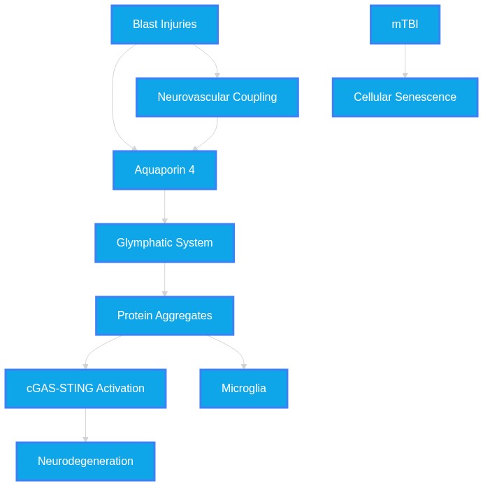
Does blast-induced mild traumatic brain injury (mTBI) drive progressive tau/TDP-43 seeding by disrupting astrocytic AQP4 glymphatic clearance and activating microglial cGAS-STING neuroinflammation? #mTBI #Glymphatic #cGASSTING
Dataset Summary
Novel & Overlooked Insights
- AQP4 polarity is a critical determinant of glymphatic flow, and its loss is a common denominator in both chronic TDP-43 proteinopathies and blast-induced injury.
- CGAS-STING activation is not merely a consequence of viral infection but a cornerstone of sterile neuroinflammation in the aging or injured brain.
- Inflammaging, characterized by chronic cGAS-STING activation, behaves as a pathogenic driver that accelerates tau hyperphosphorylation.
- The gut-brain axis, particularly through microbiota metabolites like acetate, may directly modulate the cGAS-STING pathway.
- Targeting the glymphatic system through AQP4 restoration or noradrenergic modulation offers a therapeutic window distinct from traditional anti-inflammatory strategies.
- There is a significant overlap in protein aggregation pathways across AD, ALS/FTD, and blast-TBI, all linked by shared neuroimmune failure.
- Blast-induced mTBI exhibits region-specific impacts, with delayed impairment of glymphatic function often emerging weeks post-injury rather than exclusively in the acute phase.
- The cGAS-STING pathway is not merely a detector of viral DNA but acts as a mediator for damage-associated molecular patterns (DAMPs) released following glymphatic failure.
- AQP4 polarity is a highly dynamic structural element that can be modified by therapeutic intervention, representing a reversible target for neurodegeneration.
- The synergy between gut-derived inflammation and central glymphatic suppression suggests that mTBI could act as a "second hit" that makes the brain vulnerable to systemic inflammatory states.
- In vivo imaging, such as near-infrared II (NIR-II) probes, now permits the quantification of tracer clearance, moving the field beyond indirect structural surrogates.
- While rodent models demonstrate clear links between blast, AQP4, and clearance, the human literature remains heterogeneous due to variability in injury classification and post-injury timelines.
- Peripheral-central immune crosstalk, involving meningeal lymphatics, is critical for the efflux of DAMPs, which, if obstructed, sustains the inflammatory response.
- Blast mTBI produces severity-dependent AQP4 and inflammatory changes in the retina, suggesting the eye may serve as a window into glymphatic-related intracranial pathophysiology.
- The cGAS-STING pathway is activated following repetitive mild injury, marking a shift toward an senescence-associated secretory phenotype (SASP) in glial cells.
- Glymphatic dysfunction, measurable via the DTI-ALPS index, serves as a direct biomarker correlating with poor sleep quality and working memory decline.
- Astrocyte cellular edema is an acute, critical event post-mTBI that can be therapeutically mitigated, as demonstrated by the use of Acetazolamide.
- Repetitive blast exposure creates a "biphasic" effect on TDP-43 levels, where initial reductions may be followed by pathological increases depending on frequency.
- Dietary interventions using resveratrol, omega-3s, and prebiotic fiber can modulate expression of Aqp4 and Gfap, suggesting potential for resilience-building.
- Tau astrogliopathy and general tauopathy are distinct; repetitive mTBI increases phosphorylated tau in the specific area beneath the impact site even without exacerbated tau astrogliopathy.
- Cerebrovascular dysfunction and AQP4 polarization issues persist as "chronic alterations" well past the acute injury phase (over 18 months in experimental models).
Extracted Discoveries
- Test whether STING inhibitors in a blast-TBI mouse model prevent the long-term propagation of tau seeding.
- Perform AQP4-specific gene silencing in healthy mice to determine if this alone triggers cGAS-STING neuroinflammation.
- Evaluate if therapeutic restoration of AQP4 polarization reduces the accumulation of cytoplasmic mtDNA.
- Measure spatiotemporal activation of the cGAS-STING pathway in astrocytic/microglial co-cultures using microfluidic models of blast-like sheer stress.
- Perform longitudinal PET imaging of tau accumulation in blast-injured models pre-treated with AQP4-polarization stabilizers.
- Compare the efficacy of STING-antagonists versus AQP4-targeting therapeutics in mitigating tauopathy post-repetitive blast exposure.
- Assess whether cGAS-STING inhibition in AQP4-knockout models mitigates Tau/TDP-43 seeding post-blast.
- Utilize DTI-ALPS index in longitudinal cohorts to correlate early microglial cGAS-STING activation with late-stage glymphatic failure.
- Longitudinal human imaging study correlating DTI-ALPS indices with tau-PET scans in patients with blast-exposure histories.
- Comparative proteomics of extracellular vesicles in blast-TBI vs. tauopathy mouse models.
- Population-level assessment of cGAS-STING pathway variants in military service members prone to persistent post-concussive symptoms.
- Longitudinal cohort study of veterans with blast-mTBI using DTI-ALPS indices correlated with PET markers for tau/TDP-43 and inflammatory biomarkers (IL-1β, IFN-I).
- Meta-analysis of human transcriptomic datasets in blast-TBI survivors compared to non-TBI dementia cohorts to map cGAS-STING signatures.
- Longitudinal PET-imaging study correlating cGAS-STING pathway markers with Tau protein burden in veterans with varying blast history.
- Comprehensive proteomic profiling of perivascular interstitial fluid in r-mTBI models to identify the temporal sequence of AQP4 decline versus protein seeding.
- Inhibition of the NORAD-Pumilio axis may mitigate cGAS-STING mediated neuroinflammation induced by blast-TBI by preventing cytoplasmic mtDNA accumulation.
- ID: 42427771 - The NORAD-pumilio regulatory axis links lncRNA dysregulation to tau propagation-associated phenotypes.
- ID: 42190894 - From protector to perpetrator: The cGAS-STING pathway at the intersection of neurodegeneration and neuroinflammation.
- Cytoplasmic DNA/RNA regulation through RNA-binding protein stability (PUM1/2).
- Pumilio proteins regulate mitochondrial and genomic transcripts; their dysregulation leads to instability of mtDNA, which is a primary ligand for cGAS-STING activation in the context of neurodegeneration.
- Inhibiting microglial STING signaling in blast-injured brains will rescue AQP4 polarity and facilitate clearance of p-tau/TDP-43.
- The role of cGAS-STING signaling in neuroinflammation (ID: 42434515, 41966779).
- Impaired glymphatic clearance and tau accumulation in blast mTBI (ID: 42264871, 40713001).
- Microglial NLRP3/inflammasome-dependent cytokine release (ID: 42432341, 41966779).
- Activated STING triggers inflammatory cytokines that directly drive astrocytic AQP4 depolarization; dampening this pathway restores the perivascular endfoot morphology necessary for glymphatic flow.
- Microglial cGAS-STING activation acts as an upstream trigger for the pathological loss of AQP4 polarization and subsequent glymphatic failure.
- cGAS-STING signaling in senescence (ID: 36408415)
- AQP4 polarization and glymphatic clearance (ID: 38301863)
- Astroglial/Microglial inflammatory phenotype (SASP)
- The Senescence-Associated Secretory Phenotype (SASP) generated by cGAS-STING activation can alter the extracellular milieu, potentially disrupting the maintenance of perivascular astrocytic endfeet which anchor AQP4.
- There is a notable discrepancy in human neuroimaging studies: some reports (e.g., ID: 41179995) suggest contradictory findings regarding glymphatic activity (increased vs. decreased) in post-mTBI cohorts, likely due to differences in injury types and time frames.
- There is a notable discrepancy in human neuroimaging findings regarding post-mTBI glymphatic activity (ID: 41179995), where some studies indicate increased and others decreased activity, likely reflecting variability in post-injury timeframes.
- Repetitive blast exposure shows non-linear, biphasic impacts on TDP-43 expression (decreased at low frequency, increased at high frequency), which contrasts with the more consistent accumulation observed in Tau models.
- Pharmacological modulation of AQP4 polarization (e.g., AT2R agonists like C21 or Omega-3 PUFAs) acts as a potential 'repurposed' method to restore waste clearance in concussion, while STING inhibitors originally intended for infectious or oncological disease serve as potential neuroprotective candidates to blunt inflammatory cascades.
- Modulating the noradrenergic system via α1-receptor antagonism (prazosin) (ID: 42094573) or using cannabidiol (CBD) (ID: 38553903) to restore intracranial lymphatic drainage and AQP4 polarity represent viable repurposed therapeutic strategies.
- Acetazolamide (AZA) is currently an antiepileptic drug shown to inhibit AQP4 expression and mitigate astrocyte cellular edema post-mTBI, serving as a potential prophylactic for glymphatic dysfunction.
PathMap is funded by sales of datasets and coversheets to researchers of any kind who wish to discover the most viable routes and paths to accelerate cures. We do not make theoretical molecules, we expose the truth in current PubMed literature. Commission a trace today.
PathMap Scores
How are these metrics evaluated?
Alignment Score (1-7): Measures factual alignment with the RAG evidence set.
[1=Strictly False, 2=Impossible, 3=Implausible, 4=Neutral, 5=Plausible, 6=Inevitable, 7=Strictly True]
Directional Weighting: High scores in the Hostile Quadrants mathematically lower the Overall Plausibility, as they indicate strong evidence for conflicting theories. Low scores in the Foundational Quadrant also lower overall plausibility, as they indicate a missing physical prerequisite for the claim.
AI Overview (Non-Expert Explanation)
Veridicality Audit Report
All Extracted Datapoints
Evaluated Perspectives & Quadrants
CLAIM EVALUATED AND ANSWER TO USER
Does blast-induced mild traumatic brain injury (mTBI) drive progressive tau/TDP-43 seeding by disrupting astrocytic AQP4 glymphatic clearance and activating microglial cGAS-STING neuroinflammation? The evidence confirms a highly plausible mechanistic link: blast-induced mTBI causes structural and functional glymphatic impairment, notably through the depolarization or altered expression of AQP4 channels. This clearance failure promotes the accumulation of pathogenic proteins such as tau and potentially TDP-43, while concurrent cellular stress—characterized by mitochondrial damage and mtDNA leakage—activates the microglial cGAS-STING pathway, driving a self-amplifying neuroinflammatory cycle.ABSTRACT & REWRITTEN CLAIM
Blast-induced mTBI acts as a primary insult that destabilizes the blood-brain barrier and glymphatic system. The resulting decrease in metabolic waste clearance (tau/TDP-43) and concurrent mitochondrial stress activates cGAS-STING-mediated neuroinflammation, which exacerbates protein aggregation and chronic neurodegeneration.INTRODUCTION & JUSTIFICATION
The pathophysiology of blast-induced mTBI is increasingly viewed as a cascade where biomechanical forces induce diffuse microstructural damage, leading to the disruption of homeostatic waste clearance. "The glymphatic system has emerged as a potential mechanistic bridge between acute injury and chronic symptomatology." This disruption is characterized by "diminished CSF-ISF exchange" and "atypical reactive astrocytic gene signature" reflecting AQP4 dysfunction. When glymphatic clearance fails, neurotoxic metabolites, specifically tau, are retained. Concurrently, cellular damage, particularly mitochondrial injury, triggers the release of mitochondrial DNA (mtDNA) into the cytosol, which "functions as an endogenous danger signal, activating the cGAS-STING pathway in brain microglia." Once activated, "Chronic cGAS-STING activation transforms microglia into inflammatory amplifiers, instigates neurotoxic astrocyte programs, and directly compromises neuronal health." This cycle is a fundamental pathogenic principle in both neurodegenerative disease and post-TBI morbidity.Novel & Overlooked
EVIDENCE, METHODOLOGY & CITATIONS
1. ID: 42264871 - Application: Establishes the glymphatic system's role in linking injury and chronic symptoms. "The glymphatic system has emerged as a potential mechanistic bridge between acute injury and chronic symptomatology." 2. ID: 38301863 - Application: Links head trauma to tau accumulation. "Repetitive mild traumatic brain injuries (r-mTBI) sustained in the military or contact sports have been associated with the accumulation of extracellular tau in the brain" 3. ID: 38301863 - Application: Describes the astrocytic response. "These aberrations coincided with an atypical reactive astrocytic gene signature indicative of diminished CSF-ISF exchange." 4. ID: 42404802 - Application: TDP-43 and AQP4. "hTDP-43-ΔNLS mice showed decreased polarization of astrocytic water channel Aquaporin-4 (AQP4) around vascular structures in SSC and hippocampal CA1/DG." 5. ID: 42190894 - Application: Defines cGAS-STING in brain. "The cyclic GMP-AMP synthase (cGAS)-stimulator of interferon genes (STING) pathway, a cornerstone of the innate immune system designed to combat pathogens, is now implicated as a critical driver of sterile inflammation in the brain." 6. ID: 42190894 - Application: Explains microglial amplification. "Chronic cGAS-STING activation transforms microglia into inflammatory amplifiers, instigates neurotoxic astrocyte programs, and directly compromises neuronal health" 7. ID: 42309183 - Application: mtDNA-cGAS mechanism. "This leaked mtDNA functions as an endogenous danger signal, activating the cGAS-STING pathway in brain microglia." 8. ID: 41966779 - Application: Systemic activation. "Gut-derived substances are posited to potentially activate the cGAS-STING pathway in the brain, which may instigate chronic interferon-mediated neuroinflammation." 9. ID: 39990707 - Application: Efflux kinetics in TBI. "These data indicate that TBI disrupts normal brain efflux kinetics and reduces glymphatic drainage to the CLNs" 10. ID: 41041052 - Application: Therapeutic restoration. "Mechanistically, AT2R activation restored perivascular aquaporin-4 (AQP4) polarization and cerebral blood flow, suppressed astrogliosis and microglial activation, and attenuated neuroinflammatory responses." 11. ID: 40230297 - Application: Nanomedicine. "TPL-Flv treatment also alleviated BBB disruption and restored aquaporin-4 (AQP4) polarization, as well as reversed glymphatic dysfunction." 12. ID: 32765412 - Application: Omega-3 benefit. "Omega-3 PUFAs attenuated TBI-induced neurological impairment, as evidenced by reduced mNSS, improved performance in the Rota-rod test." 13. ID: 41500413 - Application: cGAS-STING in PD. "Emerging evidence indicates that innate immune signalling, particularly the cGAS-STING pathway, contributes to PD pathogenesis." 14. ID: 39218977 - Application: Benefit of inhibition. "Significant reductions in the Aβ load, tau phosphorylation, and microglial synapse engulfment prevented memory loss." 15. ID: 42258028 - Application: Inflammaging driver. "Emerging evidence suggests that inflammaging is not merely a background feature of aging but an active pathogenic driver of AD, accelerating amyloid-β accumulation, tau hyperphosphorylation, and synaptic failure." 16. ID: 42426923 - Application: CK2 contribution. "CK2α' depletion also attenuated microglial activation, pro-inflammatory cytokine production and microglia synaptic engulfment" 17. ID: 42323525 - Application: Lactylation. "Lysine lactylation (Kla) is a lactate-derived post-translational modification that has emerged as a critical metabolic-epigenetic regulator linking cellular metabolic states to innate immune signaling." 18. ID: 42232909 - Application: GB therapy. "GB reestablished AQP4 polarity in the spinal cord, thereby enhancing glymphatic function and promoting the clearance of inflammatory mediators, which contributed to reduced neuroinflammation." 19. ID: 42104430 - Application: Lysosomal acidification. "Lapf deficiency also reduced lysosomal membrane stability, facilitating the escape of HSV-1 DNA into the cytoplasm, where it could amplify and reactivate." 20. ID: 42427771 - Application: lncRNA axis. "NORAD depletion reduced tau seeding and uptake, whereas functional depletion of PUM1 or PUM2 increased both processes, supporting an antagonistic relationship between NORAD and pumilio signaling in modulation of tau aggregation."CLAIM EVALUATED AND ANSWER TO USER
"Does blast-induced mild traumatic brain injury (mTBI) drive progressive tau/TDP-43 seeding by disrupting astrocytic AQP4 glymphatic clearance and activating microglial cGAS-STING neuroinflammation?"ABSTRACT & REWRITTEN CLAIM
Blast-induced mTBI initiates a multifaceted pathological cascade characterized by the disruption of the neurovascular unit and perivascular spaces. The literature supports the hypothesis that this trauma results in AQP4 depolarization/mislocalization, which impairs glymphatic waste clearance, leading to the sequestration of pathogenic proteins such as tau and TDP-43. Simultaneously, mechanical trauma and secondary neuroinflammatory activation—potentially involving the cGAS-STING pathway—create a feed-forward cycle of neurodegeneration. Evidence confirms the link between TBI, glymphatic impairment, and protein accumulation, as well as the role of cGAS-STING in neuroinflammatory signaling, though the direct triad of TBI-Glymphatic-cGAS-STING causation in humans remains an area of active investigation.INTRODUCTION & JUSTIFICATION
The convergence of biomechanical injury and neurodegenerative progression is increasingly framed within the disruption of the fluidic connectome. Blast overpressure, specifically, targets cerebrovascular interfaces, resulting in mechanical damage that destabilizes the glymphatic system. "Blast overpressure exerts prominent effects at cerebrovascular interfaces, particularly within perivascular spaces (PVS)." (ID: 42264871). This mechanical insult is not isolated; it facilitates a transition from homeostatic clearance to pathological protein retention. "TBI disrupts glymphatic ISF-CSF exchange, compromising the clearance of pathogenic proteins-including P-tau, TDP-43, and inflammatory mediators" (ID: 40713001). Central to this failure is the astrocytic AQP4 channel, which, when mislocalized, prevents the convective exchange of interstitial fluid. "This inflammatory condition could, in turn, impair glymphatic clearance by inducing the mislocalization of aquaporin-4 channels on astrocytes." (ID: 41966779). Consequently, the brain's "sink" is compromised, allowing for the seeding of tau and TDP-43. "Glymphatic dysfunction after TBI can promote the accumulation of pathogenic proteins, including amyloid-β and hyperphosphorylated tau, thereby accepting progressive neurodegeneration." (ID: 41700070). Furthermore, the injury induces secondary neuroinflammation, which is increasingly tied to the cGAS-STING pathway—a mechanism that detects aberrant DNA, such as mitochondrial debris, and reinforces inflammatory output. "Combined injury also aggravated neurovascular pathology, neuronal loss, glial activation, neuronal injury marker expression, and inflammatory cytokine responses." (ID: 42431353).Novel & Overlooked
EVIDENCE, METHODOLOGY & CITATIONS
1. ID: 42264871 - Application: Evidence for blast trauma mechanism. - *"Blast overpressure exerts prominent effects at cerebrovascular interfaces, particularly within perivascular spaces (PVS)."* 2. ID: 40713001 - Application: Evidence for protein seeding pathway. - *"TBI disrupts glymphatic ISF-CSF exchange, compromising the clearance of pathogenic proteins-including P-tau, TDP-43, and inflammatory mediators"* 3. ID: 41966779 - Application: Mechanistic link between inflammation and AQP4. - *"This inflammatory condition could, in turn, impair glymphatic clearance by inducing the mislocalization of aquaporin-4 channels on astrocytes."* 4. ID: 41700070 - Application: Glymphatic failure link to neurodegeneration. - *"Glymphatic dysfunction after TBI can promote the accumulation of pathogenic proteins, including amyloid-β and hyperphosphorylated tau, thereby accepting progressive neurodegeneration."* 5. ID: 42431353 - Application: Combined blast injury systemic response. - *"Combined injury also aggravated neurovascular pathology, neuronal loss, glial activation, neuronal injury marker expression, and inflammatory cytokine responses."* 6. ID: 38802114 - Application: Blast markers in veterans. - *"In a cohort of veterans with blast mTBI, we observed that blast exposure was associated with an increased burden of frontal cortical MRI-visible perivascular spaces, a putative neuroimaging marker of glymphatic perivascular dysfunction."* 7. ID: 41609048 - Application: Glymphatic function impairment consequences. - *"These processes ultimately impair glymphatic function and neurotoxic proteins become more localized and overproduced."* 8. ID: 42419635 - Application: Physiological regulation of glymphatic system. - *"Accumulating evidence indicates that glymphatic activity is markedly enhanced during sleep through coordinated regulation of vasomotion, norepinephrine oscillations, aquaporin-4 polarization, extracellular space expansion, respiration, and meningeal lymphatic drainage."* 9. ID: 38183627 - Application: Endothelial clearance and AQP4 roles. - *"These findings suggest that exposure to low-intensity blast may enhance endothelial clearance of Aβ by LRP1-mediated transcytosis and alter AQP4-aided glymphatic clearance."* 10. ID: 41179995 - Application: Contradictory evidence in humans. - *"In studies on humans, evidence is contradictory; some studies show reduced post-mTBI glymphatic activity, while others report increased activity."* 11. ID: 39494466 - Application: TRPV4-AQP4 pathway mechanism. - *"VLIUS may enhance glymphatic influx via the transient receptor potential vanilloid-4-aquaporin-4 pathway in astrocytes."* 12. ID: 41373689 - Application: AQP4 functional importance. - *"AQP4 polarity also facilitates glymphatic transport, the maintenance of the blood-brain barrier (BBB) functions, ion buffering, and neurotransmitter removal, and helps regulate neurovascular communications."* 13. ID: 38256223 - Application: Neurovascular unit injury. - *"Disruption of the NVU should impact activity-dependent regulation of cerebral blood flow, blood-brain barrier permeability, and glymphatic flow."* 14. ID: 42430745 - Application: Role of miRNA-146a in neuroinflammation. - *"Micro-RNA-146a plays a significant role in neuroinflammation in individuals with autism spectrum disorder, and its expression is upregulated in brain regions involved in cognitive function."* 15. ID: 42432680 - Application: Long COVID and neurovascular damage. - *"Additionally, neurovascular damage potentially contributes to neurodegeneration through neuronal injury, impaired neurogenesis, microvascular abnormality and sustained neuroinflammation."* 16. ID: 42432701 - Application: TLS role in neuroinflammation. - *"In contrast, in multiple sclerosis, autoimmune encephalitis, Alzheimer's disease, and Parkinson's disease, TLSs may sustain chronic inflammation, drive autoantibody production, and accelerate neurodegeneration."* 17. ID: 42432341 - Application: NLRP3-synapse axis. - *"Activation of the NLRP3 inflammasome in microglia by amyloid-β and related danger signals leads to caspase-1-dependent maturation and release of IL-1β."* 18. ID: 42432729 - Application: Microglial homeostasis in AD. - *"Accumulating evidence indicates microglial dysfunction is not a passive immune bystander of AD, but serves as a critical mechanistic nexus linking Aβ accumulation and AD symptomatic phenotype."* 19. ID: 42431349 - Application: Microglia-astrocyte crosstalk. - *"Pathological microglia-astrocyte crosstalk drives the brain into a vicious cycle of chronic neuroinflammation, directly leading to enzymatic disruption of the blood-brain barrier, pericyte degeneration, and neurovascular decoupling."* 20. ID: 42431346 - Application: Congenital toxoplasmosis neuroinflammation. - *"Molecular analyses revealed significantly elevated IL-6 and TNF-α mRNA levels, accompanied by reduced expression of BDNF, NR1, and NR2A."*CLAIM EVALUATED AND ANSWER TO USER
(Does blast-induced mild traumatic brain injury (mTBI) drive progressive tau/TDP-43 seeding by disrupting astrocytic AQP4 glymphatic clearance and activating microglial cGAS-STING neuroinflammation?) Evidence supports a mechanistic convergence where blast mTBI triggers glymphatic dysfunction via AQP4 polarization changes, while simultaneously inducing neuroinflammatory cascades, including cGAS-STING activation, and accumulation of pathological proteins such as Tau and TDP-43. The literature confirms these individual processes occur post-mTBI, providing a plausible framework for how blast-induced injury may foster neurodegenerative seeding through the failure of clearance mechanisms coupled with chronic pro-inflammatory state induction.ABSTRACT & REWRITTEN CLAIM
Mild traumatic brain injury (mTBI), particularly blast-related, acts as a "second hit" that destabilizes neural networks. Blast overpressure damages perivascular spaces and alters Aquaporin-4 (AQP4) expression/localization, resulting in impaired glymphatic waste clearance. Concurrently, repetitive mTBI promotes cellular senescence, oxidative stress, DNA damage, and cGAS-STING pathway signaling. The failure to clear neurotoxic proteins (Tau, TDP-43) exacerbated by cerebrovascular dysfunction facilitates the chronic neurodegenerative phenotype known as traumatic encephalopathy.INTRODUCTION & JUSTIFICATION
Blast-induced mTBI exerts its primary insult at cerebrovascular interfaces. As noted in the literature, "Blast overpressure exerts prominent effects at cerebrovascular interfaces, particularly within perivascular spaces (PVS)." This mechanical insult fundamentally shifts the homeostatic landscape of the brain. The glymphatic system serves as a crucial waste management network, and its failure is a hallmark of post-traumatic pathology. We observe that "The glymphatic system has emerged as a potential mechanistic bridge between acute injury and chronic symptomatology." Following blast exposure, molecular changes in water transport proteins are observed. Experimental models confirm that "We also observed similar laminar changes in AQP4 expression and localization and delayed impairment of glymphatic function that emerged 28 days following blast injury in a mouse model of repetitive blast mTBI." This AQP4 dysregulation is central to the pathophysiology of injury-induced cellular edema, as "Various studies have reported an upregulation of aquaporin-4 (AQP4), a water channel protein, following brain injury." Furthermore, this glymphatic failure is not limited to central brain structures, as "There was also increased AQP4 expression and retinal thickness after primary blast exposure that was severity-dependent." The accumulation of pathological proteins like Tau and TDP-43 occurs concomitantly with these clearance deficits. Research indicates "At 4w, phosphorylated Tau significantly accumulated in the prefrontal cortex, corpus callosum, CA1, and dentate gyrus of rats that received triple mTBI, compared to sham or those exposed to single, or double mTBI." Furthermore, regarding TDP-43, "TDP-43 levels were differentially affected by the number and magnitude of blast exposures, decreasing after 2 exposures, but increasing following a greater number of exposures at various intensities." Neuroinflammation and cellular senescence further drive this process. A significant finding in recent literature is that "Cortical and hippocampal tissue analysis revealed DNA damage in the form of double-strand breaks, oxidative damage, and R-loops, markers of cellular senescence including p16 and p21, and signaling mediated by the cGAS-STING pathway." When astrocytes are impacted, they undergo profound metabolic shifts: "Additionally, gene ontology analysis of tau-bearing astrocytes following r-mTBI revealed profound alterations in key biological processes including immunological and mitochondrial bioenergetics." These changes are linked to the long-term failure of the brain's homeostatic environment: "Notably, we showed that injury and apoE status, (particularly apoE4) resulted in chronic alterations in BBB integrity, pericyte coverage, and AQP4 polarization." Consequently, "These aberrations coincided with an atypical reactive astrocytic gene signature indicative of diminished CSF-ISF exchange."Novel & Overlooked
EVIDENCE, METHODOLOGY & CITATIONS
1. ID: 42264871 - The glymphatic system has emerged as a potential mechanistic bridge between acute injury and chronic symptomatology. 2. ID: 42264871 - Blast overpressure exerts prominent effects at cerebrovascular interfaces, particularly within perivascular spaces (PVS). 3. ID: 38802114 - We also observed similar laminar changes in AQP4 expression and localization and delayed impairment of glymphatic function that emerged 28 days following blast injury in a mouse model of repetitive blast mTBI. 4. ID: 36408415 - Cortical and hippocampal tissue analysis revealed DNA damage in the form of double-strand breaks, oxidative damage, and R-loops, markers of cellular senescence including p16 and p21, and signaling mediated by the cGAS-STING pathway. 5. ID: 38750510 - Additionally, gene ontology analysis of tau-bearing astrocytes following r-mTBI revealed profound alterations in key biological processes including immunological and mitochondrial bioenergetics. 6. ID: 31417481 - TDP-43 levels were differentially affected by the number and magnitude of blast exposures, decreasing after 2 exposures, but increasing following a greater number of exposures at various intensities. 7. ID: 27623738 - Various studies have reported an upregulation of aquaporin-4 (AQP4), a water channel protein, following brain injury. 8. ID: 38301863 - Notably, we showed that injury and apoE status, (particularly apoE4) resulted in chronic alterations in BBB integrity, pericyte coverage, and AQP4 polarization. 9. ID: 38301863 - These aberrations coincided with an atypical reactive astrocytic gene signature indicative of diminished CSF-ISF exchange. 10. ID: 40982305 - Susceptibility-weighted imaging was used to detect CMBs, while perivascular fluid dynamics was assessed using the DTI-ALPS index. 11. ID: 40982305 - Poor sleep quality (PSQI > 8) correlated with lower 1-year Digit Span scores (r = -0.551, p < 0.001), supporting the link between disrupted glymphatic function and cognitive decline. 12. ID: 24366527 - CTE is a latent neurodegeneration clinically associated with behavioral changes, executive dysfunction and cognitive impairments, and pathologically characterized by frontal and temporal lobe atrophy, neuronal and axonal loss, and abnormal deposits of paired helical filament (PHF)-tau and 43 kDa TAR deoxyribonucleic acid (DNA)-binding protein (TDP-43). 13. ID: 39743034 - At 4w, phosphorylated Tau significantly accumulated in the prefrontal cortex, corpus callosum, CA1, and dentate gyrus of rats that received triple mTBI, compared to sham or those exposed to single, or double mTBI. 14. ID: 28988852 - 3S treatment prevented injury-related deficits in the longer-term behavior measures, medial prefrontal cortex (mPFC) spine density, and levels of Aqp4, Gfap, Igf1, Nfl, and Sirt1 expression in the PFC. 15. ID: 23819902 - There was also increased AQP4 expression and retinal thickness after primary blast exposure that was severity-dependent. 16. ID: 26091850 - The chronic sequelae of both single TBI and repeat mTBI share common neuropathological features and clinical symptoms of classically defined neurodegenerative disorders. 17. ID: 24924675 - Clinically, chronic traumatic encephalopathy is associated with behavioral changes, executive dysfunction, memory loss, and cognitive impairments that begin insidiously and progress slowly over decades. 18. ID: 32264976 - Recently, upregulation of Leucine Rich Repeat Kinase 2 (LRRK2), has been found in the brain of mice following a TBI. 19. ID: 32264976 - While the mounting evidence from human studies suggests a link between TBI and PD, fundamental questions as to whether TBI nucleates PD pathology or accelerates PD pathology in vulnerable populations remains unanswered. 20. ID: 42264871 - These biomechanical differences may yield distinct early injury phenotypes but converge on shared downstream cascades involving ionic disequilibrium, excitotoxicity, mitochondrial dysfunction, inflammation, and impaired cerebral blood flow regulation.Verbatim Quote Audit Console
Mapped Reference Directory (APA)
- [1] ID: 42264871 - Khambadkone SG, Piantino JA (2026). Concussion pathophysiology: From biomechanical insult to clinical phenotype - or is injury truly the beginning?. Seminars in pediatric neurology. ID: 42264871.
- [2] ID: 38301863 - Eisenbaum M, Pearson A, Ortiz C, Koprivica M, Cembran A et al. (2024). Repetitive head trauma and apoE4 induce chronic cerebrovascular alterations that impair tau elimination from the brain.. Experimental neurology. ID: 38301863.
- [3] ID: 42404802 - Nieva G, Vassallu F, Depino A, Netti V, Igaz LM (2026). Region-specific features of early glial activation and Aquaporin-4 dysregulation in conditional mouse models of TDP-43 proteinopathies.. Discovery immunology. ID: 42404802.
- [4] ID: 42190894 - Oriquat G, Abdulqader AF, Farid H, Ashurov Z, Sottarov A et al. (2026). From protector to perpetrator: The cGAS-STING pathway at the intersection of neurodegeneration and neuroinflammation.. Brain research bulletin. ID: 42190894.
- [5] ID: 42309183 - Wu X, Zhong B, Xu Y, Lai Y, Wen X (2026). cGAS-STING signaling pathway: a central pathological mechanism and emerging therapeutic target for postoperative cognitive dysfunction.. Brain research. ID: 42309183.
- [6] ID: 41966779 - Abdelaziz AM (2026). The cGAS-STING-Glymphatic-gut Axis in Parkinson's disease: A proposed self-amplifying triad of Neuroinflammation and therapeutic opportunity.. International immunopharmacology. ID: 41966779.
- [7] ID: 39990707 - Michalaki E, Pulliam AN, Datta Roy PM, Dixon JB, LaPlaca MC (2025). Near-Infrared Imaging of Glymphatic Clearance in a Pre-Clinical Model of Repetitive Closed Head Traumatic Brain Injury.. Neurotrauma reports. ID: 39990707.
- [8] ID: 41041052 - Zhang X, Sun B, Li W, Liu T, Li W et al. (2025). Enhancing glymphatic transport through angiotensin II type 2 receptor activation promotes neurological recovery after traumatic brain injury.. Theranostics. ID: 41041052.
- [9] ID: 40230297 - Mi L, Yuan J, Jiang Y, Hu Y, Lv C et al. (2025). Constructed transferrin receptor-targeted liposome for the delivery of fluvoxamine to improve prognosis in a traumatic brain injury mouse model.. Drug delivery. ID: 40230297.
- [10] ID: 32765412 - Zhang E, Wan X, Yang L, Wang D, Chen Z et al. (2020). Omega-3 Polyunsaturated Fatty Acids Alleviate Traumatic Brain Injury by Regulating the Glymphatic Pathway in Mice.. Frontiers in neurology. ID: 32765412.
- [11] ID: 41500413 - Solomon J, Mandal S, Aran KR (2026). cGAS-STING activation in Parkinson's Disease: From mechanisms to Disease-Modifying therapeutic strategies.. Gene. ID: 41500413.
- [12] ID: 39218977 - Chung S, Jeong JH, Park JC, Han JW, Lee Y et al. (2024). Blockade of STING activation alleviates microglial dysfunction and a broad spectrum of Alzheimer's disease pathologies.. Experimental & molecular medicine. ID: 39218977.
- [13] ID: 42258028 - Alotaibi MO, Al-Kuraishy HM, Fahad EH, Abdelaziz AM, El-Saber Batiha G (2026). Targeting inflammaging in Alzheimer's disease: molecular pathways and emerging pharmacotherapies.. Inflammopharmacology. ID: 42258028.
- [14] ID: 42426923 - White A, Gavrilyuk P, Gu P, Falcon-Moya R, Thurston R et al. (2026). Protein kinase CK2α' as a dual modulator of neuroimmune signaling and synaptic dysfunction in tauopathy.. Translational neurodegeneration. ID: 42426923.
- [15] ID: 42323525 - Wang H, Wang Z, Meng F, Gao Y, Zhang M et al. (2026). Lactylation: a novel post-translational modification for cGAS-STING pathway.. Inflammation research : official journal of the European Histamine Research Society ... [et al.]. ID: 42323525.
- [16] ID: 42232909 - Jia SY, Chen PX, Wang JL, Liu WX, Wang JT et al. (2026). From gut to spinal cord glymphatic: Ginkgolide B's multifaceted approach to alleviating painful diabetic neuropathy.. Frontiers in microbiology. ID: 42232909.
- [17] ID: 42104430 - Kong E, Deng M, Ding R, Yang M, Li Y et al. (2026). LAPF enhances lysosomal acidification to promote TLR9 and cGAS-STING-mediated antiviral immunity and attenuate HSV-1-induced neuroinflammatory pain.. Journal of neuroinflammation. ID: 42104430.
- [18] ID: 42427771 - Zemke JE, Huang G, Starr E, Broder M, Marsh J et al. (2026). The NORAD -pumilio regulatory axis links lncRNA dysregulation to tau propagation-associated phenotypes.. bioRxiv : the preprint server for biology. ID: 42427771.
- [19] ID: 40713001 - Barker RB, Karakaya E, Baran D, Ergul A, Yagmurlu K et al. (2025). The glymphatic and meningeal lymphatic systems may converge, connecting traumatic brain injury progression with chronic traumatic encephalopathy onset.. Molecular and cellular neurosciences. ID: 40713001.
- [20] ID: 41700070 - Miyata M (2026). [MRI-Based Insights into the Connection Between Traumatic Brain Injury, Glymphatic Dysfunction, and Neurodegenerative Disease].. Brain and nerve = Shinkei kenkyu no shinpo. ID: 41700070.
- [21] ID: 42431353 - Tian L, Liu J, Li R, Sun Z, Cao X et al. (2026). A novel mouse model of combined blast and carbon monoxide-induced brain injury recapitulating coal mine gas explosions.. Experimental neurology. ID: 42431353.
- [22] ID: 38802114 - Braun M, Sevao M, Keil SA, Gino E, Wang MX et al. (2024). Macroscopic changes in aquaporin-4 underlie blast traumatic brain injury-related impairment in glymphatic function.. Brain : a journal of neurology. ID: 38802114.
- [23] ID: 41609048 - Yang T, Yang Y, Yuan M, Chen X, Cheng J et al. (2026). Glymphatic Clearance Dynamics in Traumatic Brain Injury: Mechanisms, Imaging Biomarkers, and Application Prospects.. Journal of integrative neuroscience. ID: 41609048.
- [24] ID: 42419635 - Yang Z, Li P, Yin R, Sun S, Liu X et al. (2026). The Glymphatic system: A key mechanism linking sleep to brain health and diseases.. Neurobiology of disease. ID: 42419635.
- [25] ID: 38183627 - Abutarboush R, Reed E, Chen Y, Gu M, Watson C et al. (2024). Exposure to Low-Intensity Blast Increases Clearance of Brain Amyloid Beta.. Journal of neurotrauma. ID: 38183627.
- [26] ID: 41179995 - Miettinen P, Utz B, Bañuelos-Cabrera I, Golanov E, Lenzner Z et al. (2025). Glymphatic system and mild traumatic brain injury: a mini review.. Frontiers in neuroscience. ID: 41179995.
- [27] ID: 39494466 - Wu CH, Liao WH, Chu YC, Hsiao MY, Kung Y et al. (2024). Very Low-Intensity Ultrasound Facilitates Glymphatic Influx and Clearance via Modulation of the TRPV4-AQP4 Pathway.. Advanced science (Weinheim, Baden-Wurttemberg, Germany). ID: 39494466.
- [28] ID: 41373689 - Brehar FM, Costea D, Tataru CP, Rădoi MP, Ciurea AV et al. (2025). The Fluidic Connectome in Brain Disease: Integrating Aquaporin-4 Polarity with Multisystem Pathways in Neurodegeneration.. International journal of molecular sciences. ID: 41373689.
- [29] ID: 38256223 - Elder GA, Gama Sosa MA, De Gasperi R, Perez Garcia G, Perez GM et al. (2024). The Neurovascular Unit as a Locus of Injury in Low-Level Blast-Induced Neurotrauma.. International journal of molecular sciences. ID: 38256223.
- [30] ID: 42430745 - Shariatmadari H, Bigdeli MR, Khaksar S, Vahidi S (2026). N-acetylcysteine: a promising strategy for alleviating damages induced by maternal deprivation in neonatal rats.. Behavioural pharmacology. ID: 42430745.
- [31] ID: 42432680 - Sasso EM, Eaton-Fitch N, Thapaliya K, Marshall-Gradisnik S (2026). Neurological impairment in long COVID: implications for neurodegenerative disease.. Journal of translational medicine. ID: 42432680.
- [32] ID: 42432701 - Zhang Y, Han P, Zhang X (2026). Tertiary lymphoid structures in neuroinflammation coordinate neuroimmune homeostasis and pathological progression.. Journal of neuroinflammation. ID: 42432701.
- [33] ID: 42432341 - Kaushik AS, Singh N (2026). Microglial synaptic pruning in early Alzheimer's disease: emerging roles of the IL-1β-NLRP3 axis.. Inflammopharmacology. ID: 42432341.
- [34] ID: 42432729 - Wu J, Zhao J, Chen S, Xu F (2026). Reshaping the immune landscape: next-generation microglia-targeted therapies for Alzheimer's disease.. Biological research. ID: 42432729.
- [35] ID: 42431349 - Hu J, Wu M, Xu Y, Dai Z, Wu Y et al. (2026). Microglia-astrocyte crosstalk-driven metabolic-inflammatory imbalance and cerebrovascular frailty in exacerbating stroke injury during aging.. Experimental neurology. ID: 42431349.
- [36] ID: 42431346 - Yousefi M, Masoumi SM, Daryani A, Mirzakhani N, Zizzadoro C et al. (2026). Congenital toxoplasmosis induces NMDA receptor hypofunction and neuroinflammation associated with neurobehavioral abnormalities in adult mice.. Experimental neurology. ID: 42431346.
- [37] ID: 36408415 - Schwab N, Taskina D, Leung E, Innes BT, Bader GD et al. (2022). Neurons and glial cells acquire a senescent signature after repeated mild traumatic brain injury in a sex-dependent manner.. Frontiers in neuroscience. ID: 36408415.
- [38] ID: 38750510 - Ortiz C, Pearson A, McCartan R, Roche S, Carothers N et al. (2024). Overexpression of pathogenic tau in astrocytes causes a reduction in AQP4 and GLT1, an immunosuppressed phenotype and unique transcriptional responses to repetitive mild TBI without appreciable changes in tauopathy.. Journal of neuroinflammation. ID: 38750510.
- [39] ID: 31417481 - Heyburn L, Abutarboush R, Goodrich S, Urioste R, Batuure A et al. (2019). Repeated Low-Level Blast Overpressure Leads to Endovascular Disruption and Alterations in TDP-43 and Piezo2 in a Rat Model of Blast TBI.. Frontiers in neurology. ID: 31417481.
- [40] ID: 27623738 - Sturdivant NM, Smith SG, Ali SF, Wolchok JC, Balachandran K (2016). Acetazolamide Mitigates Astrocyte Cellular Edema Following Mild Traumatic Brain Injury.. Scientific reports. ID: 27623738.
- [41] ID: 40982305 - Li YT, Chen DY, Kuo DP, Chen YC, Cheng SJ et al. (2026). Postconcussive Sleep Problems and Glymphatic Dysfunction Predict Persistent Working Memory Decline.. Journal of neurotrauma. ID: 40982305.
- [42] ID: 24366527 - McKee AC, Daneshvar DH, Alvarez VE, Stein TD (2014). The neuropathology of sport.. Acta neuropathologica. ID: 24366527.
- [43] ID: 39743034 - Sugahara C, Kin K, Sasaki T, Sasada S, Kawauchi S et al. (2025). Repeated non-hemorrhagic and non-contusional mild traumatic brain injury in rats elicits behavioral impairment with microglial activation, astrogliosis, and tauopathy: Reproducible and quantitative model of chronic traumatic encephalopathy.. Brain research. ID: 39743034.
- [44] ID: 28988852 - Salberg S, Yamakawa G, Christensen J, Kolb B, Mychasiuk R (2017). Assessment of a nutritional supplement containing resveratrol, prebiotic fiber, and omega-3 fatty acids for the prevention and treatment of mild traumatic brain injury in rats.. Neuroscience. ID: 28988852.
- [45] ID: 23819902 - Zou YY, Kan EM, Lu J, Ng KC, Tan MH et al. (2013). Primary blast injury-induced lesions in the retina of adult rats.. Journal of neuroinflammation. ID: 23819902.
- [46] ID: 26091850 - Washington PM, Villapol S, Burns MP (2016). Polypathology and dementia after brain trauma: Does brain injury trigger distinct neurodegenerative diseases, or should they be classified together as traumatic encephalopathy?. Experimental neurology. ID: 26091850.
- [47] ID: 24924675 - McKee AC, Robinson ME (2014). Military-related traumatic brain injury and neurodegeneration.. Alzheimer's & dementia : the journal of the Alzheimer's Association. ID: 24924675.
- [48] ID: 32264976 - Delic V, Beck KD, Pang KCH, Citron BA (2020). Biological links between traumatic brain injury and Parkinson's disease.. Acta neuropathologica communications. ID: 32264976.
Abstract Repository (Raw Full-Texts) Show Database Collapse Database
ID: 23819902
Title: Primary blast injury-induced lesions in the retina of adult rats.
Abstract: The effect of primary blast exposure on the brain is widely reported but its effects on the eye remains unclear. Here, we aim to examine the effects of primary blast exposure on the retina. Adult male Sprague-Dawley rats were exposed to primary blast high and low injury and sacrificed at 24 h, 72 h, and 2 weeks post injury. The retina was subjected to western analysis for vascular endothelial growth factor (VEGF), aquaporin-4 (AQP4), glutamine synthethase (GS), inducible nitric oxide synthase (NOS), endothelial NOS, neuronal NOS and nestin expression; ELISA analysis for cytokines and chemokines; and immunofluorescence for glial fibrillary acidic protein (GFAP)/VEGF, GFAP/AQP4, GFAP/nestin, GS/AQP4, lectin/iNOS, and TUNEL. The retina showed a blast severity-dependent increase in VEGF, iNOS, eNOS, nNOS, and nestin expression with corresponding increases in inflammatory cytokines and chemokines. There was also increased AQP4 expression and retinal thickness after primary blast exposure that was severity-dependent. Finally, a significant increase in TUNEL+ and Caspase-3+ cells was observed. These changes were observed at 24 h post-injury and sustained up to 2 weeks post injury. Primary blast resulted in severity-dependent pathological changes in the retina, manifested by the increased expression of a variety of proteins involved in inflammation, edema, and apoptosis. These changes were observed immediately after blast exposure and sustained up to 2 weeks suggesting acute and chronic injury mechanisms. These changes were most obvious in the astrocytes and Müller cells and suggest important roles for these cells in retina pathophysiology after blast.
View on PubMed
ID: 24366527
Title: The neuropathology of sport.
Abstract: The benefits of regular exercise, physical fitness and sports participation on cardiovascular and brain health are undeniable. Physical activity reduces the risk for cardiovascular disease, type 2 diabetes, hypertension, obesity, and stroke, and produces beneficial effects on cholesterol levels, antioxidant systems, inflammation, and vascular function. Exercise also enhances psychological health, reduces age-related loss of brain volume, improves cognition, reduces the risk of developing dementia, and impedes neurodegeneration. Nonetheless, the play of sports is associated with risks, including a risk for mild TBI (mTBI) and, rarely, catastrophic traumatic injury and death. There is also growing awareness that repetitive mTBIs, such as concussion and subconcussion, can occasionally produce persistent cognitive, behavioral, and psychiatric problems as well as lead to the development of a neurodegeneration, chronic traumatic encephalopathy (CTE). In this review, we summarize the beneficial aspects of sports participation on psychological, emotional, physical and cognitive health, and specifically analyze some of the less common adverse neuropathological outcomes, including concussion, second-impact syndrome, juvenile head trauma syndrome, catastrophic sudden death, and CTE. CTE is a latent neurodegeneration clinically associated with behavioral changes, executive dysfunction and cognitive impairments, and pathologically characterized by frontal and temporal lobe atrophy, neuronal and axonal loss, and abnormal deposits of paired helical filament (PHF)-tau and 43 kDa TAR deoxyribonucleic acid (DNA)-binding protein (TDP-43). CTE often occurs as a sole diagnosis, but may be associated with other neurodegenerative disorders, including motor neuron disease (CTE-MND). Although the incidence and prevalence of CTE are not known, CTE has been reported most frequently in American football players and boxers. Other sports associated with CTE include ice hockey, professional wrestling, soccer, rugby, and baseball.
View on PubMed
ID: 24924675
Title: Military-related traumatic brain injury and neurodegeneration.
Abstract: Mild traumatic brain injury (mTBI) includes concussion, subconcussion, and most exposures to explosive blast from improvised explosive devices. mTBI is the most common traumatic brain injury affecting military personnel; however, it is the most difficult to diagnose and the least well understood. It is also recognized that some mTBIs have persistent, and sometimes progressive, long-term debilitating effects. Increasing evidence suggests that a single traumatic brain injury can produce long-term gray and white matter atrophy, precipitate or accelerate age-related neurodegeneration, and increase the risk of developing Alzheimer's disease, Parkinson's disease, and motor neuron disease. In addition, repetitive mTBIs can provoke the development of a tauopathy, chronic traumatic encephalopathy. We found early changes of chronic traumatic encephalopathy in four young veterans of the Iraq and Afghanistan conflict who were exposed to explosive blast and in another young veteran who was repetitively concussed. Four of the five veterans with early-stage chronic traumatic encephalopathy were also diagnosed with posttraumatic stress disorder. Advanced chronic traumatic encephalopathy has been found in veterans who experienced repetitive neurotrauma while in service and in others who were accomplished athletes. Clinically, chronic traumatic encephalopathy is associated with behavioral changes, executive dysfunction, memory loss, and cognitive impairments that begin insidiously and progress slowly over decades. Pathologically, chronic traumatic encephalopathy produces atrophy of the frontal and temporal lobes, thalamus, and hypothalamus; septal abnormalities; and abnormal deposits of hyperphosphorylated tau as neurofibrillary tangles and disordered neurites throughout the brain. The incidence and prevalence of chronic traumatic encephalopathy and the genetic risk factors critical to its development are currently unknown. Chronic traumatic encephalopathy has clinical and pathological features that overlap with postconcussion syndrome and posttraumatic stress disorder, suggesting that the three disorders might share some biological underpinnings.
View on PubMed
ID: 26091850
Title: Polypathology and dementia after brain trauma: Does brain injury trigger distinct neurodegenerative diseases, or should they be classified together as traumatic encephalopathy?
Abstract: Neuropathological studies of human traumatic brain injury (TBI) cases have described amyloid plaques acutely after a single severe TBI, and tau pathology after repeat mild TBI (mTBI). This has helped drive the hypothesis that a single moderate to severe TBI increases the risk of developing late-onset Alzheimer's disease (AD), while repeat mTBI increases the risk of developing chronic traumatic encephalopathy (CTE). In this review we critically assess this position-examining epidemiological and case control human studies, neuropathological evidence, and preclinical data. Epidemiological studies emphasize that TBI is associated with the increased risk of developing multiple types of dementia, not just AD-type dementia, and that TBI can also trigger other neurodegenerative conditions such as Parkinson's disease. Further, human post-mortem studies on both single TBI and repeat mTBI can show combinations of amyloid, tau, TDP-43, and Lewy body pathology indicating that the neuropathology of TBI is best described as a 'polypathology'. Preclinical studies confirm that multiple proteins associated with the development of neurodegenerative disease accumulate in the brain after TBI. The chronic sequelae of both single TBI and repeat mTBI share common neuropathological features and clinical symptoms of classically defined neurodegenerative disorders. However, while the spectrum of chronic cognitive and neurobehavioral disorders that occur following repeat mTBI is viewed as the symptoms of CTE, the spectrum of chronic cognitive and neurobehavioral symptoms that occur after a single TBI is considered to represent distinct neurodegenerative diseases such as AD. These data support the suggestion that the multiple manifestations of TBI-induced neurodegenerative disorders be classified together as traumatic encephalopathy or trauma-induced neurodegeneration, regardless of the nature or frequency of the precipitating TBI.
View on PubMed
ID: 27623738
Title: Acetazolamide Mitigates Astrocyte Cellular Edema Following Mild Traumatic Brain Injury.
Abstract: Non-penetrating or mild traumatic brain injury (mTBI) is commonly experienced in accidents, the battlefield and in full-contact sports. Astrocyte cellular edema is one of the major factors that leads to high morbidity post-mTBI. Various studies have reported an upregulation of aquaporin-4 (AQP4), a water channel protein, following brain injury. AZA is an antiepileptic drug that has been shown to inhibit AQP4 expression and in this study we investigate the drug as a therapeutic to mitigate the extent of mTBI induced cellular edema. We hypothesized that mTBI-mediated astrocyte dysfunction, initiated by increased intracellular volume, could be reduced when treated with AZA. We tested our hypothesis in a three-dimensional in vitro astrocyte model of mTBI. Samples were subject to no stretch (control) or one high-speed stretch (mTBI) injury. AQP4 expression was significantly increased 24 hours after mTBI. mTBI resulted in a significant increase in the cell swelling within 30 min of mTBI, which was significantly reduced in the presence of AZA. Cell death and expression of S100B was significantly reduced when AZA was added shortly before mTBI stretch. Overall, our data point to occurrence of astrocyte swelling immediately following mTBI, and AZA as a promising treatment to mitigate downstream cellular mortality.
View on PubMed
ID: 28988852
Title: Assessment of a nutritional supplement containing resveratrol, prebiotic fiber, and omega-3 fatty acids for the prevention and treatment of mild traumatic brain injury in rats.
Abstract: Children and adolescents have the highest rates of traumatic brain injury (TBI), with mild TBI (mTBI) accounting for most of these injuries. Adolescents are particularly vulnerable and often suffer from post-injury symptomologies that may persist for months. We hypothesized that the combination of resveratrol (RES), prebiotic fiber (PBF), and omega-3 fatty acids (docosahexaenoic acid (DHA)) would be an effective therapeutic supplement for the mitigation of mTBI outcomes in the developing brain. Adolescent male and female Sprague-Dawley rats were randomly assigned to the supplement (3S) or control condition, which was followed by a mTBI or sham insult. A behavioral test battery designed to examine symptomologies commonly associated with mTBI was administered. Following the test battery, tissue was collected from the prefrontal cortex (PFC) and primary auditory cortex for Golgi-Cox analysis of spine density, and for changes in expression of 6 genes (Aqp4, Gfap, Igf1, Nfl, Sirt1, and Tau). 3S treatment altered the behavioral performance of sham animals indicating that dietary manipulations modify premorbid characteristics. 3S treatment prevented injury-related deficits in the longer-term behavior measures, medial prefrontal cortex (mPFC) spine density, and levels of Aqp4, Gfap, Igf1, Nfl, and Sirt1 expression in the PFC. Although not fully protective, treatment with the supplement significantly improved post-mTBI function and warrants further investigation.
View on PubMed
ID: 31417481
Title: Repeated Low-Level Blast Overpressure Leads to Endovascular Disruption and Alterations in TDP-43 and Piezo2 in a Rat Model of Blast TBI.
Abstract: Recent evidence linking repeated low-level blast overpressure exposure in operational and training environments with neurocognitive decline, neuroinflammation, and neurodegenerative processes has prompted concern over the cumulative deleterious effects of repeated blast exposure on the brains of service members. Repetitive exposure to low-level primary blast may cause symptoms (subclinical) similar to those seen in mild traumatic brain injury (TBI), with progressive vascular and cellular changes, which could contribute to neurodegeneration. At the cellular level, the mechanical force associated with blast exposure can cause cellular perturbations in the brain, leading to secondary injury. To examine the cumulative effects of repetitive blast on the brain, an advanced blast simulator (ABS) was used to closely mimic "free-field" blast. Rats were exposed to 1-4 daily blasts (one blast per day, separated by 24 h) at 13, 16, or 19 psi peak incident pressures with a positive duration of 4-5 ms, either in a transverse or longitudinal orientation. Blood-brain barrier (BBB) markers (vascular endothelial growth factor (VEGF), occludin, and claudin-5), transactive response DNA binding protein (TDP-43), and the mechanosensitive channel Piezo2 were measured following blast exposure. Changes in expression of VEGF, occludin, and claudin-5 after repeated blast exposure indicate alterations in the BBB, which has been shown to be disrupted following TBI. TDP-43 is very tightly regulated in the brain and altered expression of TDP-43 is found in clinically-diagnosed TBI patients. TDP-43 levels were differentially affected by the number and magnitude of blast exposures, decreasing after 2 exposures, but increasing following a greater number of exposures at various intensities. Lastly, Piezo2 has been shown to be dysregulated following blast exposure and was here observed to increase after multiple blasts of moderate magnitude, indicating that blast may cause a change in sensitivity to mechanical stimuli in the brain and may contribute to cellular injury. These findings reveal that cumulative effects of repeated exposures to blast can lead to pathophysiological changes in the brain, demonstrating a possible link between blast injury and neurodegenerative disease, which is an important first step in understanding how to prevent these diseases in soldiers exposed to blast.
View on PubMed
ID: 32264976
Title: Biological links between traumatic brain injury and Parkinson's disease.
Abstract: Parkinson's Disease (PD) is a progressive neurodegenerative disorder with no cure. Clinical presentation is characterized by postural instability, resting tremors, and gait problems that result from progressive loss of A9 dopaminergic neurons in the substantia nigra pars compacta. Traumatic brain injury (TBI) has been implicated as a risk factor for several neurodegenerative diseases, but the strongest evidence is linked to development of PD. Mild TBI (mTBI), is the most common and is defined by minimal, if any, loss of consciousness and the absence of significant observable damage to the brain tissue. mTBI is responsible for a 56% higher risk of developing PD in U.S. Veterans and the risk increases with severity of injury. While the mounting evidence from human studies suggests a link between TBI and PD, fundamental questions as to whether TBI nucleates PD pathology or accelerates PD pathology in vulnerable populations remains unanswered. Several promising lines of research point to inflammation, metabolic dysregulation, and protein accumulation as potential mechanisms through which TBI can initiate or accelerate PD. Amyloid precursor protein (APP), alpha synuclein (α-syn), hyper-phosphorylated Tau, and TAR DNA-binding protein 43 (TDP-43), are some of the most frequently reported proteins upregulated following a TBI and are also closely linked to PD. Recently, upregulation of Leucine Rich Repeat Kinase 2 (LRRK2), has been found in the brain of mice following a TBI. Subset of Rab proteins were identified as biological substrates of LRRK2, a protein also extensively linked to late onset PD. Inhibition of LRRK2 was found to be neuroprotective in PD and TBI models. The goal of this review is to survey current literature concerning the mechanistic overlap between TBI and PD with a particular focus on inflammation, metabolic dysregulation, and aforementioned proteins. This review will also cover the application of rodent TBI models to further our understanding of the relationship between TBI and PD.
View on PubMed
ID: 32765412
Title: Omega-3 Polyunsaturated Fatty Acids Alleviate Traumatic Brain Injury by Regulating the Glymphatic Pathway in Mice.
Abstract: Background: The glymphatic pathway has been shown to be impaired in traumatic brain injury (TBI). Omega-3 polysaturated fatty acids (Omega-3, PUFAs) are involved in the clearance of amyloid-ß through the glymphatic system and this effect is Aquaporin-4 (AQP4) dependent. We hypothesize that Omega-3 PUFAs can alleviate neurological impairment in TBI by protecting the glymphatic pathway. Methods: We pretreated mice with Omega-3 PUFAs rich fish oil and introduced TBI in the mice. Neurological functions were assessed through the modified neurological severity score (mNSS) system and Rota-rod test. Aß42 levels and radioisotope clearance were examined to determine the function of glymphatic system. AQP4 protein and mRNA expressions and its polarity were examined in fish oil treated TBI mice or control mice. Finally, the integrity of blood-brain barrier was determined by Evans blue extravasation and measurement of tight junction proteins (ZO-1 and Occludin) levels. Results: TBI surgery induced significant neurological functional impairment, Omega-3 PUFAs attenuated TBI-induced neurological impairment, as evidenced by reduced mNSS, improved performance in the Rota-rod test. Furthermore, Omega-3 PUFAs improved glymphatic clearance after induction of TBI in mice, reduced Aß42 accumulation, partially restored the clearance of both 3H-mannitol and 14C-Inulin. Omega-3 PUFAs also suppressed AQP4 expression and partially prevented loss of AQP4 polarity in mice undergoing TBI. Finally, Omega-3 PUFAs protected mice from TBI induced blood-brain barrier disruption. Conclusion: Omaga-3 PUFAs attenuate neurological function by partially restoring the AQP4 dependent glymphatic system in mice with TBI.
View on PubMed
ID: 36408415
Title: Neurons and glial cells acquire a senescent signature after repeated mild traumatic brain injury in a sex-dependent manner.
Abstract: Mild traumatic brain injury (mTBI) is an important public health issue, as it can lead to long-term neurological symptoms and risk of neurodegenerative disease. The pathophysiological mechanisms driving this remain unclear, and currently there are no effective therapies for mTBI. In this study on repeated mTBI (rmTBI), we have induced three mild closed-skull injuries or sham procedures, separated by 24 h, in C57BL/6 mice. We show that rmTBI mice have prolonged righting reflexes and astrogliosis, with neurological impairment in the Morris water maze (MWM) and the light dark test. Cortical and hippocampal tissue analysis revealed DNA damage in the form of double-strand breaks, oxidative damage, and R-loops, markers of cellular senescence including p16 and p21, and signaling mediated by the cGAS-STING pathway. This study identified novel sex differences after rmTBI in mice. Although these markers were all increased by rmTBI in both sexes, females had higher levels of DNA damage, lower levels of the senescence protein p16, and lower levels of cGAS-STING signaling proteins compared to their male counterparts. Single-cell RNA sequencing of the male rmTBI mouse brain revealed activation of the DNA damage response, evidence of cellular senescence, and pro-inflammatory markers reminiscent of the senescence-associated secretory phenotype (SASP) in neurons and glial cells. Cell-type specific changes were also present with evidence of brain immune activation, neurotransmission alterations in both excitatory and inhibitory neurons, and vascular dysfunction. Treatment of injured mice with the senolytic drug ABT263 significantly reduced markers of senescence only in males, but was not therapeutic in females. The reduction of senescence by ABT263 in male mice was accompanied by significantly improved performance in the MWM. This study provides compelling evidence that senescence contributes to brain dysfunction after rmTBI, but may do so in a sex-dependent manner.
View on PubMed
ID: 38183627
Title: Exposure to Low-Intensity Blast Increases Clearance of Brain Amyloid Beta.
Abstract: The long-term effects of exposure to blast overpressure are an important health concern in military personnel. Increase in amyloid beta (Aβ) has been documented after non-blast traumatic brain injury (TBI) and may contribute to neuropathology and an increased risk for Alzheimer's disease. We have shown that Aβ levels decrease following exposure to a low-intensity blast overpressure event. To further explore this observation, we examined the effects of a single 37 kPa (5.4 psi) blast exposure on brain Aβ levels, production, and clearance mechanisms in the acute (24 h) and delayed (28 days) phases post-blast exposure in an experimental rat model. Aβ and, notably, the highly neurotoxic detergent soluble Aβ42 form, was reduced at 24 h but not 28 days after blast exposure. This reduction was not associated with changes in the levels of Aβ oligomers, expression levels of amyloid precursor protein (APP), or increase in enzymes involved in the amyloidogenic cleavage of APP, the β- and ϒ-secretases BACE1 and presenilin-1, respectively. The levels of ADAM17 α-secretase (also known as tumor necrosis factor α-converting enzyme) decreased, concomitant with the reduction in brain Aβ. Additionally, significant increases in brain levels of the endothelial transporter, low-density related protein 1 (LRP1), and enhancement in co-localization of aquaporin-4 (AQP4) to perivascular astrocytic end-feet were observed 24 h after blast exposure. These findings suggest that exposure to low-intensity blast may enhance endothelial clearance of Aβ by LRP1-mediated transcytosis and alter AQP4-aided glymphatic clearance. Collectively, the data demonstrate that low-intensity blast alters enzymatic, transvascular, and perivascular clearance of Aβ.
View on PubMed
ID: 38256223
Title: The Neurovascular Unit as a Locus of Injury in Low-Level Blast-Induced Neurotrauma.
Abstract: Blast-induced neurotrauma has received much attention over the past decade. Vascular injury occurs early following blast exposure. Indeed, in animal models that approximate human mild traumatic brain injury or subclinical blast exposure, vascular pathology can occur in the presence of a normal neuropil, suggesting that the vasculature is particularly vulnerable. Brain endothelial cells and their supporting glial and neuronal elements constitute a neurovascular unit (NVU). Blast injury disrupts gliovascular and neurovascular connections in addition to damaging endothelial cells, basal laminae, smooth muscle cells, and pericytes as well as causing extracellular matrix reorganization. Perivascular pathology becomes associated with phospho-tau accumulation and chronic perivascular inflammation. Disruption of the NVU should impact activity-dependent regulation of cerebral blood flow, blood-brain barrier permeability, and glymphatic flow. Here, we review work in an animal model of low-level blast injury that we have been studying for over a decade. We review work supporting the NVU as a locus of low-level blast injury. We integrate our findings with those from other laboratories studying similar models that collectively suggest that damage to astrocytes and other perivascular cells as well as chronic immune activation play a role in the persistent neurobehavioral changes that follow blast injury.
View on PubMed
ID: 38301863
Title: Repetitive head trauma and apoE4 induce chronic cerebrovascular alterations that impair tau elimination from the brain.
Abstract: Repetitive mild traumatic brain injuries (r-mTBI) sustained in the military or contact sports have been associated with the accumulation of extracellular tau in the brain, which may contribute to the pathogenesis of neurodegenerative tauopathies. The expression of the apolipoprotein E4 (apoE4) isoform has been associated with higher levels of tau in the brain, and worse clinical outcomes after r-mTBI, though the influence of apoE genotype on extracellular tau dynamics in the brain is poorly understood. We recently demonstrated that extracellular tau can be eliminated across blood-brain barrier (BBB), which is progressively impaired following r-mTBI. The current studies investigated the influence of repetitive mild TBI (r-mTBI) and apoE genotype on the elimination of extracellular solutes from the brain. Following intracortical injection of biotin-labeled tau into humanized apoE-Tr mice, the levels of exogenous tau residing in the brain of apoE4 mice were elevated compared to other isoforms, indicating reduced tau elimination. Additionally, we found exposure to r-mTBI increased tau residence in apoE2 mice, similar to our observations in E2FAD animals. Each of these findings may be the result of diminished tau efflux via LRP1 at the BBB, as LRP1 inhibition significantly reduced tau uptake in endothelial cells and decreased tau transit across an in vitro model of the BBB (basolateral-to-apical). Notably, we showed that injury and apoE status, (particularly apoE4) resulted in chronic alterations in BBB integrity, pericyte coverage, and AQP4 polarization. These aberrations coincided with an atypical reactive astrocytic gene signature indicative of diminished CSF-ISF exchange. Our work found that CSF movement was reduced in the chronic phase following r-mTBI (>18 months post injury) across all apoE genotypes. In summary, we show that apoE genotype strongly influences cerebrovascular homeostasis, which can lead to age-dependent deficiencies in the elimination of toxic proteins from the brain, like tau, particularly in the aftermath of head trauma.
View on PubMed
ID: 38750510
Title: Overexpression of pathogenic tau in astrocytes causes a reduction in AQP4 and GLT1, an immunosuppressed phenotype and unique transcriptional responses to repetitive mild TBI without appreciable changes in tauopathy.
Abstract: Epidemiological studies have unveiled a robust link between exposure to repetitive mild traumatic brain injury (r-mTBI) and elevated susceptibility to develop neurodegenerative disorders, notably chronic traumatic encephalopathy (CTE). The pathogenic lesion in CTE cases is characterized by the accumulation of hyperphosphorylated tau in neurons around small cerebral blood vessels which can be accompanied by astrocytes that contain phosphorylated tau, the latter termed tau astrogliopathy. However, the contribution of tau astrogliopathy to the pathobiology and functional consequences of r-mTBI/CTE or whether it is merely a consequence of aging remains unclear. We addressed these pivotal questions by utilizing a mouse model harboring tau-bearing astrocytes, GFAPP301L mice, subjected to our r-mTBI paradigm. Despite the fact that r-mTBI did not exacerbate tau astrogliopathy or general tauopathy, it increased phosphorylated tau in the area underneath the impact site. Additionally, gene ontology analysis of tau-bearing astrocytes following r-mTBI revealed profound alterations in key biological processes including immunological and mitochondrial bioenergetics. Moreover, gene array analysis of microdissected astrocytes accrued from stage IV CTE human brains revealed an immunosuppressed astroglial phenotype similar to tau-bearing astrocytes in the GFAPP301L model. Additionally, hippocampal reduction of proteins involved in water transport (AQP4) and glutamate homeostasis (GLT1) was found in the mouse model of tau astrogliopathy. Collectively, these findings reveal the importance of understanding tau astrogliopathy and its role in astroglial pathobiology under normal circumstances and following r-mTBI. The identified mechanisms using this GFAPP301L model may suggest targets for therapeutic interventions in r-mTBI pathogenesis in the context of CTE.
View on PubMed
ID: 38802114
Title: Macroscopic changes in aquaporin-4 underlie blast traumatic brain injury-related impairment in glymphatic function.
Abstract: Mild traumatic brain injury (mTBI) has emerged as a potential risk factor for the development of neurodegenerative conditions such as Alzheimer's disease and chronic traumatic encephalopathy. Blast mTBI, caused by exposure to a pressure wave from an explosion, is predominantly experienced by military personnel and has increased in prevalence and severity in recent decades. Yet the underlying pathology of blast mTBI is largely unknown. We examined the expression and localization of AQP4 in human post-mortem frontal cortex and observed distinct laminar differences in AQP4 expression following blast exposure. We also observed similar laminar changes in AQP4 expression and localization and delayed impairment of glymphatic function that emerged 28 days following blast injury in a mouse model of repetitive blast mTBI. In a cohort of veterans with blast mTBI, we observed that blast exposure was associated with an increased burden of frontal cortical MRI-visible perivascular spaces, a putative neuroimaging marker of glymphatic perivascular dysfunction. These findings suggest that changes in AQP4 and delayed glymphatic impairment following blast injury may render the post-traumatic brain vulnerable to post-concussive symptoms and chronic neurodegeneration.
View on PubMed
ID: 39218977
Title: Blockade of STING activation alleviates microglial dysfunction and a broad spectrum of Alzheimer's disease pathologies.
Abstract: Abnormal glial activation promotes neurodegeneration in Alzheimer's disease (AD), the most common cause of dementia. Stimulation of the cGAS-STING pathway induces microglial dysfunction and sterile inflammation, which exacerbates AD. We showed that inhibiting STING activation can control microglia and ameliorate a wide spectrum of AD symptoms. The cGAS-STING pathway is required for the detection of ectopic DNA and the subsequent immune response. Amyloid-β (Aβ) and tau induce mitochondrial stress, which causes DNA to be released into the cytoplasm of microglia. cGAS and STING are highly expressed in Aβ plaque-associated microglia, and neuronal STING is upregulated in the brains of AD model animals. The presence of the APOE ε4 allele, an AD risk factor, also upregulated both proteins. STING activation was necessary for microglial NLRP3 activation, proinflammatory responses, and type-I-interferon responses. Pharmacological STING inhibition reduced a wide range of AD pathogenic features in AppNL-G-F/hTau double-knock-in mice. An unanticipated transcriptome shift in microglia reduced gliosis and cerebral inflammation. Significant reductions in the Aβ load, tau phosphorylation, and microglial synapse engulfment prevented memory loss. To summarize, our study describes the pathogenic mechanism of STING activation as well as its potential as a therapeutic target in AD.
View on PubMed
ID: 39494466
Title: Very Low-Intensity Ultrasound Facilitates Glymphatic Influx and Clearance via Modulation of the TRPV4-AQP4 Pathway.
Abstract: Recently, the glymphatic system has been proposed as a mechanism for waste clearance from the brain parenchyma. Glymphatic dysfunction has previously been shown to be associated with several neurological diseases, including Alzheimer's disease, traumatic brain injury, and stroke. As such, it may serve as an important target for therapeutic interventions. In the present study, very low-intensity ultrasound (VLIUS) (center frequency, 1 MHz; pulse repetition frequency, 1 kHz; duty factor, 1%; spatial peak temporal average intensity [Ispta] = 3.68 mW cm2; and duration, 5 min) is found to significantly enhance the influx of cerebrospinal fluid tracers into the paravascular spaces of the brain, and further facilitate interstitial substance clearance from the brain parenchyma, including exogenous β-amyloid. Notably, no evidence of brain damage is observed following VLIUS stimulation. VLIUS may enhance glymphatic influx via the transient receptor potential vanilloid-4-aquaporin-4 pathway in astrocytes. This mechanism may provide insights into VLIUS-regulated glymphatic function that modifies the natural course of central nervous system disorders related to waste clearance dysfunction.
View on PubMed
ID: 39743034
Title: Repeated non-hemorrhagic and non-contusional mild traumatic brain injury in rats elicits behavioral impairment with microglial activation, astrogliosis, and tauopathy: Reproducible and quantitative model of chronic traumatic encephalopathy.
Abstract: Chronic traumatic encephalopathy (CTE) has attracted attention due to sports-related head trauma or repetitive mild traumatic brain injury (mTBI). However, the pathology of CTE remains underexplored. Reproducible and quantitative model of CTE has yet to be established. The aim of this study is to establish a highly reproducible model of CTE with behavioral and histological manifestations. First, the pathological symptoms of mTBI with no intracranial hemorrhage or contusion using the weight drop model of 52 g ball from a height of 30 cm was determined using hematoxylin and eosin staining. Adult rats that received single, double, or triple head impacts were compared with sham behaviorally and histologically. Results revealed that rats exposed to repetitive mTBI showed motor impairment with gradual recovery over time, which was prolonged as the number of head impact increased. Similarly, cognitive function was impaired by repetitive mTBI and the recovery depended on the number of head impact. Histologically, GFAP positive astrocytes increased with repetitive mTBI, although Iba-1 positive microglial aggregation was limited. At 4w, phosphorylated Tau significantly accumulated in the prefrontal cortex, corpus callosum, CA1, and dentate gyrus of rats that received triple mTBI, compared to sham or those exposed to single, or double mTBI. This repetitive mTBI rat model provides a highly reproducible and quantifiable brain and behavioral pathology reminiscent of CTE.
View on PubMed
ID: 39990707
Title: Near-Infrared Imaging of Glymphatic Clearance in a Pre-Clinical Model of Repetitive Closed Head Traumatic Brain Injury.
Abstract: Traumatic brain injury (TBI) is a major health disorder for which there are few treatments. The glymphatic system is the brain's inbuilt lymphatic-like system that is thought to be responsible for clearing waste products from the brain to the lymph nodes. Although there is evidence that glymphatic drainage is crucial for brain homeostasis, its role in TBI pathogenesis remains elusive. Here, we investigated how glymphatic clearance is altered following TBI in rats using real-time non-invasive imaging. Twenty-four hours following repetitive closed-head TBI or sham conditions, we injected infrared dye intraventricularly and used near-infrared (NIR) imaging to quantify signal intensity, intensity over time, and appearance time of NIR dye in different brain regions. TBI yielded a lower NIR signal and lower rate of NIR dye change in the lateral ventricle and surrounding parietal cortex compared with sham conditions, indicating reduced cerebrospinal fluid perfusion. NIR dye appearance took significantly longer to reach the anterior regions of the brain, while perfusion to the posterior of the brain was faster in TBI compared with sham animals. Aquaporin-4 (AQP4) expression was reduced 24 h after TBI across all cortical regions examined in the posterior of the brain and in the ventral cortex at all coronal levels, suggesting a complex relationship between AQP4 and glymph function. Furthermore, NIR imaging revealed that NIR dye was detectable in the cervical lymph nodes (CLNs) of sham animals but not in TBI animals, yet there was evidence of blood accumulation in the CLNs of TBI animals, suggesting that TBI-related extravascular blood is removed through the glymph system. These data indicate that TBI disrupts normal brain efflux kinetics and reduces glymphatic drainage to the CLNs, demonstrating that restoring glymphatic function may be a promising therapeutic target.
View on PubMed
ID: 40230297
Title: Constructed transferrin receptor-targeted liposome for the delivery of fluvoxamine to improve prognosis in a traumatic brain injury mouse model.
Abstract: The dysregulation of blood-brain barrier (BBB) activates pathological mechanisms such as neuroinflammation after traumatic brain injury (TBI), and glymphatic system dysfunction accelerates toxic waste accumulation after TBI. It is essential to find an effective way to inhibit inflammation and repair BBB and glymphatic system after TBI; however, effective and lasting drug therapy remains challenging because BBB severely prevents drugs from being delivered to central nervous system. Transferrin receptors (TfRs) are mainly expressed on brain capillary endothelial cells. Here, we report a TfR-targeted nanomedicine for TBI treatment by penetrating BBB and delivering fluvoxamine (Flv). The TfR-targeted polypeptide liposome loaded with Flv (TPL-Flv) implements cell targeting ability on human umbilical vein endothelial cells (HUVECs) in vitro detected by flow cytometry, and drug safety was proved through cell viability analysis and blood routine and biochemistry analysis. Afterwards, we established a controlled cortical impact model to explore TPL-Flv administration effects on TBI mice. We confirmed that TPL-Flv could stimulate CXCR4/SDF-1 signaling pathway, activate Treg cells, and inhibit inflammation after TBI. TPL-Flv treatment also alleviated BBB disruption and restored aquaporin-4 (AQP4) polarization, as well as reversed glymphatic dysfunction. Furthermore, TPL-Flv accomplished remarkable improvement of motor and cognitive functions. These findings demonstrate that TPL-Flv can effectively cross BBB and achieve drug delivery to cerebral tissue, validating its potential to improve therapeutic outcomes for TBI.
View on PubMed
ID: 40713001
Title: The glymphatic and meningeal lymphatic systems may converge, connecting traumatic brain injury progression with chronic traumatic encephalopathy onset.
Abstract: Chronic traumatic encephalopathy (CTE), a progressive neurodegenerative disease marked by perivascular deposition of hyperphosphorylated tau (P-tau), is strongly linked to repetitive concussive traumatic brain injuries (TBIs). Emerging evidence implicates disruptions in the clearance of interstitial fluid (ISF) and cerebrospinal fluid (CSF) from the brain-specifically within the glymphatic and meningeal lymphatic systems-as a pivotal driver of disease onset and progression. TBI disrupts glymphatic ISF-CSF exchange, compromising the clearance of pathogenic proteins-including P-tau, TDP-43, and inflammatory mediators-while promoting perivascular accumulation and neuroinflammation. Simultaneously, meningeal lymphatic dysfunction impedes CSF drainage and sustains neuroimmune activation, further amplifying glymphatic failure. Developmental trajectories of these systems suggest age-dependent susceptibilities to injury, potentially shaping both acute outcomes and long-term neurodegenerative risk. Species-specific differences between rodents and humans in brain fluid clearance pathways add translational complexity, emphasizing the need for refined models. This review reconceptualizes CTE as a disorder driven by disrupted brain fluid clearance, highlighting the convergent roles of glymphatic and meningeal lymphatic dysfunction in linking TBI to chronic neurodegeneration and identifying therapeutic targets to restore clearance and resilience.
View on PubMed
ID: 40982305
Title: Postconcussive Sleep Problems and Glymphatic Dysfunction Predict Persistent Working Memory Decline.
Abstract: Persistent working memory decline (PWMD) is a common sequela of mild traumatic brain injury (mTBI), yet reliable biomarkers for predicting long-term working memory outcomes remain lacking. The glymphatic system, a brain-wide waste clearance network, plays a crucial role in cognitive recovery. The diffusion tensor imaging analysis along the perivascular space (DTI-ALPS) index, a noninvasive magnetic resonance imaging (MRI)-based technique, offers a promising approach to evaluate perivascular fluid dynamics-a key component of glymphatic function. However, its role in long-term working memory dysfunction remains underexplored, particularly in the presence of traumatic cerebral microbleeds (CMBs) and poor sleep quality-as measured by Pittsburgh Sleep Quality Index (PSQI)-both of which have been suggested to disrupt glymphatic clearance, exacerbate neurovascular impairment, and contribute to cognitive decline. This study aims to investigate the interplay between CMBs, sleep quality, and perivascular fluid dynamics in predicting PWMD after mTBI. We further assess the feasibility of a machine learning-based approach to enhance individualized working memory outcome prediction. Between September 2015 and October 2022, 3,068 patients presenting with concussion were screened, and 471 met the inclusion criteria for mTBI. A total of 184 patients provided informed consent, and 61 completed both baseline and 1-year follow-up assessments. In addition, 61 demographically matched healthy controls were recruited. Susceptibility-weighted imaging was used to detect CMBs, while perivascular fluid dynamics was assessed using the DTI-ALPS index. Sleep quality was evaluated using the PSQI, and working memory was measured with the Digit Span test at baseline and 1-year post-injury. Mediation analysis was conducted to examine the indirect effects of perivascular fluid dynamics on cognitive outcomes, and a machine learning model incorporating DTI-ALPS, CMBs, sleep quality, and baseline cognitive scores was developed for individualized prediction. CMBs were present in 29.5% of mTBI patients and were associated with significantly lower DTI-ALPS index values (p < 0.001), suggesting compromised perivascular fluid dynamics and glymphatic impairment. Poor sleep quality (PSQI > 8) correlated with lower 1-year Digit Span scores (r = -0.551, p < 0.001), supporting the link between disrupted glymphatic function and cognitive decline. Mediation analysis revealed that the DTI-ALPS index partially mediated the relationship between CMBs and PWMD (Sobel test, p = 0.031). Machine learning-based predictive modeling achieved a high accuracy in forecasting 1-year working memory outcomes (R2 = 0.78). These findings highlight the potential of noninvasive MRI-based assessment of perivascular fluid dynamics as an early biomarker for PWMD. Given the essential role of the glymphatic system in sleep and memory, integrating DTI-ALPS with CMB detection and sleep quality evaluation may enhance prognostic accuracy and inform personalized rehabilitation strategies for mTBI patients.
View on PubMed
ID: 41041052
Title: Enhancing glymphatic transport through angiotensin II type 2 receptor activation promotes neurological recovery after traumatic brain injury.
Abstract: Background: Traumatic brain injury (TBI) may impair the function of the glymphatic system, leading to diminished metabolic waste clearance and aggravated neurological deficits. While angiotensin II type 2 receptor (AT2R) activation has demonstrated neuroprotective effects, its specific impact on the glymphatic system following TBI remains uncharacterized. Methods: We utilized near-infrared II (NIR-II) probes with distinct protein-binding capacities to visualize glymphatic transport in TBI mice and investigate how compound 21 (C21)-mediated AT2R activation modulates post-traumatic glymphatic function. Perivascular aquaporin-4 (AQP4) polarization was analyzed by immunofluorescence. RNA sequencing was performed to explore the C21-induced dynamic immune modulation. β-amyloid clearance efficiency and phosphorylated tau accumulation were quantified in mouse brain tissue. Motor and cognitive functions were comprehensively evaluated through standardized behavioral tests. Results: Our results demonstrate that C21-mediated AT2R activation enhanced glymphatic influx and promoted glymphatic clearance after TBI. Mechanistically, AT2R activation restored perivascular aquaporin-4 (AQP4) polarization and cerebral blood flow, suppressed astrogliosis and microglial activation, and attenuated neuroinflammatory responses. Furthermore, AT2R activation enhanced β-amyloid clearance efficiency and reduced phosphorylated tau accumulation, thereby promoting motor and cognitive functional recovery. Conclusion: By employing non-invasive or minimally invasive NIR-II imaging, our study highlights the protective effects of AT2R activation on the glymphatic system following TBI, revealing its potential as a promising therapeutic strategy for mitigating TBI-induced damage and improving neurological outcomes.
View on PubMed
ID: 41179995
Title: Glymphatic system and mild traumatic brain injury: a mini review.
Abstract: Since the discovery of the glymphatic system in 2012, research on this brain-wide fluid exchange pathway has focused on understanding its role in different neurological diseases. Mild traumatic brain injury (mTBI) is a prevalent, yet often undiagnosed, condition that increases the risk of developing debilitating neurodegenerative diseases. mTBI may lead to impaired glymphatic system function and, therefore, accumulation of metabolic waste in the brain. In this review, we summarize 24 studies (10 rodent, 13 human, 1 both) published during 2013-2025, reporting post-mTBI changes in the glymphatic system. According to pre-clinical models, potential post-mTBI drivers of glymphatic dysfunction include depolarization of aquaporin 4 water channels and sleep deprivation. In studies on humans, evidence is contradictory; some studies show reduced post-mTBI glymphatic activity, while others report increased activity. However, these studies used different patient populations, which were likely exposed to different mTBI types and post-injury time frames. Furthermore, studies on humans used non-invasive imaging techniques, which only indirectly measure glymphatic activity. Taken together, these inconsistencies point to major gaps in the field, highlighting the need for standardized injury classification and post-injury time frames, and more direct measurements of glymphatic activity in humans. Notably, sleep deprivation, post-concussive symptoms, and cognitive impairment have often been linked to post-injury glymphatic dysfunction. Nevertheless, to better understand mTBI implications on glymphatic system functioning, further research is needed. Such research could help develop novel diagnostics or treatment strategies for mTBI and potentially mitigate the long-term risks of developing neurodegenerative disorders.
View on PubMed
ID: 41373689
Title: The Fluidic Connectome in Brain Disease: Integrating Aquaporin-4 Polarity with Multisystem Pathways in Neurodegeneration.
Abstract: The way in which Aquaporin-4 (AQP4) is localized on the astrocytes' surface-i.e., with AQP4 channels predominantly located on the endfeet of astrocytes near the blood vessels-represents an important structural element for maintaining brain fluid homeostasis. In addition to this structural function, AQP4 polarity also facilitates glymphatic transport, the maintenance of the blood-brain barrier (BBB) functions, ion buffering, and neurotransmitter removal, and helps regulate neurovascular communications. The growing body of literature suggests that the loss of AQP4 polarity-a loss in the organization of AQP4 channels to the perivascular membrane-is associated with increased vascular, inflammatory, and metabolic disturbances in the context of many neurological diseases. As a result, this review attempts to synthesize both experimental and clinical studies to highlight that AQP4 depolarization often occurs in conjunction with early signs of neurodegeneration and neuroinflammation; however, we are aware that the loss of AQP4 polarity is only one factor in a complex pathophysiological environment. This review examines the molecular structure responsible for maintaining the polarity of AQP4-such as dystrophin-syntrophin complexes, orthogonal particle arrays, lipid microdomains, trafficking pathways, and transcriptional regulators-and describes how the vulnerability of these systems to various types of vascular stress, inflammatory signals, energy deficits, and mechanical injury can lead to a loss of AQP4 polarity. Furthermore, we will explore how a loss of AQP4 polarity can lead to the disruption of perivascular fluid movement, changes in blood-brain barrier morphology, enhanced neuroimmune activity, changes in ionic and metabolic balance, and disruptions in the global neural network synchronization. Importantly, we recognize that each of these disruptions will likely occur in concert with other disease-specific mechanisms. Alterations in AQP4 polarity have been observed in a variety of neurological disorders including Alzheimer's disease, Parkinson's disease, multiple sclerosis, traumatic brain injury, and glioma; however, we also observe that the same alterations in fluid regulation occur across all of these different diseases, but that no single upstream event accounts for the alteration in polarity. Ultimately, we will outline emerging therapeutic avenues to restore perivascular fluid transport, and will include molecular-based therapeutic agents designed to modify the anchoring of AQP4, methods designed to modulate the state of astrocytes, biomaterials-based drug delivery systems, and therapeutic methods that leverage dynamic modulation of the neurovascular interface. Future advances in multi-omic profiling, spatial proteomics, glymphatic imaging, and artificial intelligence will allow for earlier identification of AQP4 polarity disturbances and potentially allow for the development of more personalized treatment plans. Ultimately, by linking these concepts together, this review aims to frame AQP4 polarity as a modifiable aspect of the "fluidic connectome", and highlight its importance in maintaining overall brain health across disease states.
View on PubMed
ID: 41500413
Title: cGAS-STING activation in Parkinson's Disease: From mechanisms to Disease-Modifying therapeutic strategies.
Abstract: Parkinson's disease (PD) is a progressive degenerative neuronal disorder that involves the selective loss of dopaminergic neurons in the substantia nigra, resulting in severe motor and non-motor impairments. Key pathological hallmarks include the accumulation of misfolded α-synuclein and mitochondrial dysfunction. Emerging evidence indicates that innate immune signalling, particularly the cGAS-STING pathway, contributes to PD pathogenesis. It acts as a cytosolic DNA sensor; cGAS can recognise genomic instability or mitochondrial damage by generating an IFN-I response through STING activation. Persistent stimulation of the cGAS-STING pathway in microglia promotes chronic neuroinflammation and contributes to dopaminergic neuronal loss. Mitochondrial dysfunction, impaired DNA repair, and α-Synuclein aggregation may converge to sustain pathway activation, establishing a self-reinforcing cycle of inflammation and neurodegeneration. Understanding the interaction of cGAS-STING signalling, mitochondrial integrity, and protein aggregation offers important mechanistic insights into PD pathology. It suggests meaningful targets for disease-modifying therapeutic approaches for PD that address neuroinflammation and neuronal survival.
View on PubMed
ID: 41609048
Title: Glymphatic Clearance Dynamics in Traumatic Brain Injury: Mechanisms, Imaging Biomarkers, and Application Prospects.
Abstract: The pathological increase in brain catabolites after traumatic brain injury strongly correlates with a higher risk of neurodegenerative disease. This review examines the pathogenic role of glymphatic clearance dysfunction in that process. The glymphatic network enables cerebrospinal and interstitial fluid exchange and paracellular flow. These processes are mediated by astrocytic aquaporin-4. Glymphatic function is regulated by arterial pulsatility, sleep-wake cycles, and intramural periarterial drainage, with meningeal lymphatic vessels acting as the final drainage site. Mechanical trauma causes aquaporin-4 depolarization and mislocalization; it also triggers neuroinflammatory activation and blood-brain barrier disruption. These processes ultimately impair glymphatic function and neurotoxic proteins become more localized and overproduced. Previous studies have linked clearance defects to secondary neuron injury. Current evidence in humans has come mostly from pilot studies. Recent advances in neuroimaging provide new assessment tools. Dynamic contrast-enhanced magnetic resonance imaging (MRI) reveals delayed tracer clearance. Diffusion tensor imaging along perivascular spaces shows abnormalities in key parameters. These imaging findings preliminarily associate with fluctuations in cerebrospinal fluid catabolites. Therapeutic research suggests several reparative strategies. Physical exercise improves aquaporin-4 polarization integrity. Cannabidiol administration in experimental models increases meningeal lymphatic drainage and reduces tau pathology. Angiotensin II type 1 receptor antagonists may indirectly improve clearance by stabilizing the blood-brain barrier. Lymphatic pathways have been used as therapeutic targets for cannabidiol. Biological evidence also supports their role in traumatic brain injury progression. Further investigation is needed to validate whether these represent independent contributing processes. Multimodal imaging, novel biomarker assays, and chronobiological modulation strategies are improving visualization. Microfluidic modeling could clarify the glymphatic-biomarker relationship; it may also advance precision medicine approaches for traumatic brain injury.
View on PubMed
ID: 41700070
Title: [MRI-Based Insights into the Connection Between Traumatic Brain Injury, Glymphatic Dysfunction, and Neurodegenerative Disease].
Abstract: Traumatic brain injury (TBI) is a recognized risk factor for dementia and other neurodegenerative disorders in the chronic phase. Growing evidence indicates that dysfunction of the glymphatic system, which is a cerebrospinal fluid-driven waste-clearance pathway, may contribute to this association. Glymphatic dysfunction after TBI can promote the accumulation of pathogenic proteins, including amyloid-β and hyperphosphorylated tau, thereby accepting progressive neurodegeneration. This review synthesizes current knowledge on the link between TBI-induced glymphatic dysfunction and subsequent neurodegeneration. Particular emphasis is placed on recent advances in magnetic resonance imaging (MRI) that enable in vivo evaluation of glymphatic function and related structural changes. Key MRI approaches include contrast-enhanced including, diffusion tensor imaging-derived analysis along the perivascular space (ALPS) index, and volumetric evaluation of the enlarged perivascular spaces and the choroid plexus. These MRI biomarkers enable noninvasive measurement of glymphatic dysfunction and their potential contribution to neurodegenerative processes. By integrating evidence from preclinical models and clinical studies, this review highlights the role of glymphatic dysfunction in the link between TBI and neurodegeneration. This underscores the utility of MRI-based markers for early detection, mechanistic insight, and the development of targeted interventions for TBI-associated neurodegenerative disorders.
View on PubMed
ID: 41966779
Title: The cGAS-STING-Glymphatic-gut Axis in Parkinson's disease: A proposed self-amplifying triad of Neuroinflammation and therapeutic opportunity.
Abstract: Parkinson's disease (PD) is increasingly recognized not as a disorder of a single brain region but as a systems-level failure involving both peripheral and central networks. This review summarizes emerging evidence across these domains to propose an innovative integrative model: the vicious triad of PD pathogenesis. The idea suggests that inflammatory signals from the gut, alongside pathologic protein aggregates such as misfolded α-synuclein, may trigger and sustain a central innate immune response. Gut-derived substances are posited to potentially activate the cGAS-STING pathway in the brain, which may instigate chronic interferon-mediated neuroinflammation. This inflammatory condition could, in turn, impair glymphatic clearance by inducing the mislocalization of aquaporin-4 channels on astrocytes. The inability to eliminate cytotoxic waste, such as mitochondrial DNA and α-synuclein clumps, establishes a continual reservoir of damage-associated molecular patterns. These agonists may then enhance cGAS-STING signaling, potentially creating a self-sustaining, closed-loop pathogenic cycle that could mechanistically link gut dysbiosis, neuroinflammation, and glymphatic dysfunction. While each pairwise interaction within this triad is supported by indicative experimental evidence from human, animal, and cellular studies, direct proof of their triadic causation within a single system remains an unresolved issue requiring thorough confirmation. Moving beyond linear or parallel models, this framework reinterprets PD as a dynamic systems disorder fueled by this feed-forward circuit. The triad model explains the disease's chronic progression, the stereotypical spread of pathology, and its clinical heterogeneity. Crucially, this framework necessitates a paradigm shift from monotherapeutic approaches to combinatorial strategies that simultaneously target all three components: the peripheral trigger (gut), the central immune amplifier (cGAS-STING), and the clearance sink (glymphatics). Promising therapies consistent with this concept include microbiota engineering, STING antagonists, astrocyte manipulation, and sleep therapy. A framework for biomarker-driven, individualized clinical studies aimed at testing this notion is delineated. The vicious triad theory presents a framework for significant disease change in PD by integrating the gut-brain axis, neuroinflammation, and waste clearance into a cohesive pathogenic cycle.
View on PubMed
ID: 42104430
Title: LAPF enhances lysosomal acidification to promote TLR9 and cGAS-STING-mediated antiviral immunity and attenuate HSV-1-induced neuroinflammatory pain.
Abstract: Postherpetic neuralgia (PHN) is characterized by neural injury and neuroinflammation resulting from viral infection and reactivation. Herpes simplex virus type 1 (HSV-1) is capable of inducing virus-associated PHN-like neuropathic pain and has been widely used as a model for studying virus-induced neuroinflammatory pain. However, the immune mechanisms underlying virus-induced neuroinflammation and pain remain incompletely understood. In this study, we used an HSV-1-induced neuroinflammatory pain model and observed reduced Lapf expression following HSV-1 infection through transcriptome sequencing, which was further confirmed to be localized in microglia of the spinal dorsal horn by immunofluorescence staining. Lapf microglia-specific deficiency aggravated neuroinflammation and promoted mechanical allodynia by impairing antiviral innate immunity both in vivo and in vitro. Overexpression of Lapf in microglia strengthened antiviral innate immunity and suppressed HSV-1 replication. Mechanistically, transcriptome sequencing of Lapf microglia-specific deficient mice identified lysosomal endocytosis as a critical pathway in LAPF-mediated antiviral innate immunity. Lapf deficiency decreased lysosomal acidity, resulting in reduced TLR9 activation, thereby impairing viral DNA sensing and IFN-I production. Lapf deficiency also reduced lysosomal membrane stability, facilitating the escape of HSV-1 DNA into the cytoplasm, where it could amplify and reactivate. Conversely, Lapf overexpression enhanced lysosomal acidity and membrane stability, promoting TLR9 activation and antiviral innate immunity. Furthermore, Lapf deficiency markedly reduced the phosphorylation of STING, TBK1, and IRF3, whereas Lapf overexpression restored cGAS-STING signaling. This effect was abolished by lysosomal acidification inhibitor chloroquine (CQ), supporting that LAPF promotes lysosomal acidification-dependent antiviral immunity via TLR9 and cGAS-STING pathways. Pharmacological enhancement of LAPF activity using the dephosphorylation inhibitor SHP099 alleviated neuroinflammation and mechanical allodynia in HSV-1-induced neuroinflammatory pain model mice, suggesting potential therapeutic implications. In conclusion, our findings demonstrate that LAPF enhances lysosomal acidification to promote dual antiviral innate immune responses via TLR9 and cGAS-STING pathways in HSV-1 infection, thereby attenuating HSV-1-induced neuroinflammatory pain. These results provide mechanistic insights and potential therapeutic targets for virus-associated neuroinflammatory pain.
View on PubMed
ID: 42190894
Title: From protector to perpetrator: The cGAS-STING pathway at the intersection of neurodegeneration and neuroinflammation.
Abstract: The cyclic GMP-AMP synthase (cGAS)-stimulator of interferon genes (STING) pathway, a cornerstone of the innate immune system designed to combat pathogens, is now implicated as a critical driver of sterile inflammation in the brain. This review synthesizes compelling evidence that in the aging and diseased central nervous system, endogenous cytosolic DNA, sourced from genomic instability, mitochondrial dysfunction, and activated retrotransposons, hijacks this pathway. Chronic cGAS-STING activation transforms microglia into inflammatory amplifiers, instigates neurotoxic astrocyte programs, and directly compromises neuronal health, creating a self-perpetuating cycle of neuroinflammation. We dissect the cell-type specific consequences within the neurovascular unit and establish the pathway's role in the pathogenesis of ALS/FTD, Alzheimer's, Parkinson's, and Huntington's diseases. Crucially, we evaluate the therapeutic potential of targeting this axis, discussing small-molecule inhibitors, oligonucleotide therapies, and upstream interventions to quell the source of immunogenic DNA. We also explicitly examine contradictory preclinical data, including the retracted PINK1-Parkin-STING report and context-dependent neurovascular findings, to provide a balanced appraisal of STING biology in the CNS. By reconciling its dual protective and pathogenic roles, this review posits cGAS-STING as a pivotal mechanism-based therapeutic node for halting the progression of neurodegenerative disorders.
View on PubMed
ID: 42232909
Title: From gut to spinal cord glymphatic: Ginkgolide B's multifaceted approach to alleviating painful diabetic neuropathy.
Abstract: Painful diabetic neuropathy (PDN) is a common complication of type 2 diabetes, characterized by neuropathic pain and inflammation. Its pathogenesis involves oxidative stress, inflammatory responses, and dysfunction of the spinal cord glymphatic system. This study aimed to investigate the protective effects of Ginkgolide B (GB) in alleviating PDN, with a particular focus on its roles in modulating the gut microbiota and enhancing glymphatic function in the spinal cord. A PDN model was established in male Sprague-Dawley rats to evaluate the therapeutic effects of GB. GB was administered to assess its impact on gut microbiota composition, intestinal barrier integrity, and inflammation in both the intestine and spinal cord. Additionally, the effect of GB on aquaporin-4 (AQP4) polarization in the spinal cord glymphatic system was examined to determine its role in facilitating the clearance of inflammatory mediators. GB treatment significantly alleviated hallmark features of PDN, including neuropathic pain and spinal cord inflammation. It modulated the gut microbiota, restored intestinal barrier function, and reduced intestinal inflammation. Moreover, GB reestablished AQP4 polarity in the spinal cord, thereby enhancing glymphatic function and promoting the clearance of inflammatory mediators, which contributed to reduced neuroinflammation. These findings suggest that Ginkgolide B may represent a multifaceted therapeutic strategy for PDN. By regulating the microbiota-gut-spinal cord glymphatic axis, improving glymphatic function, and alleviating PDN symptoms, GB shows promise as a novel treatment targeting both metabolic and neuroinflammatory components of the disease.
View on PubMed
ID: 42258028
Title: Targeting inflammaging in Alzheimer's disease: molecular pathways and emerging pharmacotherapies.
Abstract: Alzheimer's disease (AD), the leading cause of dementia, is intrinsically linked to the aging process. A central mechanism driving this association is inflammaging, a state of chronic, low-grade inflammation resulting from innate immune dysregulation. Emerging evidence suggests that inflammaging is not merely a background feature of aging but an active pathogenic driver of AD, accelerating amyloid-β accumulation, tau hyperphosphorylation, and synaptic failure. This review synthesizes the molecular circuitry connecting inflammaging to AD, detailing the synergistic roles of the NLRP3 inflammasome, impaired autophagy, TREM2 signaling, and the cGAS-STING pathway. Furthermore, we critically evaluate pharmacological strategies designed to disrupt these cascades, including specific NLRP3 inhibitors, senolytic agents, and autophagy enhancers. We propose that these therapies offer a vital complementary approach to amyloid-targeting treatments, potentially modifying disease progression by extinguishing the persistent inflammatory milieu of the aging brain.
View on PubMed
ID: 42264871
Title: Concussion pathophysiology: From biomechanical insult to clinical phenotype - or is injury truly the beginning?
Abstract: Mild traumatic brain injury (mTBI) is associated with substantial morbidity worldwide. Emerging evidence demonstrates that both impact- and blast-related mTBI produce diffuse microstructural and functional alterations, e.g., diffuse axonal injury, astroglial and microglial activation, cerebrovascular dysfunction, and neurometabolic disturbance. Rotational acceleration in impact injury preferentially induces white matter shear. Blast overpressure exerts prominent effects at cerebrovascular interfaces, particularly within perivascular spaces (PVS). These biomechanical differences may yield distinct early injury phenotypes but converge on shared downstream cascades involving ionic disequilibrium, excitotoxicity, mitochondrial dysfunction, inflammation, and impaired cerebral blood flow regulation. The glymphatic system has emerged as a potential mechanistic bridge between acute injury and chronic symptomatology. Experimental and clinical studies demonstrate altered aquaporin-4 polarization, increased PVS burden, impaired solute clearance, elevated inflammatory markers, and tau pathology following mTBI. Increased PVS burden has been linked to persistent cognitive deficits and overall post-concussive symptom burden, suggesting a role of glymphatic dysfunction in long-term outcomes. However, biomarker-phenotype correlations remain modest across cognitive, headache, sleep, and affective domains. In this review, we summarize the existing evidence for biological alterations following mTBI and discuss a systems-based framework in which mTBI functions as a "second hit," destabilizing neural networks with variable pre-injury vulnerability. Under this model, chronic phenotypes may reflect interactions between injury-induced pathophysiology and host-specific risk factors, rather than direct linear effects of tissue damage alone. Integrating advanced neuroimaging, molecular biomarkers, and longitudinal phenotyping may clarify mechanistic pathways and inform targeted resilience-building interventions.
View on PubMed
ID: 42309183
Title: cGAS-STING signaling pathway: a central pathological mechanism and emerging therapeutic target for postoperative cognitive dysfunction.
Abstract: Postoperative cognitive dysfunction (POCD) is a prevalent neurological complication in older patients following surgery. However, the upstream molecular triggers of perioperative neuroinflammation, a key factor in its pathogenesis, remain insufficiently understood. This review systematically examines the emerging evidence implicating the cGAS-STING signaling pathway as a potentially central mediator in the pathological progression of POCD. Integrating recent advancements, we outline a critical pathological cascade in POCD: perioperative stressors, including anesthesia and surgical trauma, induce mitochondrial injury, resulting in the release of mitochondrial DNA (mtDNA) into the cytosol. This leaked mtDNA functions as an endogenous danger signal, activating the cGAS-STING pathway in brain microglia. Activation of this pathway drives neuroinflammation, characterized by proinflammatory (M1-like) microglial polarization, regulated cell death (e.g., pyroptosis), and a self-perpetuating cycle of mitochondrial dysfunction, ultimately leading to neuronal damage and cognitive decline. We propose the mtDNA-cGAS-STING axis as a candidate pivotal link between perioperative stress and the neuropathology of POCD, based on converging preclinical evidence. Therapeutic strategies targeting this pathway, such as cGAS-STING inhibition or the promotion of mitophagy, have shown significant neuroprotective effects in preclinical studies. These findings offer promising avenues for the prevention and treatment of POCD and highlight potential implications for perioperative neuroprotection in older adults.
View on PubMed
ID: 42323525
Title: Lactylation: a novel post-translational modification for cGAS-STING pathway.
Abstract: Lysine lactylation (Kla) is a lactate-derived post-translational modification that has emerged as a critical metabolic-epigenetic regulator linking cellular metabolic states to innate immune signaling. The cGAS-STING pathway, a central cytosolic DNA-sensing mechanism essential for antiviral defense, antitumor immunity, and inflammatory regulation, is profoundly influenced by the metabolic milieu. However, the precise role of lactylation in modulating this pathway remains to be systematically synthesized. This review aims to comprehensively analyze the molecular mechanisms by which lysine lactylation regulates the cGAS-STING signaling axis, and to discuss the pathophysiological implications and therapeutic potential of targeting this modification in diseases ranging from autoimmunity and neuroinflammation to cancer. A comprehensive review of the relevant literature was conducted to summarize the biochemical basis of lactylation (including writers, erasers, and readers) and to systematically examine emerging evidence demonstrating direct and indirect regulation of cGAS-STING components by lactylation. Studies involving site-specific modifications, disease models, and therapeutic interventions were collated and analyzed. Lactylation directly targets core pathway components-cGAS at residues such as K21, K131, K156, K162, K275, and K409, and STING-altering their stability, enzymatic activity, DNA-binding capacity, phase separation, and downstream signaling outputs. Depending on context, lactylation exerts dual effects: it stabilizes cGAS and amplifies type I interferon responses in autoimmune diseases (systemic lupus erythematosus, rheumatoid arthritis) and hypoxic-ischemic encephalopathy, but promotes cGAS degradation or suppresses STING activity in cancer (lung adenocarcinoma, glioblastoma) and neuropathic pain, thereby facilitating immune evasion or pain sensitization. Indirectly, lactylation modulates cytosolic DNA ligand availability by influencing mitochondrial DNA release (via HMGB1, VDAC1, Arg1, DRP1) or DNA repair (via KU70). The discovery of specific lactyltransferases (AARS1/2, p300) and delactylases (SIRT1-3, HDAC1-3) establishes lactylation as a dynamic, enzymatically controlled process. Lactylation functions as a pivotal metabolic-immune checkpoint that fine-tunes cGAS-STING signaling in a cell-type- and disease-specific manner. Targeting the lactylation regulatory axis-by inhibiting pathogenic lactylation to restore anti-tumor immunity or enhancing it to dampen deleterious inflammation-offers a novel immunometabolic therapeutic strategy for autoimmune disorders, chronic infections, neurodegeneration, and cancer.
View on PubMed
ID: 42404802
Title: Region-specific features of early glial activation and Aquaporin-4 dysregulation in conditional mouse models of TDP-43 proteinopathies.
Abstract: Aggregation and cytoplasmic mislocalization of TDP-43 are key features of several neurodegenerative diseases, including amyotrophic lateral sclerosis (ALS) and frontotemporal dementia (FTD). Neuroinflammatory processes mediated by glial cells play crucial roles in the pathophysiology of these and other diseases, defined as TDP-43 proteinopathies. Here, we characterized region-specific glial activation in two conditional mouse models: hTDP-43-WT (overexpressing nuclear wild-type human TDP-43) and hTDP-43-ΔNLS (expressing cytoplasmic TDP-43 with altered nuclear localization signal) following 1 month of transgene expression. Immunofluorescence analysis revealed distinct patterns of microglial activation across brain regions. hTDP-43-WT mice exhibited significant microgliosis in motor (MC) and somatosensory (SSC) cortices and hippocampal dentate gyrus (DG) with pronounced morphological alterations (i.e. increased soma size). Sholl analysis demonstrated reduced branching length and complexity in MC, SSC, and hippocampal subfields. hTDP-43-ΔNLS mice displayed more pronounced microglial activation in hippocampal regions (CA1, DG) compared to cortical areas, with significant increases in microglial density. Additionally, we observed region-specific cortical astrocytosis in both models, suggesting coordinated glial reactivity. hTDP-43-ΔNLS mice showed decreased polarization of astrocytic water channel Aquaporin-4 (AQP4) around vascular structures in SSC and hippocampal CA1/DG. The changes in AQP4 localization, which is critical for glymphatic function, support the hypothesis that this waste clearance system for the brain is altered in TDP-43 proteinopathies. These findings demonstrate that these different animal models of ALS/FTD induce distinct neuroinflammatory signatures, potentially contributing to the region-specific vulnerability observed in these diseases. Our data provide insights into early glial-mediated pathogenic mechanisms that could guide targeted therapeutic strategies for TDP-43 proteinopathies.
View on PubMed
ID: 42419635
Title: The Glymphatic system: A key mechanism linking sleep to brain health and diseases.
Abstract: Sleep is increasingly recognized as a fundamental regulator of brain homeostasis, yet the mechanisms linking sleep to neurological health have only recently begun to emerge. The glymphatic system, a brain-wide perivascular transport network, has provided a mechanistic framework connecting sleep physiology with brain health and disease. Accumulating evidence indicates that glymphatic activity is markedly enhanced during sleep through coordinated regulation of vasomotion, norepinephrine oscillations, aquaporin-4 polarization, extracellular space expansion, respiration, and meningeal lymphatic drainage. Conversely, chronic sleep disruption impairs glymphatic transport, promotes the accumulation of neurotoxic metabolites, and contributes to neuroinflammation, thereby accelerating the progression of diverse neurological disorders. In this review, we integrate recent advances in glymphatic biology from structural organization and transport mechanisms to sleep-dependent regulation and emerging neuroimaging approaches. We critically evaluate current evidence supporting glymphatic dysfunction in neurodegenerative diseases, traumatic brain injury, cerebrovascular disorders, psychiatric disorders, brain tumors, and ocular diseases, highlighting sleep-related impairment as a common mechanistic denominator. Particular emphasis is placed on the translational potential and limitations of non-invasive imaging biomarkers, including DTI-ALPS, dynamic contrast-enhanced MRI, diffusion MRI, PET, and emerging multimodal techniques. We also discuss major controversies surrounding glymphatic physiology, including the relative contributions of bulk flow and diffusion, species-specific differences, and the challenges of validating human imaging biomarkers. Finally, we propose a conceptual sleep-glymphatic-disease axis that integrates current mechanistic knowledge with clinical translation. Understanding how sleep regulates glymphatic function may provide new opportunities for disease prevention, biomarker development, and therapeutic intervention across a broad spectrum of brain disorders.
View on PubMed
ID: 42426923
Title: Protein kinase CK2α' as a dual modulator of neuroimmune signaling and synaptic dysfunction in tauopathy.
Abstract: Tauopathies are a group of neurodegenerative diseases characterized by tau accumulation, neuroinflammation, and synaptic dysfunction, yet effective treatments remain elusive. Protein kinase CK2 is a holoenzyme composed of two regulatory (CK2β) and two catalytic subunits (CK2α and CK2α') and has been linked to multiple aspects of tau pathology. However, genetic evidence defining the specific contributions of CK2 subunits to tau phosphorylation and tauopathy remains lacking. Elucidating subunit-specific roles is critical for the rational development of CK2-targeted therapies. To investigate the impact of CK2 in tauopathy, Neuro-2a and primary cell cultures expressing mutant tau were treated with siRNAs targeting the two catalytic subunits of CK2, CK2α and CK2α'. In addition, the PS19 mouse model of tauopathy was bred to be haploinsufficient for the catalytic subunit CK2α'. Changes in pathology and symptomatology were analyzed via immunohistochemistry, immunoblotting, RNA-sequencing, in situ hybridization, electrophysiology, and Barnes Maze. We found that the expression of the catalytic subunit CK2α', but not catalytic CK2α or regulatory CK2β subunits, was elevated in postmortem brains of dementia patients and in the hippocampus of PS19 tauopathy mice, especially in neurons and microglia. Using a haploinsufficient model of CK2α' in PS19 mice, we demonstrated that the PS19:CK2α'(+/-) mice had significantly decreased phosphorylated tau and total tau burden in the hippocampus and cortex. CK2α' depletion also attenuated microglial activation, pro-inflammatory cytokine production and microglia synaptic engulfment, and enhanced synaptic gene expression, synaptic density, and long-term potentiation. Importantly, CK2α' haploinsufficiency rescued cognitive deficits assessed in the Barnes maze. Here, we show CK2α', one of the two catalytic subunits of CK2, as a novel regulator of tau-mediated neurodegeneration. These effects appear to be mediated through both neuronal and glial functions and may involve CK2α'-dependent modulation of tau phosphorylation as well as neuroinflammatory and immune signaling pathways. These findings identify CK2α' as a mechanistically defined and potentially druggable target for therapeutic strategies aimed at modifying tau-driven neurodegeneration.
View on PubMed
ID: 42427771
Title: The NORAD -pumilio regulatory axis links lncRNA dysregulation to tau propagation-associated phenotypes.
Abstract: Long non-coding RNAs (lncRNAs) are increasingly implicated in neurodegenerative disease, yet their roles in tauopathy remain poorly understood. Here, we defined the lncRNA landscape across iPSC-derived neurons, astrocytes, and microglia harboring the frontotemporal dementia-associated MAPT IVS10+16 mutation and investigated how lncRNA dysregulation interfaces with tau pathology. Transcriptomic analyses revealed extensive cell-type specific lncRNA expression changes, with neurons exhibiting the greatest degree of mutation-associated remodeling. Comparative analyses with MAPT IVS10+16 patient brain tissue identified NORAD and MIR22HG as lncRNAs significantly dysregulated across all three cell types and human brains. NORAD was also altered in Alzheimer's disease and Parkinson's disease brains, suggesting a broader role in neurodegenerative disease. Mechanistically, NORAD -associated protein networks converged on pathways related to RNA regulation, cytoskeletal organization, proteostasis, and tau interaction networks. Given the established role of NORAD in regulating PUM1 and PUM2 RNA-binding (pumilio) proteins, we examined the NORAD -pumilio axis and identified enrichment of pumilio-associated pathways linked to autophagy, endocytosis, proteostasis, and cytoskeletal regulation. NORAD depletion reduced tau seeding and uptake, whereas functional depletion of PUM1 or PUM2 increased both processes, supporting an antagonistic relationship between NORAD and pumilio signaling in modulation of tau aggregation. Together, these findings identify widespread lncRNA dysregulation across neural cell types in the setting of a MAPT mutation and nominate the NORAD -pumilio axis as a regulatory pathway linking RNA homeostasis and tau propagation biology.
View on PubMed
ID: 42430745
Title: N-acetylcysteine: a promising strategy for alleviating damages induced by maternal deprivation in neonatal rats.
Abstract: Maternal deprivation in the postnatal period triggers complex conditions along with impairment in brain development. Research indicates that N-acetyl-L-cysteine (NAC), a nootropic agent, restores glutathione levels for antioxidant protection in neurons. It also balances neurotransmitters and alleviates irritability and anxiety symptoms by reducing oxidative damage. Micro-RNA-146a plays a significant role in neuroinflammation in individuals with autism spectrum disorder, and its expression is upregulated in brain regions involved in cognitive function. This study assessed the effects of NAC on autistic-like behaviors and miRNA146a gene expression in an animal model of maternal deprivation. Rats were divided into four groups: control, NAC-treated, maternal deprivation model, and maternal deprivation model treated with NAC. Rats in the maternal deprivation model groups were deprived of their mothers for 10 consecutive days (3 h/day), starting at postnatal day 1 (PND1) or 24 h after birth. From PND30, the treated groups received gastric gavage of NAC at 150 mg/kg body weight for 30 days. Behavioral tests were performed at PND61, and brain tissue samples were collected to assess miRNA146a gene expression levels using real time PCR. This study indicates that NAC treatment alleviated repetitive and anxiety-like behaviors and improved exploration and sociability in the maternal deprivation model group. It also significantly reduced the overexpression of miRNA146a gene. These findings suggest that NAC may be a promising dietary supplement or therapeutic candidate for behavioral disorders caused by maternal deprivation. The protective effect of NAC likely occurred through the downregulation of miRNA146a gene expression.
View on PubMed
ID: 42431346
Title: Congenital toxoplasmosis induces NMDA receptor hypofunction and neuroinflammation associated with neurobehavioral abnormalities in adult mice.
Abstract: Maternal infection with Toxoplasma gondii can disrupt fetal brain development, yet the mechanisms underlying the long-term neurobehavioral consequences of congenital toxoplasmosis remain incompletely understood. In this study, we investigated the effects of congenital toxoplasmosis on adult offspring behavior, with particular emphasis on how the gestational timing of maternal infection and offspring sex influence the nature and severity of these alterations. We also evaluated neuroinflammation, neurotrophism, and N-methyl-d-aspartate receptor (NMDAR) subunit expression. Pregnant dams were infected with T. gondii tachyzoites on gestational days (GD) 5, 12, or 17, and offspring of both sexes were assessed in early adulthood (8 weeks) using the open-field, elevated plus maze, Y-maze, and marble burying tests. Brain mRNA expression levels of interleukin-6 (IL-6), tumor necrosis factor-α (TNF-α), brain-derived neurotrophic factor (BDNF), and the NMDAR subunits NR1 and NR2A were also quantified. Congenital infection induced hyperactivity, increased anxiety-like behavior, impaired spatial working memory, and enhanced repetitive behaviors. Molecular analyses revealed significantly elevated IL-6 and TNF-α mRNA levels, accompanied by reduced expression of BDNF, NR1, and NR2A. These effects were most pronounced following early- (GD-5) and mid-gestational (GD-12) infection, which were also associated with greater brain cyst burden and more severe neuroinflammation. Male offspring exhibited more pronounced neuroinflammatory and behavioral alterations than females infected at the same gestational stage. Taken together, these findings demonstrate that congenital toxoplasmosis produces behavioral and molecular abnormalities in adult mice and suggest that gestational timing and sex are important determinants of severity and long-term neurodevelopmental outcomes.
View on PubMed
ID: 42431349
Title: Microglia-astrocyte crosstalk-driven metabolic-inflammatory imbalance and cerebrovascular frailty in exacerbating stroke injury during aging.
Abstract: The severity of ischemic stroke damage increases markedly with age, which is closely tied to the physical and functional deterioration of the neurovascular unit. In this review, we discuss how the bidirectional microglia-astrocyte interactions essentially dictate this age-associated vascular frailty. Distinct from previous reviews that separately summarize post-ischemic microglia-astrocyte crosstalk or senescent microglia biology, this review focuses on the aging ischemic brain and integrates these two fields within the framework of neurovascular unit frailty. With sustained metabolic pressure, microglia undergo an irreversible immunometabolic shift toward senescence, pivoting into active drivers of inflammation. These dysfunctional microglia induce neighboring astrocytes into a neurotoxic state by releasing senescence-associated secretory phenotype factors. Pathological microglia-astrocyte crosstalk drives the brain into a vicious cycle of chronic neuroinflammation, directly leading to enzymatic disruption of the blood-brain barrier, pericyte degeneration, and neurovascular decoupling. Ultimately, these cellular abnormalities manifest as clinical outcomes such as impaired microvascular recanalization and progressive white matter damage. Therefore, targeted intervention strategies centered on clearing senescent cells and intervening in metabolic reprogramming hold promise as a new therapeutic pathway to alleviate neuroinflammation and salvage cerebral vascular function.
View on PubMed
ID: 42431353
Title: A novel mouse model of combined blast and carbon monoxide-induced brain injury recapitulating coal mine gas explosions.
Abstract: Coal mine gas explosions expose victims to concurrent blast-wave injury and carbon monoxide poisoning, producing complex brain damage that is not well captured by existing animal models. Here, we established a mouse model combining methane-air blast exposure in a closed shock tube with acute systemic carbon monoxide administration. Male C57BL/6 mice were assigned to normal control, blast-wave injury (BW), carbon monoxide poisoning (CO), or combined BW + CO injury groups. Behavioral testing, histology, injury biomarker analysis, inflammatory assays, and RNA sequencing were used to compare single and combined insults. Compared with either BW or CO alone, BW + CO injury produced broader and more persistent deficits in anxiety-like behavior, spatial learning and memory, working memory, and motor coordination. Combined injury also aggravated neurovascular pathology, neuronal loss, glial activation, neuronal injury marker expression, and inflammatory cytokine responses. RNA sequencing at 24 h revealed region-selective transcriptomic profiles. Hippocampal responses were enriched for synaptic/neuropeptide signaling and extracellular-matrix changes, whereas cortical responses showed metabolic reprogramming, synaptic pathway alterations, and immune-pathway modulation. Together, these findings indicate that combined blast and CO exposure induces a distinct pathological state consistent with a biologically interactive or non-additive combined effect, although formal interaction modeling was not performed. This model provides a controlled platform for studying acute and subacute mechanisms of complex CNS injury relevant to coal mine gas explosions and for testing targeted therapeutic strategies.
View on PubMed
ID: 42432341
Title: Microglial synaptic pruning in early Alzheimer's disease: emerging roles of the IL-1β-NLRP3 axis.
Abstract: Alzheimer's disease is a progressive neurodegenerative disorder characterized by early synaptic dysfunction that precedes overt neuronal loss and cognitive decline. While amyloid-β and tau pathologies have long dominated disease models, growing evidence highlights neuroinflammation as a critical driver of early pathological changes. In particular, microglia-mediated inflammatory signaling has emerged as a key regulator of synaptic integrity. This review focuses on the interleukin-1β (IL-1β)-NLRP3 inflammasome axis as a central mechanism linking innate immune activation to aberrant synaptic pruning in early Alzheimer's disease. Activation of the NLRP3 inflammasome in microglia by amyloid-β and related danger signals leads to caspase-1-dependent maturation and release of IL-1β. Elevated IL-1β amplifies inflammatory signaling, alters microglial phenotype, and promotes complement-mediated tagging of synapses, resulting in excessive elimination of functional synaptic connections. Experimental evidence from in vitro systems, transgenic mouse models, and pharmacological inhibition studies supports a causal role for this axis in synapse loss, impaired synaptic plasticity, and cognitive deficits. Importantly, these inflammatory and synaptic alterations occur at early disease stages, underscoring their relevance to disease initiation rather than late-stage neurodegeneration. The review further discusses the impact of IL-1β-NLRP3 signaling on neuronal network function, hippocampal plasticity, and cognitive performance, as well as its translational implications. Therapeutic strategies targeting inflammasome activation or IL-1β signaling show promise in preserving synaptic function in preclinical models. Overall, the IL-1β-NLRP3-synapse axis represents a compelling framework for understanding early Alzheimer's disease pathology and offers a rational target for early intervention strategies to slow disease progression.
View on PubMed
ID: 42432680
Title: Neurological impairment in long COVID: implications for neurodegenerative disease.
Abstract: It has been six years since the COVID-19 pandemic and, despite substantial advances in management, the disease sequelae known as long COVID continues to represent a significant medical and societal burden. Long COVID is characterised by persistent neurological and neurocognitive symptoms, including brain fog, memory deficits, attention impairments, and fatigue, lasting for months after acute SARS-CoV-2 infection. In this review, we collated emerging neurological findings related to long COVID, discussing neurodegenerative processes associated with long COVID, potential clinical implications and research limitations. Neurological and neurocognitive manifestations arise through multiple mechanisms, including direct SARS-CoV-2 invasion of the central nervous system and peripheral lymphocyte infiltration. Additionally, neurovascular damage potentially contributes to neurodegeneration through neuronal injury, impaired neurogenesis, microvascular abnormality and sustained neuroinflammation. Understanding the mechanisms underlying neurological and neurocognitive symptoms is essential for developing long-term monitoring strategies and targeted interventions to mitigate neurocognitive decline in individuals with long COVID.
View on PubMed
ID: 42432701
Title: Tertiary lymphoid structures in neuroinflammation coordinate neuroimmune homeostasis and pathological progression.
Abstract: The central nervous system (CNS) has long been considered immune privilege due to the blood-brain barrier, lack of traditional lymphatic drainage, and unique immune microenvironment. However, recent neuroimmunology research has demonstrated that the CNS maintains continuous communication with the peripheral immune system via meningeal lymphatic vessels, lymphoid systems, and border-associated macrophages. This paradigm shift has brought tertiary lymphoid structures (TLSs), ectopic lymphoid aggregates induced by chronic inflammation, infection, or tumors, into focus as key players in neuroimmune interactions. TLSs exert a dual effect in neuroinflammation. In infectious diseases like viral encephalitis, they promote local antibody production and T cell responses, aiding pathogen clearance. In contrast, in multiple sclerosis, autoimmune encephalitis, Alzheimer's disease, and Parkinson's disease, TLSs may sustain chronic inflammation, drive autoantibody production, and accelerate neurodegeneration. This review systematically summarizes the composition, induction mechanisms, and functional heterogeneity of TLSs across neurological diseases. We discuss their protective versus pathogenic roles in neuroinflammation and highlight their diagnostic value and therapeutic potential, aiming to provide new insights for precision intervention in neuroimmunological disorders.
View on PubMed
ID: 42432729
Title: Reshaping the immune landscape: next-generation microglia-targeted therapies for Alzheimer's disease.
Abstract: Alzheimer's disease (AD) is a significant global health challenge characterized as a multifactorial neurodegenerative disorder, involving amyloid-β (Aβ) and Tau aggregation, neuroinflammation and progressive neuronal injury. While Amyloid-targeted therapies have achieved a breakthrough in prevention of Aβ aggregation, the strategies face notable limitations in achieving curative outcomes and management of amyloid-independent central nervous system (CNS) dysfunction. Consequently, targeting microglia, the central immune cells of the brain, has emerged as a promising strategy to enhance the specificity and efficacy of AD interventions. Accumulating evidence indicates microglial dysfunction is not a passive immune bystander of AD, but serves as a critical mechanistic nexus linking Aβ accumulation and AD symptomatic phenotype. This review critically examines the "next generation" of microglial therapeutics, moving beyond broad immunosuppression to precision phenotype modulation. We highlight breakthrough strategies in recent years including immune reconstitution, metabolic reprogramming, nanomaterial-mediated drug delivery, and the revolutionary potential of iPSC-derived microglia replacement. By elucidating the rationale underlying the specific strategies based on microglial biofunction and potential molecular mechanism in AD pathology, we provide an overview of current development of clinical trials and cutting-edge modalities aimed at restoring microglial homeostasis, affording an opportunity to alter the AD trajectory. This review aims to delineate the path from bench to bedside and propose promising pathways to overcome current bottlenecks in AD drug development.
View on PubMed
Investigator Profile