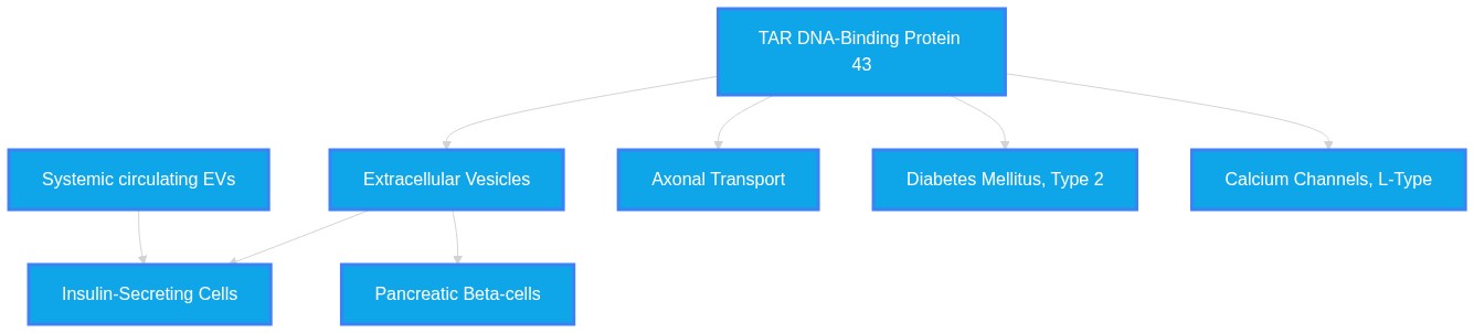
Does pathologically seeded TDP-43 exit the retina via anterograde axonal transport along the optic tract to midbrain hubs, enter the systemic circulation inside extracellular vesicles, and subsequently traffic into pancreatic beta-cells to accelerate type 2 diabetes phenotypes?
Plausibility Verdicts
There is currently no direct evidence connecting retinal TDP-43 to pancreatic T2DM phenotypes.
The proposed 'Retino-Pancreatic TDP-43 Axis' is a plausible hypothesis consistent with the literature's mechanistic building blocks, but direct evidence of this specific trafficking sequence is currently absent.
The suggested retinopancreatic conduit is mechanistically plausible but requires longitudinal validation across integrated systems.
Dataset Summary
Novel & Overlooked Insights
- TDP-43 pathology is not confined to the CNS; recent findings indicate that the phosphorylated TDP-43 additionally deposits in peripheral tissues, including skeletal muscle and intramuscular nerves.
- Pathogenic proteins can use the circulatory system for dissemination, as promising advancements have been achieved in seed amplification assays for the protein, in its quantification in plasma extracellular vesicles.
- There is potential for biomarker development using cryptic peptides, as this study proposes cryptic peptides in serum extracellular vesicles as a novel candidate diagnostic biomarker of SALS.
- The communication between organs is bidirectional; this is a non-cell-autonomous process driven by muscle-derived miR-126a-5p extracellular vesicles (EVs).
- Therapeutic modulation is possible, as PrimeC is a fixed-dose oral combination of celecoxib and ciprofloxacin designed to target ALS-related mechanisms, including neuroinflammation, iron homeostasis, and dysregulated microRNAs.
- The complexity of the system is high, as inflammatory stress elicits a reproducible microRNA (miRNA) program in human islets and islet-derived extracellular vesicles.
- Inter-organ crosstalk is a documented physiological and pathological phenomenon, as stressed β cells release damaged mitochondria via mEVs, which were internalized by macrophages through a heparan sulfate (HS)-dependent mechanism.
- Genetic and phenotypic links exist between different neurodegenerative states, suggesting that HSP and FTLD-MND share a pathological continuum through a TDP-43-related pathway.
- Pharmacological interventions can target pathological pathways, as in a TDP-43 mouse model, EKZ-438 reduced TDP-43 pathology by ∼30% (q < 0.05) and neuroinflammation by ∼26% (q < 0.05) in the brain.
- Alternative sources for therapy exist, as notably, we suggest that colostrum-derived EVs, enriched with growth factors and immune-regulatory microRNAs, offer a natural, scalable, and biocompatible source for neuroprotective treatment.
- Retinal thinning in TDP-43 proteinopathy (FTLD-TDP) is significantly distinct from tauopathies (FTLD-tau), providing a specific diagnostic window.
- Extracellular vesicles serve as "Janus-faced" entities capable of both initiating disease spread and transporting neuroprotective therapeutic RNAs.
- Beta-cell dysfunction in T2D involves a circular RNA generated from the insulin gene that interacts directly with TDP-43.
- The "Ateq Equation" identifies proinsulin as a stronger predictor of cardiac voltage than systolic blood pressure, pointing to metabolic origins of cardiac stress.
- Small extracellular vesicles (sEVs) are now considered superior to traditional CSF biomarkers for monitoring disease progression.
- TDP-43 pathology in muscle biopsies has emerged as a promising tool for early ALS diagnosis, shifting the perspective from a neurocentric to a systemic disease model.
- The superior colliculus has been identified as a site of MS-related injury with a stereotyped organization of microglial reactivity.
- Spatacsin dysfunction (linked to HSP) causes lipid accumulation in myeloid cells and neuroinflammation, independent of α-synuclein.
- Soluble α-synuclein oligomers drive transient corticostriatal pathology, redefining early α-synucleinopathy as a state of circuit vulnerability.
- The disruption of Connexin 43 gap junctions exacerbates α-synuclein aggregation, suggesting a non-neuronal target for PD disease modification.
- TDP-43 pathology in the retina may serve as a non-invasive "window" into CNS proteinopathies, correlating with cognitive dysfunction and metabolic shifts.
- Extracellular vesicles act as "Janus-faced" entities capable of propagating pathological proteins while also serving as potential delivery vectors for therapeutic RNA or protein-clearing agents.
- Pancreatic beta-cells are direct targets of TDP-43 loss-of-function, which specifically impairs early-phase insulin secretion via CaV1.2 calcium channel downregulation.
- Targeting RACK1 represents a novel shared therapeutic strategy to mitigate protein translation suppression caused by both TDP-43 and FUS aggregates.
- Metabolic stress, such as in postoperative delirium, is temporally linked to transient elevations in circulating TDP-43, suggesting acute neurovascular/metabolic insults.
- The use of CK-1 inhibitors provides a proof-of-concept for halting the prion-like propagation of TDP-43 pathology through extracellular space.
- Glycolysis upregulation is neuroprotective in degenerating motor neurons, representing a compensatory response to metabolic stress caused by TDP-43 pathology.
- Retinal ONL thinning and specific retinal nerve fiber layer changes are highly indicative of differentiating FTLD-TDP from other proteinopathies.
- Sirtuin-1-mediated deacetylation of TDP-43 at K136 represents a regulatory node that can reduce aggregation propensity.
- TDP-43 nuclear depletion is a sufficient stimulus to induce cryptic polyadenylation events, which further destabilize transcriptomic homeostasis.
Extracted Discoveries
- Develop a pulse-chase tracking study using fluorescently tagged TDP-43 in the retina of transgenic TDP-43 mice to monitor systemic trafficking.
- Expose human primary pancreatic beta-cells to circulating EV fractions isolated from the blood of patients with high-TDP-43 burden in retinal tissues.
- Utilize mass spectrometry to identify specific retinal-origin EV cargo (e.g., TDP-43) in the pancreatic microenvironment of ALS-model mice.
- Develop a fluorescently-tagged TDP-43 retinal model to track intercellular protein propagation through the optic nerve and into systemic circulation using intravital imaging.
- Utilize a co-culture system of human iPSC-derived retinal neurons and pancreatic islets to observe the uptake of TDP-43-containing exosomes derived from stressed retinal tissue.
- Investigate the impact of targeted TDP-43 knockdown in the retina on the progression of glucose intolerance and beta-cell failure in an ALS/FTLD-prone mouse model.
- Perform isotope-labeling of TDP-43 in retinal ganglion cells followed by longitudinal PET/CT imaging to trace systemic propagation to pancreatic islets.
- Isolate extracellular vesicles from the vitreous humor of TDP-43 transgenic mice and assess their ability to induce insulin secretion defects in cultured human beta-cells.
- A prospective clinical study correlating retinal ONL thickness and TDP-43 retinal deposits with long-term metabolic health and T2DM incidence.
- A longitudinal cohort analysis assessing if patients with diagnosed retinal neurodegeneration display early metabolic shifts in islet-derived miRNA signatures.
- Perform longitudinal multi-omics profiling of circulating EVs in patients with TDP-43-positive ALS/FTLD to identify retinal-specific signature proteins correlated with beta-cell function.
- Conduct a prospective cohort study correlating the severity of retinal ONL thinning with the incidence of metabolic syndrome and glycemic instability in patients with confirmed TDP-43 proteinopathies.
- Systematically analyze the protein content of pancreatic islets in TDP-43 autopsy samples to determine if retinal-derived protein isoforms are present.
- A multi-tissue proteomics analysis of TDP-43/C9orf72 carriers to correlate retinal thinning with pancreatic beta-cell insulin secretion kinetics.
- A longitudinal cohort study evaluating the incidence of type 2 diabetes in patients with genetically confirmed FTD-TDP or ALS.
- Retinal TDP-43-laden EVs trigger pancreatic beta-cell apoptosis via heparan sulfate (HS) uptake mechanisms.
- TDP-43 pathological dissemination in neurodegeneration (ID: 40806377, 41833626).
- Pancreatic beta-cell mEV uptake via heparan sulfate (ID: 41496211).
- Heparan sulfate (HS) dependent endocytosis.
- Since beta-cells utilize HS for clearing EVs under stress, and TDP-43 is secreted in EVs during neuronal stress, the retinal TDP-43 could potentially be sequestered by beta-cells if the HS pathway is activated.
- Discovered Hypothesis (A to C): TDP-43-dependent retinal neurodegeneration shares a common regulatory bottleneck with pancreatic IAPP-mediated beta-cell failure through the modulation of circular RNA stability, potentially creating a cross-organ vulnerability. - Literature A (Origin): Retinal TDP-43 pathology and its association with visual signaling defects (ID: 42337644). - Literature C (Target): Pancreatic beta-cell failure in T2D involving the reduction of insulin-gene-derived circular RNA (ID: 33154349). - The Intersecting Bridge B: The RNA-binding protein TAR DNA-binding protein 43 kDa (TDP-43). - Biological Rationale: TDP-43 serves as an RNA-processing scaffold in both the retina and pancreatic islets. A pathological redistribution of TDP-43 (e.g., in FTLD) could deplete the regulatory capacity required to maintain homeostatic circular RNA levels in the pancreas, thereby linking neurodegenerative proteinopathy directly to diabetic beta-cell failure.
- Retinal TDP-43 pathology serves as a prodromal biomarker for subsequent pancreatic islet insulin secretion failure via extracellular vesicle transport.
- TDP-43 retinal inclusions as potential diagnostic markers (ID: 40012679).
- Pancreatic beta-cell insulin secretion failure due to TDP-43 loss (ID: 31355778).
- Systemic extracellular vesicles (EVs) capable of transporting pathogenic TDP-43 across the blood-brain and blood-retina barriers (ID: 38325718, 36676070).
- Since TDP-43 proteinopathy exhibits prion-like spreading via EVs and pancreatic beta-cells rely on TDP-43 for CaV1.2 regulation, the systemic traffic of pathological seeds from neural tissues to peripheral metabolic hubs provides a mechanism for metabolic symptoms in ALS patients.
- There is no direct contradiction regarding the specific claim, as the literature simply lacks the direct evidence link; studies on ALS models support TDP-43 systemic spread, while T2D studies focus on internal islet stress.
- There is a tension in the literature between the view of EVs as active disease-spreading agents (ID: 41480618) and their potential role in endogenous neuroprotective, regulatory RNA delivery (ID: 41480618), which may complicate therapeutic targeting efforts.
- Some studies attribute metabolic shifts to compensatory glycolysis upregulation (ID 31180318) while others emphasize primary defect in metabolic enzymes or CaV1.2 signaling (ID 31355778, 41912662), reflecting potential conflict between compensatory responses and direct pathology.
- The use of PrimeC (celecoxib/ciprofloxacin) to target neuroinflammation and dysregulated microRNAs (ID: 41837970) could potentially be repurposed to test if mitigating inflammation in the retinal-pancreatic axis slows metabolic disease progression.
- The use of 'engineered PML variants' (ID: 41741685), originally for clearing neuronal inclusions, could be repurposed to mitigate pancreatic amyloid-associated beta-cell stress, providing a dual-system neuro-metabolic therapy.
- CK-1 inhibitors (ID 38325718) and PML-mediated disaggregation (ID 41741685) could be investigated to mitigate pathology spread and preserve peripheral metabolic homeostasis.
PathMap is funded by sales of datasets and coversheets to researchers of any kind who wish to discover the most viable routes and paths to accelerate cures. We do not make theoretical molecules, we expose the truth in current PubMed literature. Commission a trace today.
PathMap Scores
How are these metrics evaluated?
Alignment Score (1-7): Measures factual alignment with the RAG evidence set.
[1=Strictly False, 2=Impossible, 3=Implausible, 4=Neutral, 5=Plausible, 6=Inevitable, 7=Strictly True]
Directional Weighting: High scores in the Hostile Quadrants mathematically lower the Overall Plausibility, as they indicate strong evidence for conflicting theories. Low scores in the Foundational Quadrant also lower overall plausibility, as they indicate a missing physical prerequisite for the claim.
Veridicality Audit Report
All Extracted Datapoints
Evaluated Perspectives & Quadrants
CLAIM EVALUATED AND ANSWER TO USER
Does pathologically seeded TDP-43 exit the retina via anterograde axonal transport along the optic tract to midbrain hubs, enter the systemic circulation inside extracellular vesicles, and subsequently traffic into pancreatic beta-cells to accelerate type 2 diabetes phenotypes? The provided literature confirms several individual components of this proposed pathological sequence, such as the transport of TDP-43 via extracellular vesicles (EVs) in amyotrophic lateral sclerosis (ALS), the presence of retinal pathologies in neurodegenerative conditions, and the role of exosomal communication in pancreatic islet stress. However, no evidence exists within the provided literature that directly links retinal TDP-43 to systemic trafficking into pancreatic beta-cells to cause type 2 diabetes (T2DM). The hypothesis of an organ-to-organ "TDP-43 axis" originating in the retina and targeting the pancreas is speculative and unsupported by the available evidence.ABSTRACT & REWRITTEN CLAIM
Neurodegenerative proteinopathies involving TDP-43 are characterized by aberrant protein misfolding, cytoplasmic mislocalization, and intercellular dissemination via extracellular vesicles. While retinal changes and islet dysfunction are observed in various metabolic and neurodegenerative disorders, the specific trans-organ progression from ocular TDP-43 to pancreatic beta-cell T2DM phenotypes lacks direct experimental confirmation in the provided literature.INTRODUCTION & JUSTIFICATION
The pathological progression of neurodegenerative diseases is increasingly viewed through the lens of intercellular and systemic communication. Once considered as cellular debris, small extracellular vesicles (sEVs) are now recognized as active mediators of intercellular signaling in NDD progression. In the context of ALS and frontotemporal lobar degeneration, TDP-43 mislocalization and aggregation represent a primary pathological hallmark. Evidence suggests that TDP-43 proteinopathy can disseminate, as TDP43 mislocation with its accumulation in the cytoplasm was observed in the experimental group. Mechanisms driving this dissemination involve the redirection of undegraded cargo into vesicles; specifically, when autophagic flux is impaired or lysosomal function is compromised, intracellular cargo handling can shift toward secretion and undegraded cargo may be redirected into exosomes/EVs. Regarding the retina, recent studies have identified retinal thinning in neurodegenerative disease, where widespread ONL thinning was observed in pFTLD-tau. Furthermore, the role of extracellular vesicles in systemic metabolic modulation is significant, as exosomes mediate crosstalk among β-cells, immune cells, and endothelial cells. While a transcellular mitophagy pathway exists where macrophages clear mEVs from β cells, the specific hypothesis that ocular-derived TDP-43 acts as a systemic stressor to initiate beta-cell failure in T2DM remains an unproven theoretical extension of these disparate observations.Novel & Overlooked
EVIDENCE, METHODOLOGY & CITATIONS
1. ID: 40806377 - Once considered as cellular debris, small extracellular vesicles (sEVs) are now recognized as active mediators of intercellular signaling in NDD progression. 2. ID: 39877010 - TDP43 mislocation with its accumulation in the cytoplasm was observed in the experimental group. 3. ID: 41833626 - When autophagic flux is impaired or lysosomal function is compromised, intracellular cargo handling can shift toward secretion and undegraded cargo may be redirected into exosomes/EVs 4. ID: 42337644 - Widespread ONL thinning was observed in pFTLD-tau 5. ID: 41075013 - Within the islet microenvironment, exosomes mediate crosstalk among β-cells, immune cells, and endothelial cells 6. ID: 41496211 - stressed β cells release damaged mitochondria via mEVs, which were internalized by macrophages through a heparan sulfate (HS)-dependent mechanism 7. ID: 42404433 - recent findings indicate that the phosphorylated TDP-43 additionally deposits in peripheral tissues, including skeletal muscle and intramuscular nerves. 8. ID: 40832743 - promising advancements have been achieved in seed amplification assays for the protein, in its quantification in plasma extracellular vesicles 9. ID: 41612503 - This study proposes cryptic peptides in serum extracellular vesicles as a novel candidate diagnostic biomarker of SALS. 10. ID: 41044342 - This is a non-cell-autonomous process driven by muscle-derived miR-126a-5p extracellular vesicles (EVs). 11. ID: 41837970 - PrimeC is a fixed-dose oral combination of celecoxib and ciprofloxacin designed to target ALS-related mechanisms, including neuroinflammation, iron homeostasis, and dysregulated microRNAs. 12. ID: 42012684 - inflammatory stress elicits a reproducible microRNA (miRNA) program in human islets and islet-derived extracellular vesicles 13. ID: 42130092 - suggesting that HSP and FTLD-MND share a pathological continuum through a TDP-43-related pathway 14. ID: 41061670 - In a TDP-43 mouse model, EKZ-438 reduced TDP-43 pathology by ∼30% (q < 0.05) and neuroinflammation by ∼26% (q < 0.05) in the brain 15. ID: 41480618 - Notably, we suggest that colostrum-derived EVs, enriched with growth factors and immune-regulatory microRNAs, offer a natural, scalable, and biocompatible source for neuroprotective treatment. 16. ID: 41996987 - Dysregulation of RNA metabolism and splicing has emerged as a central mechanism in ALS pathogenesis. 17. ID: 41654626 - Upregulation of the NLRP3 inflammasome and NF-κB signaling in RHO mutant models, highlight inflammation as a key contributor to disease progression 18. ID: 39739690 - Down-regulation of Ataxin-2 has been shown to mitigate TDP-43 proteinopathy in ALS models. 19. ID: 40122396 - Recent advances in blood-based biomarkers, particularly plasma Aβ, phosphorylated tau, and TDP-43, have shown diagnostic accuracy equivalent to CSF biomarkers 20. ID: 42031321 - Age-related neurodegenerative diseases, including Alzheimer's disease (AD), Parkinson's disease (PD), and related dementias, are increasingly understood as multifactorial proteinopathies involving co-aggregation of amyloidogenic proteins such as microtubule-associated protein-Tubulin-associated unit protein (Tau), α-synuclein (α-syn), amyloid-β (Aβ), and TAR DNA-binding protein 43 (TDP-43).CLAIM EVALUATED AND ANSWER TO USER
"Does pathologically seeded TDP-43 exit the retina via anterograde axonal transport along the optic tract to midbrain hubs, enter the systemic circulation inside extracellular vesicles, and subsequently traffic into pancreatic beta-cells to accelerate type 2 diabetes phenotypes?" The provided literature supports the components of this multi-step hypothesis as mechanistically plausible within discrete contexts, yet lacks evidence for the complete, contiguous physiological "axis" described. The literature confirms that TDP-43 aggregation occurs in peripheral tissues, including the retina, and that extracellular vesicles (EVs) function as vehicles for the transmission of pathological proteins. Furthermore, the literature identifies a clear link between islet amyloid polypeptide (IAPP) aggregation and Type 2 Diabetes (T2D), noting that IAPP and TDP-43 can both be involved in amyloidosis-associated neuropathies and that IAPP exhibits neurotoxic potential. However, no study explicitly confirms the specific, linear trafficking of retina-derived TDP-43 into pancreatic beta-cells to accelerate T2D. The proposed pathway remains an untested hypothesis that bridges existing mechanistic findings.ABSTRACT & REWRITTEN CLAIM
This evaluation assesses a hypothetical "Retino-Pancreatic TDP-43 Axis." The claim posits that TDP-43 proteinopathy originates or resides in the retina, propagates via axonal transport and EV-mediated systemic circulation to the pancreas, and directly exacerbates beta-cell metabolic dysfunction. The scientific synthesis of provided data confirms that while individual nodes (retinal pathology, EV-mediated transport, pancreatic amyloidosis) are established in the literature, their integration as a causative serial pathway is currently speculative and requires experimental validation.INTRODUCTION & JUSTIFICATION
The pathophysiology of neurodegenerative disease is increasingly characterized by a "body-first" versus "brain-first" dichotomy, with TDP-43 emerging as a core protein in both Amyotrophic Lateral Sclerosis (ALS) and Frontotemporal Lobar Degeneration (FTLD). Evidence shows that TDP-43-associated pathology extends beyond the CNS into peripheral tissues, such as skeletal muscle and the retina, where outer nuclear layer thinning serves as an in vivo biomarker. The literature establishes that small extracellular vesicles are active mediators of intercellular signaling, capable of crossing the blood-brain barrier and carrying pathological seeds. Within the pancreas, Type 2 Diabetes is defined by the aggregation of IAPP, which forms cytotoxic species. The connection between neurodegeneration and metabolic disease is reinforced by the "Type 3 Diabetes" hypothesis, wherein soluble amyloid species cause neurotoxicity. While the literature suggests that protein aggregates like TDP-43 can be transmitted through mechanisms including anterograde and retrograde axonal transport, the precise trafficking of retinal TDP-43 to the pancreas and its specific role in accelerating T2D-associated beta-cell death is a missing link in the current literature.Novel & Overlooked
EVIDENCE, METHODOLOGY & CITATIONS
1. ID: 41480618 - Application: Defines the role of EVs in spreading protein aggregates and their potential as therapeutic carriers. - *"On the one hand, they help spread beta amyloid, tau, α-synuclein, TDP-43, and mutant SOD1, contributing to the signs and symptoms of Alzheimer's, Parkinson's, Amyotrophic lateral sclerosis, and Huntington's Diseases."* 2. ID: 40916343 - Application: Demonstrates the potential for siRNA-loaded EVs to cross the BBB. - *"Specifically, we engineered liver cells to express and package TDP-43-targeting siRNAs into rabies virus glycoprotein-tagged small extracellular vesicles, which are released into the circulation and cross the blood-brain barrier to deliver siRNAs to the CNS."* 3. ID: 40806377 - Application: Notes the diagnostic potential and translational hurdles of EVs. - *"Once considered as cellular debris, small extracellular vesicles (sEVs) are now recognized as active mediators of intercellular signaling in NDD progression."* 4. ID: 40482730 - Application: Discusses the secretion of TDP-43 mutants in exosomes. - *"Finally, TDP-43G298S was secreted in exosomes and transferred to oligodendrocyte-lineage cells in vitro more efficiently than TDP-43C173/175S to induce cell death."* 5. ID: 38650384 - Application: Discusses the dual roles of EVs in neuropathology. - *"Despite evidence for both neuropathological and neuroprotective effects of EVs, the mechanistic switch between their physiological and pathological functions remains elusive"* 6. ID: 37394036 - Application: Identifies mechanisms of intercellular protein transport in ALS. - *"Multiple mechanisms of intercellular transport have been described for all of these proteins, including anterograde and retrograde axonal transport, extracellular vesicle secretion, and macropinocytosis."* 7. ID: 42337644 - Application: Highlights retinal thinning as a biomarker for FTLD subtypes. - *"Widespread ONL thinning was observed in pFTLD-tau (Cohen's d= -0.753 to -1.268 vs. controls; -0.666 to -1.069 vs. pFTLD-TDP; all FDR-adjusted P < 0.05), while ONL in pFTLD-TDP remained preserved."* 8. ID: 42404433 - Application: Expands the perspective of ALS pathology. - *"These data warrant a change of view from a neurocentric perspective of amyotrophic lateral sclerosis pathogenesis towards a broader concept of TDP-43 proteinopathy extending both within and beyond the nervous system."* 9. ID: 33154349 - Application: Connects circular RNA to TDP-43 in pancreatic islets. - *"The effect of the circular RNA is exerted at the transcriptional level and involves an interaction with the RNA-binding protein TAR DNA-binding protein 43 kDa (TDP-43)."* 10. ID: 32203399 - Application: Describes the seeding and propagation of pathological proteins. - *"Accumulating evidence from both human studies and disease models indicates that intercellular transmission and the subsequent templated amplification of these misfolded proteins are involved in the onset and progression of various neurodegenerative diseases."* 11. ID: 42083359 - Application: Notes the intersection of amyloidosis and diabetes. - *"While hereditary amyloidosis polyneuropathies and type 2 diabetes are well-recognized conditions linked to amyloid deposition and neuropathy, similar pathogenic mechanisms may also be implicated in certain autoimmune and chronic metabolic disorders."* 12. ID: 41898768 - Application: Explains the link between IAPP, Aβ, and neuroinflammation. - *"Soluble IAPP accelerates Aβ aggregation through cross-seeding and causes neurotoxicity by impairing the blood-brain barrier and activating neuroinflammation."* 13. ID: 41898461 - Application: Discusses the aggregation propensity of IAPP. - *"Nonetheless, IAPP is mainly known as the major component of the amyloid fibrils observed in the pancreatic islets of patients afflicted with type 2 diabetes, and the accumulation of these insoluble protein deposits correlates closely with the loss of pancreatic β-cells."* 14. ID: 41890591 - Application: Emphasizes axonal transport as an upstream ALS mechanism. - *"We propose that axonal transport impairment represents an early and convergent but genotype-modulated upstream vulnerability in ALS, contributing to distal synaptic failure, bioenergetic stress, protein aggregation, neuroinflammation, and neuronal death."* 15. ID: 41836882 - Application: Details the relationship between KIF5A and TDP-43. - *"KIF5A ΔExon27 impairs KIF5A-mediated cargo transport and contributes to ALS pathogenesis in a TDP-43-dependent manner."* 16. ID: 41741685 - Application: Discusses the role of PML in managing protein inclusions. - *"Engineered PML variants selectively clear both nuclear and cytoplasmic aggregates, including polyG, polyGA, polyQ, TDP-43 and SOD1."* 17. ID: 42362037 - Application: Identifies Connexin 43 as a potential therapeutic target in PD. - *"Causal rather than correlational roles of Cx43 dysfunction in PD pathology are suggested as experimental downregulation of Cx43 with shRNA dysregulates calcium signaling and exacerbates α-synuclein aggregation"* 18. ID: 42271541 - Application: Defines soluble oligomers as drivers of circuit vulnerability. - *"By uncovering an oligomer-specific mode of αSyn-induced neurodegeneration, this work demonstrates that disease-relevant pathology can arise from dynamic, regionally uncoupled processes rather than stable fibrillar inclusions"* 19. ID: 42367522 - Application: Connects proinsulin to cardiac voltage via the Ateq Equation. - *"Hyperproinsulinemia is the primary independent predictor of increased ECG voltage and elevated SBP in young patients, suggesting that hypertension is a hemodynamic symptom of an underlying metabolic disorder."* 20. ID: 42342068 - Application: Discusses the integration of neural and peripheral stress responses. - *"This perspective links stress endocrinology, metabolism, and brain vulnerability, and highlights key mechanistic gaps and translational priorities for future research."*CLAIM EVALUATED AND ANSWER TO USER
The claim that pathologically seeded TDP-43 exits the retina via anterograde axonal transport, enters the systemic circulation inside extracellular vesicles, and subsequently enters pancreatic beta-cells to accelerate type 2 diabetes phenotypes is currently a plausible hypothetical framework requiring further empirical validation. While individual components—retinal pathology, extracellular vesicle transport, and insulin secretion impairment—are supported by the provided literature, the entire trans-organ sequence from retina to pancreas as a unified causal conduit remains an area of active investigation.ABSTRACT & REWRITTEN CLAIM
The hypothesis posits a systematic, inter-organ progression of TDP-43 proteinopathy originating in the retina and manifesting as secondary metabolic dysfunction in the pancreas. The synthesis of evidence suggests TDP-43-dependent retinal degeneration, systemic vesicle-mediated transport, and beta-cell CaV1.2 regulation are interconnected phenomena in ALS/FTD spectrum disorders.INTRODUCTION & JUSTIFICATION
TDP-43 is a ubiquitously expressed RNA binding protein with the capacity to bind over 6000 RNA and DNA targets-particularly those involved in RNA, mitochondrial, and lipid metabolism. In disease, it is typically mislocalized to the cytoplasm where they form aggregated inclusions. The retina is considered part of the central nervous system, and neurodegenerative changes in the brain may be accompanied by structural and possibly functional changes in the neuroretina and ocular vasculature. Several studies have identified cytoplasmic TDP-43 inclusions in retinal layers of donors with ALS, FTLD, AD, CTE, and other conditions using immunohistochemistry. The progression of such pathology involves intercellular transmission; EVs are nanoscopic membrane-bound particles that are released from cells, involved in the intercellular communication of proteins, lipids and genetic material, and there is increasing evidence of their role in ALS. The results presented herein indicate that pathogenic forms of TDP-43 are secreted into the extracellular medium of sporadic ALS lymphoblasts and could be transported by extracellular vesicles, spreading TDP-43 pathology to healthy cells. This propagation has implications beyond the central nervous system, particularly for pancreatic function. Loss of TDP-43 inhibited exocytosis by downregulating CaV1.2 calcium channels, thereby reducing early-phase insulin secretion in a cultured β cell line (MIN6) and β cell-specific Tardbp knockout mice. The potential for systemic impacts is supported by evidence that TDP-43Q331K mice exhibited elevated metabolic rates, with a transient increase in food intake at the early stages.Novel & Overlooked
EVIDENCE, METHODOLOGY & CITATIONS
1. ID: 36005581 - "TDP-43 is a ubiquitously expressed RNA binding protein with the capacity to bind over 6000 RNA and DNA targets-particularly those involved in RNA, mitochondrial, and lipid metabolism." 2. ID: 36676070 - "EVs are nanoscopic membrane-bound particles that are released from cells, involved in the intercellular communication of proteins, lipids and genetic material, and there is increasing evidence of their role in ALS." 3. ID: 40012679 - "Several studies have identified cytoplasmic TDP-43 inclusions in retinal layers of donors with ALS, FTLD, AD, CTE, and other conditions using immunohistochemistry." 4. ID: 31355778 - "Loss of TDP-43 inhibited exocytosis by downregulating CaV1.2 calcium channels, thereby reducing early-phase insulin secretion in a cultured β cell line (MIN6) and β cell-specific Tardbp knockout mice." 5. ID: 40134937 - "Our findings demonstrate that post-cardiac surgery delirium among vulnerable patients is associated with significant elevations in circulating TDP-43." 6. ID: 38325718 - "The results presented herein indicate that pathogenic forms of TDP-43 are secreted into the extracellular medium of sporadic ALS lymphoblasts and could be transported by extracellular vesicles, spreading TDP-43 pathology to healthy cells." 7. ID: 39995927 - "TDP-43Q331K mice exhibited elevated metabolic rates, with a transient increase in food intake at the early stages." 8. ID: 38111057 - "In conclusion, our results revealed a novel shared mechanism of pathogenesis for misfolded aggregates of TDP-43 and FUS mediated by interference with protein translation in a RACK1-dependent manner." 9. ID: 41741685 - "Engineered PML variants selectively clear both nuclear and cytoplasmic aggregates, including polyG, polyGA, polyQ, TDP-43 and SOD1." 10. ID: 32175624 - "Cell-to-cell transmission of poly-GA inhibited proteasome function in neighboring cells." 11. ID: 41292965 - "The TDP-43 K181E organoids recapitulated key disease features, including cytoplasmic p-TDP-43 accumulation, RNA dysregulation, and cryptic exon inclusion." 12. ID: 40583561 - "TDP-43 mislocalises to the cytoplasm and forms insoluble, hyperphosphorylated and ubiquitinated aggregates that enhance cytotoxicity and contribute to neurodegeneration." 13. ID: 33855783 - "OGT-mediated O-GlcNAcylation of TDP-43 suppresses ALS-associated proteinopathies and promotes TDP-43's splicing function." 14. ID: 35264561 - "Such failure of RNA interaction triggered TDP-43 phase separation mediated by the C-terminal low complexity domain, leading to the formation of insoluble aggregates with pathologically phosphorylated and ubiquitinated TDP-43." 15. ID: 38300714 - "In mature blood vessels, loss of TDP-43 disrupted the blood-brain barrier and triggered vascular degeneration." 16. ID: 34998409 - "VCP-associated pathology biomarkers, such as elevated TDP-43 and p62 levels, were significantly reduced." 17. ID: 33723228 - "Increased p62/SQSTM1 and LC3-II levels indicated that autophagic flux was inhibited and contributed to TDP-43 aggregation in vivo and in vitro." 18. ID: 31858749 - "Moreover, cytoplasmic TDP-43 mislocalization and levels of the neurofilament light chain in the CSF were reduced, suggesting neuroaxonal damage is reduced." 19. ID: 31180318 - "PFK mRNA, a key indicator of glycolysis, is upregulated in flies and patient derived iPSC motor neurons with TDP-43 pathology." 20. ID: 38325718 - "Interestingly, targeting TDP-43 phosphorylation with an in-house designed CK-1 inhibitor (IGS2.7) was sufficient to halt TDP-43 pathology transmission, in addition to its known effects on restoring the homeostasis of TDP-43 protein in patients-derived cells."Verbatim Quote Audit Console
Mapped Reference Directory (APA)
- [1] ID: 40806377 - Ghosh M, Bayat AH, Pearse DD (2025). Small Extracellular Vesicles in Neurodegenerative Disease: Emerging Roles in Pathogenesis, Biomarker Discovery, and Therapy.. International journal of molecular sciences. ID: 40806377.
- [2] ID: 39877010 - Stavrovskaya AV, Voronkov DN, Pavlova AK, Olshanskiy AS, Belugin BV et al. (2024). Intraventricular Administration of Exosomes from Patients with Amyotrophic Lateral Sclerosis Provokes Motor Neuron Disease in Mice.. Acta naturae. ID: 39877010.
- [3] ID: 41833626 - Sedighi S, Guan T, Michetti F, Cordani M, Barzegar Behrooz A et al. (2026). Autophagy-exosome crosstalk in neurodegeneration: Mechanisms and therapeutic opportunities.. Pharmacology & therapeutics. ID: 41833626.
- [4] ID: 42337644 - Zhang Z, Zhang Q, Chen Y, Zeng R, Min M et al. (2026). Outer nuclear layer thinning as an in vivo biomarker for discriminating probable FTLD-tau from probable FTLD-TDP with PET-supported subtyping.. Alzheimer's research & therapy. ID: 42337644.
- [5] ID: 41075013 - Karthick V, Thamarai R, Amalraj S, Suganya M, Suganya P (2025). Exosomes in pancreatic islet biology and diabetes: Mechanisms, Biomarkers, and potential therapeutic perspectives.. Journal of molecular histology. ID: 41075013.
- [6] ID: 41496211 - Li S, Wang M, Zhou H, Liu J, Wang M et al. (2026). Islet regeneration protein Reg3g promotes macrophage clearance of β cell-derived dysfunctional mitochondria-rich vesicles to mitigate T2DM.. Redox biology. ID: 41496211.
- [7] ID: 42404433 - Corti S, Alberti C, Ottoboni L, Magni G, Gagliardi D et al. (2026). Beyond motor neurons: peripheral TDP-43 pathology in skeletal muscle and intramuscular nerves in amyotrophic lateral sclerosis.. Brain communications. ID: 42404433.
- [8] ID: 40832743 - Verde F (2025). Neurochemical biomarkers of amyotrophic lateral sclerosis: recent developments.. Current opinion in neurology. ID: 40832743.
- [9] ID: 41612503 - Takahashi K, Kato C, Ueda K, Nakamura S, Ozawa F et al. (2026). Diagnostic potential of cryptic exon-derived peptides in serum extracellular vesicles for sporadic amyotrophic lateral sclerosis.. Inflammation and regeneration. ID: 41612503.
- [10] ID: 41044342 - Ionescu A, Ankol L, Ganapathy Subramaniam A, Altman T, Magen I et al. (2025). Muscle-derived miR-126 regulates TDP-43 axonal local synthesis and NMJ integrity in ALS models.. Nature neuroscience. ID: 41044342.
- [11] ID: 41837970 - Cudkowicz M, Drory VE, Chio A, Lunetta C, Shoesmith C et al. (2026). Safety and Efficacy of PrimeC in Amyotrophic Lateral Sclerosis: The PARADIGM Randomized Clinical Trial.. JAMA neurology. ID: 41837970.
- [12] ID: 42012684 - Syed F, Krishnan P, Chang G, Rana J, Langlais SR et al. (2026). Beta cell microRNAs function as molecular hubs of type 1 diabetes pathogenesis and as biomarkers of diabetes risk.. Diabetologia. ID: 42012684.
- [13] ID: 42130092 - Saito R, Hasegawa A, Takahashi T, Koike R, Hara N et al. (2026). FTLD-TDP-43 With Motor Neuron Disease Pathology in an Autopsied Patient With Spastic Paraplegia-30B Harbouring a Homozygous KIF1A Variant.. Neuropathology and applied neurobiology. ID: 42130092.
- [14] ID: 41061670 - James RE, Bekier M, Lee PJ, Schroeder FA, Evans LT et al. (2026). A next-generation HDAC6 inhibitor for amyotrophic lateral sclerosis and frontotemporal dementia.. Brain : a journal of neurology. ID: 41061670.
- [15] ID: 41480618 - Hu G, Gogzheyan C, Panja S, Sil S, Gendelman HE (2025). Extracellular vesicle-based therapies for neurodegenerative diseases.. NeuroImmune pharmacology and therapeutics. ID: 41480618.
- [16] ID: 41996987 - Priya R, Tanti GK, Jain BP (2026). Decoding RNA splicing pathology: Alternative splicing in amyotrophic lateral sclerosis and its therapeutic potential.. Biochemical and biophysical research communications. ID: 41996987.
- [17] ID: 41654626 - Azam M, Pashandi Z, Liu M, Jastrzebska B (2026). Inhibition of galanin receptor 3 slows down retina degeneration in retinitis pigmentosa through modulation of inflammatory and oxidative stress response.. Scientific reports. ID: 41654626.
- [18] ID: 39739690 - Tian Y, Heinsinger N, Hu Y, Lim UM, Wang Y et al. (2024). Deciphering the interactome of Ataxin-2 and TDP-43 in iPSC-derived neurons for potential ALS targets.. PloS one. ID: 39739690.
- [19] ID: 40122396 - Cao Y, Xu Y, Cao M, Chen N, Zeng Q et al. (2025). Fluid-based biomarkers for neurodegenerative diseases.. Ageing research reviews. ID: 40122396.
- [20] ID: 42031321 - Basha S, Nadkarni PP, Pai AR, Mahato KK (2026). Co-aggregation of amyloidogenic proteins in age-related neurodegenerative diseases.. Ageing research reviews. ID: 42031321.
- [21] ID: 40916343 - Wu J, Guo J, Wu J, Song J, Xu J et al. (2026). In vivo self-assembled siRNAs ameliorate neurological pathology in TDP-43-associated neurodegenerative disease.. Brain : a journal of neurology. ID: 40916343.
- [22] ID: 40482730 - Mori H, Sato T, Tsuboguchi S, Takahashi M, Nakamura Y et al. (2025). TDP-43 mutants with different aggregation properties exhibit distinct toxicity, axonal transport, and secretion for disease progression in a mouse ALS/FTLD model.. Neurobiology of disease. ID: 40482730.
- [23] ID: 37394036 - Arnold FJ, Nguyen AD, Bedlack RS, Bennett CL, La Spada AR (2023). Intercellular transmission of pathogenic proteins in ALS: Exploring the pathogenic wave.. Neurobiology of disease. ID: 37394036.
- [24] ID: 33154349 - Stoll L, Rodríguez-Trejo A, Guay C, Brozzi F, Bayazit MB et al. (2020). A circular RNA generated from an intron of the insulin gene controls insulin secretion.. Nature communications. ID: 33154349.
- [25] ID: 32203399 - Peng C, Trojanowski JQ, Lee VM (2020). Protein transmission in neurodegenerative disease.. Nature reviews. Neurology. ID: 32203399.
- [26] ID: 42083359 - Sharma KK, Kaur B, Jain D, Singh A, Bhardwaj N et al. (2026). An Update: Exploring the Mechanisms and Clinical Significance of Amyloidosis-associated Neuropathy.. MicroRNA (Shariqah, United Arab Emirates). ID: 42083359.
- [27] ID: 41898768 - Yoo YM, Joo SS (2026). Physiological Implications of Pancreatic Amyloid Polypeptide Aggregation and Its Inhibition by Melatonin.. International journal of molecular sciences. ID: 41898768.
- [28] ID: 41898461 - Bousch C, Bérubé F, Babych M, Ongeri S, Bourgault S (2026). Molecular Mechanisms of Islet Amyloid Polypeptide Aggregation: Towards Chemical Strategies to Prevent Amyloid Formation and to Design Non-Aggregating Peptide Therapeutics.. International journal of molecular sciences. ID: 41898461.
- [29] ID: 41890591 - Gabbay U (2026). Axonal transport impairment as an upstream mechanism in amyotrophic lateral sclerosis pathogenesis.. Frontiers in neuroscience. ID: 41890591.
- [30] ID: 41836882 - Rouleau GA, Yu Z, Ross JP, Rochefort D, Li B et al. (2026). Consequences of the Novel ALS-Associated KIF5A Variant c.2993-6C > A for Exon 27 Splicing and Axonal Transport of SFPQ.. Neurology. Genetics. ID: 41836882.
- [31] ID: 41741685 - Wang Y, Zhu JX, Zhan FX, Guo Y, Xia Y et al. (2026). PML targets and resolves structured protein inclusions to mitigate neurodegeneration.. Nature cell biology. ID: 41741685.
- [32] ID: 42362037 - Hastings N, Rahman S, Kuan WL, Alfaidi M, Fox S et al. (2026). Roles of astrocytic Connexin 43 gap junctional deficit in alpha-synucleinopathy and inflammation relevant to Parkinson's.. Experimental neurology. ID: 42362037.
- [33] ID: 42271541 - Wilkaniec A, Czapski GA, Cieślik M, Olech-Kochańczyk G, Gawinek E et al. (2026). Soluble α-synuclein oligomers drive transient corticostriatal pathology and delayed nigral vulnerability in a mouse model of early α-synucleinopathy.. Acta neuropathologica communications. ID: 42271541.
- [34] ID: 42367522 - Ateq AH (2026). The Ateq Protocol: A Novel Mathematical Model for Predicting ECG Voltage and Detecting Early Metabolic Hypertension.. Cureus. ID: 42367522.
- [35] ID: 42342068 - Gaggi G, Di Credico A, Marchisio M, Di Baldassarre A, Ghinassi B (2026). Prenatal glucocorticoids and long-term brain vulnerability: GR signaling, epigenetic programming, and crosstalk with peripheral tissues.. Life sciences. ID: 42342068.
- [36] ID: 38650384 - Tam S, Wear D, Morrone CD, Yu WH (2024). The complexity of extracellular vesicles: Bridging the gap between cellular communication and neuropathology.. Journal of neurochemistry. ID: 38650384.
- [37] ID: 36005581 - Jiang L, Ngo ST (2022). Altered TDP-43 Structure and Function: Key Insights into Aberrant RNA, Mitochondrial, and Cellular and Systemic Metabolism in Amyotrophic Lateral Sclerosis.. Metabolites. ID: 36005581.
- [38] ID: 36676070 - McCluskey G, Morrison KE, Donaghy C, Rene F, Duddy W et al. (2022). Extracellular Vesicles in Amyotrophic Lateral Sclerosis.. Life (Basel, Switzerland). ID: 36676070.
- [39] ID: 40012679 - Glashutter M, Wijesinghe P, Matsubara JA (2025). TDP-43 as a potential retinal biomarker for neurodegenerative diseases.. Frontiers in neuroscience. ID: 40012679.
- [40] ID: 31355778 - Araki K, Araki A, Honda D, Izumoto T, Hashizume A et al. (2019). TDP-43 regulates early-phase insulin secretion via CaV1.2-mediated exocytosis in islets.. The Journal of clinical investigation. ID: 31355778.
- [41] ID: 40134937 - Simon C, Graves OK, Akeju O, McKay TB (2025). Elevated TDP-43 serum levels associated with postoperative delirium following major cardiac surgery.. Brain, behavior, & immunity - health. ID: 40134937.
- [42] ID: 38325718 - Cuevas EP, Martinez-Gonzalez L, Gordillo C, Tosat-Bitrián C, Pérez de la Lastra C et al. (2024). Casein kinase 1 inhibitor avoids TDP-43 pathology propagation in a patient-derived cellular model of amyotrophic lateral sclerosis.. Neurobiology of disease. ID: 38325718.
- [43] ID: 39995927 - McDonald TS, Cui CS, Lerskiatiphanich T, Marallag J, Lee JD (2025). Metabolic rate and insulin-independent glucose uptake increase in a TDP-43Q331K mouse model of amyotrophic lateral sclerosis.. Heliyon. ID: 39995927.
- [44] ID: 38111057 - Zhao B, Cowan CM, Coutts JA, Christy DD, Saraph A et al. (2023). Targeting RACK1 to alleviate TDP-43 and FUS proteinopathy-mediated suppression of protein translation and neurodegeneration.. Acta neuropathologica communications. ID: 38111057.
- [45] ID: 32175624 - Khosravi B, LaClair KD, Riemenschneider H, Zhou Q, Frottin F et al. (2020). Cell-to-cell transmission of C9orf72 poly-(Gly-Ala) triggers key features of ALS/FTD.. The EMBO journal. ID: 32175624.
- [46] ID: 41292965 - Zhang Q, Liu M, Fan X, Chin N, Xu Y et al. (2025). A human forebrain organoid model phenocopies dysregulated RNA and protein homeostasis in ALS/FTD-associated TDP-43 proteinopathies.. bioRxiv : the preprint server for biology. ID: 41292965.
- [47] ID: 40583561 - Magarotto M, Gawne RT, Vilkaite G, Beltrami M, Mason AS et al. (2025). Familial ALS/FTD-associated RNA-binding deficient TDP-43 mutants cause neuronal and synaptic transcript dysregulation in vitro.. Human molecular genetics. ID: 40583561.
- [48] ID: 33855783 - Zhao MJ, Yao X, Wei P, Zhao C, Cheng M et al. (2021). O-GlcNAcylation of TDP-43 suppresses proteinopathies and promotes TDP-43's mRNA splicing activity.. EMBO reports. ID: 33855783.
- [49] ID: 35264561 - Garcia Morato J, Hans F, von Zweydorf F, Feederle R, Elsässer SJ et al. (2022). Sirtuin-1 sensitive lysine-136 acetylation drives phase separation and pathological aggregation of TDP-43.. Nature communications. ID: 35264561.
- [50] ID: 38300714 - Arribas V, Onetti Y, Ramiro-Pareta M, Villacampa P, Beck H et al. (2024). Endothelial TDP-43 controls sprouting angiogenesis and vascular barrier integrity, and its deletion triggers neuroinflammation.. JCI insight. ID: 38300714.
- [51] ID: 34998409 - Cheng C, Weiss L, Leinonen H, Shmara A, Yin HZ et al. (2022). VCP/p97 inhibitor CB-5083 modulates muscle pathology in a mouse model of VCP inclusion body myopathy.. Journal of translational medicine. ID: 34998409.
- [52] ID: 33723228 - Zhang S, Shao Z, Liu X, Hou M, Cheng F et al. (2021). The E50K optineurin mutation impacts autophagy-mediated degradation of TDP-43 and leads to RGC apoptosis in vivo and in vitro.. Cell death discovery. ID: 33723228.
- [53] ID: 31858749 - Zhou Q, Mareljic N, Michaelsen M, Parhizkar S, Heindl S et al. (2020). Active poly-GA vaccination prevents microglia activation and motor deficits in a C9orf72 mouse model.. EMBO molecular medicine. ID: 31858749.
- [54] ID: 31180318 - Manzo E, Lorenzini I, Barrameda D, O'Conner AG, Barrows JM et al. (2019). Glycolysis upregulation is neuroprotective as a compensatory mechanism in ALS.. eLife. ID: 31180318.
Abstract Repository (Raw Full-Texts) Show Database Collapse Database
ID: 31180318
Title: Glycolysis upregulation is neuroprotective as a compensatory mechanism in ALS.
Abstract: Amyotrophic Lateral Sclerosis (ALS), is a fatal neurodegenerative disorder, with TDP-43 inclusions as a major pathological hallmark. Using a Drosophila model of TDP-43 proteinopathy we found significant alterations in glucose metabolism including increased pyruvate, suggesting that modulating glycolysis may be neuroprotective. Indeed, a high sugar diet improves locomotor and lifespan defects caused by TDP-43 proteinopathy in motor neurons or glia, but not muscle, suggesting that metabolic dysregulation occurs in the nervous system. Overexpressing human glucose transporter GLUT-3 in motor neurons mitigates TDP-43 dependent defects in synaptic vesicle recycling and improves locomotion. Furthermore, PFK mRNA, a key indicator of glycolysis, is upregulated in flies and patient derived iPSC motor neurons with TDP-43 pathology. Surprisingly, PFK overexpression rescues TDP-43 induced locomotor deficits. These findings from multiple ALS models show that mechanistically, glycolysis is upregulated in degenerating motor neurons as a compensatory mechanism and suggest that increased glucose availability is protective.
View on PubMed
ID: 31355778
Title: TDP-43 regulates early-phase insulin secretion via CaV1.2-mediated exocytosis in islets.
Abstract: TAR DNA-binding protein 43 kDa (TDP-43), encoded by TARDBP, is an RNA-binding protein, the nuclear depletion of which is the histopathological hallmark of amyotrophic lateral sclerosis (ALS), a fatal neurodegenerative disorder affecting both upper and lower motor neurons. Besides motor symptoms, patients with ALS often develop nonneuronal signs including glucose intolerance, but the underlying pathomechanism is still controversial, i.e., whether it is impaired insulin secretion and/or insulin resistance. Here, we showed that ALS subjects reduced early-phase insulin secretion and that the nuclear localization of TDP-43 was lost in the islets of autopsied ALS pancreas. Loss of TDP-43 inhibited exocytosis by downregulating CaV1.2 calcium channels, thereby reducing early-phase insulin secretion in a cultured β cell line (MIN6) and β cell-specific Tardbp knockout mice. Overexpression of CaV1.2 restored early-phase insulin secretion in Tardbp knocked-down MIN6 cells. Our findings suggest that TDP-43 regulates cellular exocytosis mediated by L-type voltage-dependent calcium channels and thus plays an important role in the early phase of insulin secretion by pancreatic islets. Thus, nuclear loss of TDP-43 is implicated in not only the selective loss of motor neurons but also in glucose intolerance due to impaired insulin secretion at an early stage of ALS.
View on PubMed
ID: 31858749
Title: Active poly-GA vaccination prevents microglia activation and motor deficits in a C9orf72 mouse model.
Abstract: The C9orf72 repeat expansion is the most common genetic cause of amyotrophic lateral sclerosis (ALS) and/or frontotemporal dementia (FTD). Non-canonical translation of the expanded repeat results in abundant poly-GA inclusion pathology throughout the CNS. (GA)149 -CFP expression in mice triggers motor deficits and neuroinflammation. Since poly-GA is transmitted between cells, we investigated the therapeutic potential of anti-GA antibodies by vaccinating (GA)149 -CFP mice. To overcome poor immunogenicity, we compared the antibody response of multivalent ovalbumin-(GA)10 conjugates and pre-aggregated carrier-free (GA)15 . Only ovalbumin-(GA)10 immunization induced a strong anti-GA response. The resulting antisera detected poly-GA aggregates in cell culture and patient tissue. Ovalbumin-(GA)10 immunization largely rescued the motor function in (GA)149 -CFP transgenic mice and reduced poly-GA inclusions. Transcriptome analysis showed less neuroinflammation in ovalbumin-(GA)10 -immunized poly-GA mice, which was corroborated by semiquantitative and morphological analysis of microglia/macrophages. Moreover, cytoplasmic TDP-43 mislocalization and levels of the neurofilament light chain in the CSF were reduced, suggesting neuroaxonal damage is reduced. Our data suggest that immunotherapy may be a viable primary prevention strategy for ALS/FTD in C9orf72 mutation carriers.
View on PubMed
ID: 32175624
Title: Cell-to-cell transmission of C9orf72 poly-(Gly-Ala) triggers key features of ALS/FTD.
Abstract: The C9orf72 repeat expansion causes amyotrophic lateral sclerosis and frontotemporal dementia, but the poor correlation between C9orf72-specific pathology and TDP-43 pathology linked to neurodegeneration hinders targeted therapeutic development. Here, we addressed the role of the aggregating dipeptide repeat proteins resulting from unconventional translation of the repeat in all reading frames. Poly-GA promoted cytoplasmic mislocalization and aggregation of TDP-43 non-cell-autonomously, and anti-GA antibodies ameliorated TDP-43 mislocalization in both donor and receiver cells. Cell-to-cell transmission of poly-GA inhibited proteasome function in neighboring cells. Importantly, proteasome inhibition led to the accumulation of TDP-43 ubiquitinated within the nuclear localization signal (NLS) at lysine 95. Mutagenesis of this ubiquitination site completely blocked poly-GA-dependent mislocalization of TDP-43. Boosting proteasome function with rolipram reduced both poly-GA and TDP-43 aggregation. Our data from cell lines, primary neurons, transgenic mice, and patient tissue suggest that poly-GA promotes TDP-43 aggregation by inhibiting the proteasome cell-autonomously and non-cell-autonomously, which can be prevented by inhibiting poly-GA transmission with antibodies or boosting proteasome activity with rolipram.
View on PubMed
ID: 32203399
Title: Protein transmission in neurodegenerative disease.
Abstract: Most neurodegenerative diseases are characterized by the intracellular or extracellular aggregation of misfolded proteins such as amyloid-β and tau in Alzheimer disease, α-synuclein in Parkinson disease, and TAR DNA-binding protein 43 in amyotrophic lateral sclerosis. Accumulating evidence from both human studies and disease models indicates that intercellular transmission and the subsequent templated amplification of these misfolded proteins are involved in the onset and progression of various neurodegenerative diseases. The misfolded proteins that are transferred between cells are referred to as 'pathological seeds'. Recent studies have made exciting progress in identifying the characteristics of different pathological seeds, particularly those isolated from diseased brains. Advances have also been made in our understanding of the molecular mechanisms that regulate the transmission process, and the influence of the host cell on the conformation and properties of pathological seeds. The aim of this Review is to summarize our current knowledge of the cell-to-cell transmission of pathological proteins and to identify key questions for future investigation.
View on PubMed
ID: 33154349
Title: A circular RNA generated from an intron of the insulin gene controls insulin secretion.
Abstract: Fine-tuning of insulin release from pancreatic β-cells is essential to maintain blood glucose homeostasis. Here, we report that insulin secretion is regulated by a circular RNA containing the lariat sequence of the second intron of the insulin gene. Silencing of this intronic circular RNA in pancreatic islets leads to a decrease in the expression of key components of the secretory machinery of β-cells, resulting in impaired glucose- or KCl-induced insulin release and calcium signaling. The effect of the circular RNA is exerted at the transcriptional level and involves an interaction with the RNA-binding protein TAR DNA-binding protein 43 kDa (TDP-43). The level of this circularized intron is reduced in the islets of rodent diabetes models and of type 2 diabetic patients, possibly explaining their impaired secretory capacity. The study of this and other circular RNAs helps understanding β-cell dysfunction under diabetes conditions, and the etiology of this common metabolic disorder.
View on PubMed
ID: 33723228
Title: The E50K optineurin mutation impacts autophagy-mediated degradation of TDP-43 and leads to RGC apoptosis in vivo and in vitro.
Abstract: The glaucoma-associated E50K mutation in optineurin (OPTN) is known to affect autophagy and cause the apoptosis of retinal ganglion cells (RGCs), but the pathogenic mechanism remains unclear. In this study, we investigated whether the OPTN (E50K) mutation caused TDP-43 aggregation by disrupting autophagy in vivo and in vitro. OPTN (E50K) mutant mice were generated and analysed for genotype and phenotype. Adeno-associated virus type 2 vectors containing either GFP only, GFP-tagged wild-type OPTN or GFP-tagged E50K-mutated OPTN were used to transfect R28 cells. Loss of RGCs decreased retinal thickness and visual impairment were observed in OPTN (E50K) mice compared with WT mice. Moreover, overexpression of E50K OPTN induced R28 cell apoptosis. Increased p62/SQSTM1 and LC3-II levels indicated that autophagic flux was inhibited and contributed to TDP-43 aggregation in vivo and in vitro. We found that rapamycin effectively reduced the aggregation of TDP-43 in OPTN (E50K) mice and decreased the protein levels of p62/SQSTM1 and the autophagic marker LC3-II. Moreover, rapamycin increased the RGC number and visual function of E50K mice. In addition, we also observed increased cytoplasmic TDP-43 in the spinal cord and motor dysfunction in 24-month-old OPTN (E50K) mice, indicating that TDP-43 accumulation may be the common pathological mechanism of glaucoma and amyotrophic lateral sclerosis (ALS). In conclusion, the disruption of autophagy by OPTN (E50K) affected the degradation of TDP-43 and may play an important role in OPTN (E50K)-mediated glaucomatous retinal neurodegeneration.
View on PubMed
ID: 33855783
Title: O-GlcNAcylation of TDP-43 suppresses proteinopathies and promotes TDP-43's mRNA splicing activity.
Abstract: Pathological TDP-43 aggregation is characteristic of several neurodegenerative diseases, including amyotrophic lateral sclerosis (ALS) and frontotemporal lobar degeneration (FTLD-TDP); however, how TDP-43 aggregation and function are regulated remain poorly understood. Here, we show that O-GlcNAc transferase OGT-mediated O-GlcNAcylation of TDP-43 suppresses ALS-associated proteinopathies and promotes TDP-43's splicing function. Biochemical and cell-based assays indicate that OGT's catalytic activity suppresses TDP-43 aggregation and hyperphosphorylation, whereas abolishment of TDP-43 O-GlcNAcylation impairs its RNA splicing activity. We further show that TDP-43 mutations in the O-GlcNAcylation sites improve locomotion defects of larvae and adult flies and extend adult life spans, following TDP-43 overexpression in Drosophila motor neurons. We finally demonstrate that O-GlcNAcylation of TDP-43 promotes proper splicing of many mRNAs, including STMN2, which is required for normal axonal outgrowth and regeneration. Our findings suggest that O-GlcNAcylation might be a target for the treatment of TDP-43-linked pathogenesis.
View on PubMed
ID: 34998409
Title: VCP/p97 inhibitor CB-5083 modulates muscle pathology in a mouse model of VCP inclusion body myopathy.
Abstract: Pathogenic gain of function variants in Valosin-containing protein (VCP) cause a unique disease characterized by inclusion body myopathy with early-onset Paget disease of bone and frontotemporal dementia (also known as Multisystem proteinopathy (MSP)). Previous studies in drosophila models of VCP disease indicate treatment with VCP inhibitors mitigates disease pathology. Earlier-generation VCP inhibitors display off-target effects and relatively low therapeutic potency. New generation of VCP inhibitors needs to be evaluated in a mouse model of VCP disease. In this study, we tested the safety and efficacy of a novel and potent VCP inhibitor, CB-5083 using VCP patient-derived myoblast cells and an animal model of VCP disease. First, we analyzed the effect of CB-5083 in patient-derived myoblasts on the typical disease autophagy and TDP-43 profile by Western blot. Next, we determined the maximum tolerated dosage of CB-5083 in mice and treated the 2-month-old VCPR155H/R155H mice for 5 months with 15 mg/kg CB-5083. We analyzed motor function monthly by Rotarod; and we assessed the end-point blood toxicology, and the muscle and brain pathology, including autophagy and TDP-43 profile, using Western blot and immunohistochemistry. We also treated 12-month-old VCPR155H/+ mice for 6 months and performed similar analysis. Finally, we assessed the potential side effects of CB-5083 on retinal function, using electroretinography in chronically treated VCPR155H/155H mice. In vitro analyses using patient-derived myoblasts confirmed that CB-5083 can modulate expression of the proteins in the autophagy pathways. We found that chronic CB-5083 treatment is well tolerated in the homozygous mice harboring patient-specific VCP variant, R155H, and can ameliorate the muscle pathology characteristic of the disease. VCP-associated pathology biomarkers, such as elevated TDP-43 and p62 levels, were significantly reduced. Finally, to address the potential adverse effect of CB-5083 on visual function observed in a previous oncology clinical trial, we analyzed retinal function in mice treated with moderate doses of CB-5083 for 5 months and documented the absence of permanent ocular toxicity. Altogether, these findings suggest that long-term use of CB-5083 by moderate doses is safe and can improve VCP disease-associated muscle pathology. Our results provide translationally relevant evidence that VCP inhibitors could be beneficial in the treatment of VCP disease.
View on PubMed
ID: 35264561
Title: Sirtuin-1 sensitive lysine-136 acetylation drives phase separation and pathological aggregation of TDP-43.
Abstract: Trans-activation response DNA-binding protein of 43  kDa (TDP-43) regulates RNA processing and forms neuropathological aggregates in patients with amyotrophic lateral sclerosis and frontotemporal lobar degeneration. Investigating TDP-43 post-translational modifications, we discovered that K84 acetylation reduced nuclear import whereas K136 acetylation impaired RNA binding and splicing capabilities of TDP-43. Such failure of RNA interaction triggered TDP-43 phase separation mediated by the C-terminal low complexity domain, leading to the formation of insoluble aggregates with pathologically phosphorylated and ubiquitinated TDP-43. Introduction of acetyl-lysine at the identified sites via amber suppression confirmed the results from site-directed mutagenesis. K84-acetylated TDP-43 showed cytoplasmic mislocalization, and the aggregation propensity of K136-acetylated TDP-43 was confirmed. We generated antibodies selective for TDP-43 acetylated at these lysines, and found that sirtuin-1 can potently deacetylate K136-acetylated TDP-43 and reduce its aggregation propensity. Thus, distinct lysine acetylations modulate nuclear import, RNA binding and phase separation of TDP-43, suggesting regulatory mechanisms for TDP-43 pathogenesis.
View on PubMed
ID: 36005581
Title: Altered TDP-43 Structure and Function: Key Insights into Aberrant RNA, Mitochondrial, and Cellular and Systemic Metabolism in Amyotrophic Lateral Sclerosis.
Abstract: Amyotrophic lateral sclerosis (ALS) is a progressive and fatal neuromuscular disorder with no cure available and limited treatment options. ALS is a highly heterogeneous disease, whereby patients present with vastly different phenotypes. Despite this heterogeneity, over 97% of patients will exhibit pathological TAR-DNA binding protein-43 (TDP-43) cytoplasmic inclusions. TDP-43 is a ubiquitously expressed RNA binding protein with the capacity to bind over 6000 RNA and DNA targets-particularly those involved in RNA, mitochondrial, and lipid metabolism. Here, we review the unique structure and function of TDP-43 and its role in affecting the aforementioned metabolic processes in ALS. Considering evidence published specifically in TDP-43-relevant in vitro, in vivo, and ex vivo models we posit that TDP-43 acts in a positive feedback loop with mRNA transcription/translation, stress granules, cytoplasmic aggregates, and mitochondrial proteins causing a relentless cycle of disease-like pathology eventuating in neuronal toxicity. Given its undeniable presence in ALS pathology, TDP-43 presents as a promising target for mechanistic disease modelling and future therapeutic investigations.
View on PubMed
ID: 36676070
Title: Extracellular Vesicles in Amyotrophic Lateral Sclerosis.
Abstract: Amyotrophic Lateral Sclerosis is a progressive neurodegenerative disease and is the most common adult motor neuron disease. The disease pathogenesis is complex with the perturbation of multiple pathways proposed, including mitochondrial dysfunction, RNA processing, glutamate excitotoxicity, endoplasmic reticulum stress, protein homeostasis and endosomal transport/extracellular vesicle (EV) secretion. EVs are nanoscopic membrane-bound particles that are released from cells, involved in the intercellular communication of proteins, lipids and genetic material, and there is increasing evidence of their role in ALS. After discussing the biogenesis of EVs, we review their roles in the propagation of pathological proteins in ALS, such as TDP-43, SOD1 and FUS, and their contribution to disease pathology. We also discuss the ALS related genes which are involved in EV formation and vesicular trafficking, before considering the EV protein and RNA dysregulation found in ALS and how these have been investigated as potential biomarkers. Finally, we highlight the potential use of EVs as therapeutic agents in ALS, in particular EVs derived from mesenchymal stem cells and EVs as drug delivery vectors for potential treatment strategies.
View on PubMed
ID: 37394036
Title: Intercellular transmission of pathogenic proteins in ALS: Exploring the pathogenic wave.
Abstract: In patients with amyotrophic lateral sclerosis (ALS), disease symptoms and pathology typically spread in a predictable spatiotemporal pattern beginning at a focal site of onset and progressing along defined neuroanatomical tracts. Like other neurodegenerative diseases, ALS is characterized by the presence of protein aggregates in postmortem patient tissue. Cytoplasmic, ubiquitin-positive aggregates of TDP-43 are observed in approximately 97% of sporadic and familial ALS patients, while SOD1 inclusions are likely specific to cases of SOD1-ALS. Additionally, the most common subtype of familial ALS, caused by a hexanucleotide repeat expansion in the first intron of the C9orf72 gene (C9-ALS), is further characterized by the presence of aggregated dipeptide repeat proteins (DPRs). As we will describe, cell-to-cell propagation of these pathological proteins tightly correlates with the contiguous spread of disease. While TDP-43 and SOD1 are capable of seeding protein misfolding and aggregation in a prion-like manner, C9orf72 DPRs appear to induce (and transmit) a 'disease state' more generally. Multiple mechanisms of intercellular transport have been described for all of these proteins, including anterograde and retrograde axonal transport, extracellular vesicle secretion, and macropinocytosis. In addition to neuron-to-neuron transmission, transmission of pathological proteins occurs between neurons and glia. Given that the spread of ALS disease pathology corresponds with the spread of symptoms in patients, the various mechanisms by which ALS-associated protein aggregates propagate through the central nervous system should be closely examined.
View on PubMed
ID: 38111057
Title: Targeting RACK1 to alleviate TDP-43 and FUS proteinopathy-mediated suppression of protein translation and neurodegeneration.
Abstract: TAR DNA-binding protein 43 (TDP-43) and Fused in Sarcoma/Translocated in Sarcoma (FUS) are ribonucleoproteins associated with pathogenesis of amyotrophic lateral sclerosis (ALS) and frontotemporal lobar degeneration (FTLD). Under physiological conditions, TDP-43 and FUS are predominantly localized in the nucleus, where they participate in transcriptional regulation, RNA splicing and metabolism. In disease, however, they are typically mislocalized to the cytoplasm where they form aggregated inclusions. A number of shared cellular pathways have been identified that contribute to TDP-43 and FUS toxicity in neurodegeneration. In the present study, we report a novel pathogenic mechanism shared by these two proteins. We found that pathological FUS co-aggregates with a ribosomal protein, the Receptor for Activated C-Kinase 1 (RACK1), in the cytoplasm of spinal cord motor neurons of ALS, as previously reported for pathological TDP-43. In HEK293T cells transiently transfected with TDP-43 or FUS mutant lacking a functional nuclear localization signal (NLS; TDP-43ΔNLS and FUSΔNLS), cytoplasmic TDP-43 and FUS induced co-aggregation with endogenous RACK1. These co-aggregates sequestered the translational machinery through interaction with the polyribosome, accompanied by a significant reduction of global protein translation. RACK1 knockdown decreased cytoplasmic aggregation of TDP-43ΔNLS or FUSΔNLS and alleviated associated global translational suppression. Surprisingly, RACK1 knockdown also led to partial nuclear localization of TDP-43ΔNLS and FUSΔNLS in some transfected cells, despite the absence of NLS. In vivo, RACK1 knockdown alleviated retinal neuronal degeneration in transgenic Drosophila melanogaster expressing hTDP-43WT or hTDP-43Q331K and improved motor function of hTDP-43WT flies, with no observed adverse effects on neuronal health in control knockdown flies. In conclusion, our results revealed a novel shared mechanism of pathogenesis for misfolded aggregates of TDP-43 and FUS mediated by interference with protein translation in a RACK1-dependent manner. We provide proof-of-concept evidence for targeting RACK1 as a potential therapeutic approach for TDP-43 or FUS proteinopathy associated with ALS and FTLD.
View on PubMed
ID: 38300714
Title: Endothelial TDP-43 controls sprouting angiogenesis and vascular barrier integrity, and its deletion triggers neuroinflammation.
Abstract: TAR DNA-binding protein 43 (TDP-43) is a DNA/RNA-binding protein that regulates gene expression, and its malfunction in neurons has been causally associated with multiple neurodegenerative disorders. Although progress has been made in understanding the functions of TDP-43 in neurons, little is known about its roles in endothelial cells (ECs), angiogenesis, and vascular function. Using inducible EC-specific TDP-43-KO mice, we showed that TDP-43 is required for sprouting angiogenesis, vascular barrier integrity, and blood vessel stability. Postnatal EC-specific deletion of TDP-43 led to retinal hypovascularization due to defects in vessel sprouting associated with reduced EC proliferation and migration. In mature blood vessels, loss of TDP-43 disrupted the blood-brain barrier and triggered vascular degeneration. These vascular defects were associated with an inflammatory response in the CNS with activation of microglia and astrocytes. Mechanistically, deletion of TDP-43 disrupted the fibronectin matrix around sprouting vessels and reduced β-catenin signaling in ECs. Together, our results indicate that TDP-43 is essential for the formation of a stable and mature vasculature.
View on PubMed
ID: 38325718
Title: Casein kinase 1 inhibitor avoids TDP-43 pathology propagation in a patient-derived cellular model of amyotrophic lateral sclerosis.
Abstract: Amyotrophic lateral sclerosis is a fatal neurodegenerative disease without a cure to reverse its progression. Its main hallmark is the nuclear protein TDP-43, which undergoes different post-translational modifications leading to a loss of function in the nucleus and an increase in toxicity in the cytoplasm. Previous reports have indicated that pathogenic TDP-43 exhibits prion-like propagation in various contexts. With the aim of advancing therapeutics focused on preventing the propagation of TDP-43 pathology, we studied the potential role of pathogenic TDP-43 in lymphoblasts from sporadic ALS patients. We used lymphoblastoid cell lines from sporadic ALS patients as a source of pathogenic forms of TDP-43, and healthy human cells (lymphoblasts, myoblasts, neuroblastoma SH-SY5Y, or osteosarcoma U2OS) as recipient cells to investigate the seeding and spread of TDP-43 proteinopathy. Furthermore, we evaluated the potential of targeting TDP-43 phosphorylation with a CK-1 inhibitor to prevent the propagation of the pathology. The results presented herein indicate that pathogenic forms of TDP-43 are secreted into the extracellular medium of sporadic ALS lymphoblasts and could be transported by extracellular vesicles, spreading TDP-43 pathology to healthy cells. Moreover, tunneling nanotubes have also been discovered in pathological cells and may be involved in the transport of TDP-43. Interestingly, targeting TDP-43 phosphorylation with an in-house designed CK-1 inhibitor (IGS2.7) was sufficient to halt TDP-43 pathology transmission, in addition to its known effects on restoring the homeostasis of TDP-43 protein in patients-derived cells.
View on PubMed
ID: 38650384
Title: The complexity of extracellular vesicles: Bridging the gap between cellular communication and neuropathology.
Abstract: Brain-derived extracellular vesicles (EVs) serve a prominent role in maintaining homeostasis and contributing to pathology in health and disease. This review establishes a crucial link between physiological processes leading to EV biogenesis and their impacts on disease. EVs are involved in the clearance and transport of proteins and nucleic acids, responding to changes in cellular processes associated with neurodegeneration, including autophagic disruption, organellar dysfunction, aging, and other cell stresses. In neurodegenerative disorders (e.g., Alzheimer's disease, Parkinson's disease, etc.), EVs contribute to the spread of pathological proteins like amyloid β, tau, ɑ-synuclein, prions, and TDP-43, exacerbating neurodegeneration and accelerating disease progression. Despite evidence for both neuropathological and neuroprotective effects of EVs, the mechanistic switch between their physiological and pathological functions remains elusive, warranting further research into their involvement in neurodegenerative disease. Moreover, owing to their innate ability to traverse the blood-brain barrier and their ubiquitous nature, EVs emerge as promising candidates for novel diagnostic and therapeutic strategies. The review uniquely positions itself at the intersection of EV cell biology, neurophysiology, and neuropathology, offering insights into the diverse biological roles of EVs in health and disease.
View on PubMed
ID: 39739690
Title: Deciphering the interactome of Ataxin-2 and TDP-43 in iPSC-derived neurons for potential ALS targets.
Abstract: Ataxin-2 is a protein containing a polyQ extension and intermediate length of polyQ extensions increases the risk of Amyotrophic Lateral Sclerosis (ALS). Down-regulation of Ataxin-2 has been shown to mitigate TDP-43 proteinopathy in ALS models. To identify alternative therapeutic targets that can mitigate TDP-43 toxicity, we examined the interaction between Ataxin-2 and TDP-43. Co-immunoprecipitation demonstrated that Ataxin-2 and TDP-43 interact, that their interaction is mediated through the RNA recognition motif (RRM) of TDP-43, and knocking down Ataxin-2 or mutating the RRM domains rescued TDP-43 toxicity in an iPSC-derived neuronal model with TDP-43 overexpression. To decipher the Ataxin-2 and TDP-43 interactome, we used co-immunoprecipitation followed by mass spectrometry to identify proteins that interacted with Ataxin-2 and TDP-43 under conditions of endogenous or overexpressed TDP-43 in iPSC-derived neurons. Multiple interactome proteins were differentially regulated by TDP-43 overexpression and toxicity, including those involved in RNA regulation, cell survival, cytoskeleton reorganization, protein modification, and diseases. Interestingly, the RNA-binding protein (RBP), TAF15 which has been implicated in ALS was identified as a strong binder of Ataxin-2 in the condition of TDP-43 overexpression. Together, this study provides a comprehensive annotation of the Ataxin-2 and TDP-43 interactome and identifies potential therapeutic pathways and targets that could be modulated to alleviate Ataxin-2 and TDP-43 interaction-induced toxicity in ALS.
View on PubMed
ID: 39877010
Title: Intraventricular Administration of Exosomes from Patients with Amyotrophic Lateral Sclerosis Provokes Motor Neuron Disease in Mice.
Abstract: Amyotrophic lateral sclerosis (ALS) is a severe disease of the central nervous system (CNS) characterized by motor neuron damage leading to death from respiratory failure. The neurodegenerative process in ALS is characterized by an accumulation of aberrant proteins (TDP-43, SOD1, etc.) in CNS cells. The trans-synaptic transmission of these proteins via exosomes may be one of the mechanisms through which the pathology progresses. The aim of this work was to study the effect of an intraventricular injection of exosomes obtained from the cerebrospinal fluid (CSF) of ALS patients on the motor activity and CNS pathomorphology of mice. The exosomes were obtained from two ALS patients and a healthy donor. Exosome suspensions at high and low concentrations were injected into the lateral brain ventricles of male BALB/c mice (n = 45). Motor activity and physiological parameters were evaluated twice a month; morphological examination of the spinal cord was performed 14 months after the start of the experiment. Nine months after administration of exosomes from the ALS patients, the animals started exhibiting a pathological motor phenotype; i.e., altered locomotion with paresis of hind limbs, coordination impairment, and increasing episodes of immobility. The motor symptoms accelerated after administration of a higher concentration of exosomes. The experimental group showed a significant decrease in motor neuron density in the ventral horns of the spinal cord, a significant increase in the number of microglial cells, and microglia activation. The TDP43 protein in the control animals was localized in the nuclei of motor neurons. TDP43 mislocation with its accumulation in the cytoplasm was observed in the experimental group. Thus, the triggering effect of the exosomal proteins derived from the CSF of ALS patients in the development of a motor neuron pathology in the experimental animals was established. This confirms the pathogenetic role of exosomes in neurodegenerative progression and makes it possible to identify a new target for ALS therapy.
View on PubMed
ID: 39995927
Title: Metabolic rate and insulin-independent glucose uptake increase in a TDP-43Q331K mouse model of amyotrophic lateral sclerosis.
Abstract: Impaired glucose regulation is increasingly recognised in amyotrophic lateral sclerosis (ALS), yet the precise mechanisms remain unclear. Here, we investigated energy balance and glucose control in TAR DNA-binding protein 43 (TDP-43)Q331K mice, a model of ALS, at both the early and late symptomatic stages of disease. Mutant TDP-43Q331K mice and non-transgenic controls underwent indirect calorimetry, as well as intraperitoneal glucose, insulin, and glucagon tolerance testing. We also examined plasma hormone levels and quantified α- and β-cell areas in pancreatic islets. Throughout disease progression, TDP-43Q331K mice exhibited elevated metabolic rates, with a transient increase in food intake at the early stages. At the later stages of disease, heightened glucose uptake was observed despite unchanged insulin secretion or tolerance, indicating mechanisms independent of insulin. Notably, TDP-43Q331K mice maintained fasting blood glucose levels even when circulating glucagon levels were reduced, suggesting that alternative pathways contribute to preserving euglycemia. These findings reveal a distinct metabolic profile in TDP-43Q331K mice, underscoring the complexity of glucose dyshomeostasis in ALS.
View on PubMed
ID: 40012679
Title: TDP-43 as a potential retinal biomarker for neurodegenerative diseases.
Abstract: TDP-43 proteinopathies are a spectrum of neurodegenerative diseases (NDDs) characterized by the pathological cytoplasmic aggregation of the TDP-43 protein. These include amyotrophic lateral sclerosis (ALS), frontotemporal lobar degeneration (FTLD), Alzheimer's disease (AD), chronic traumatic encephalopathy (CTE), and others. TDP-43 in the eye shows promise as a biomarker for these NDDs. Several studies have identified cytoplasmic TDP-43 inclusions in retinal layers of donors with ALS, FTLD, AD, CTE, and other conditions using immunohistochemistry. Our findings suggest that pathological aggregates of TDP-43 in the human retina are most prevalent in FTLD-TDP, ALS, and CTE, suggesting these diseases may provide the most reliable context for studying the potential of TDP-43 as a retinal biomarker. Animal model studies have been pivotal in exploring TDP-43's roles in the retina, including its nuclear and cytoplasmic localization, RNA binding properties, and interactions with other proteins. Despite these advances, more research is needed to develop therapeutic strategies. A major limitation of human autopsy studies is the lack of corresponding brain pathology assessments to confirm TDP-43 proteinopathy diagnosis and staging. Other limitations include small sample sizes, lack of antemortem eye pathology and clinical histories, and limited comparisons across multiple NDDs. Future directions for the TDP-43 as a retinal biomarker for NDDs include retinal tracers, hyperspectral imaging, oculomics, and machine learning development.
View on PubMed
ID: 40122396
Title: Fluid-based biomarkers for neurodegenerative diseases.
Abstract: Neurodegenerative diseases, such as Alzheimer's Disease (AD), Multiple Sclerosis (MS), Parkinson's Disease (PD), and Amyotrophic Lateral Sclerosis (ALS) are increasingly prevalent as global populations age. Fluid biomarkers, derived from cerebrospinal fluid (CSF), blood, saliva, urine, and exosomes, offer a promising solution for early diagnosis, prognosis, and disease monitoring. These biomarkers can reflect critical pathological processes like amyloid-beta (Aβ) deposition, tau protein hyperphosphorylation, α-syn misfolding, TDP-43 mislocalization and aggregation, and neuronal damage, enabling detection long before clinical symptoms emerge. Recent advances in blood-based biomarkers, particularly plasma Aβ, phosphorylated tau, and TDP-43, have shown diagnostic accuracy equivalent to CSF biomarkers, offering more accessible testing options. This review discusses the current challenges in fluid biomarker research, including variability, standardization, and sensitivity issues, and explores how combining multiple biomarkers with clinical symptoms improves diagnostic reliability. Ethical considerations, future directions involving extracellular vehicles (EVs), and the integration of artificial intelligence (AI) are also highlighted. Continued research efforts will be key to overcoming these obstacles, enabling fluid biomarkers to become crucial tools in personalized medicine for neurodegenerative diseases.
View on PubMed
ID: 40134937
Title: Elevated TDP-43 serum levels associated with postoperative delirium following major cardiac surgery.
Abstract: Postoperative delirium is a recurring complication among vulnerable patients undergoing major cardiac surgery. While delirium has been associated with prodromal dementia, there is minimal evidence to support the causality of this nuanced relationship. Clarification as to how postoperative delirium might lead to neurodegenerative dementias, perhaps through evidence of contemporaneous biomarkers, would heighten the plausibility of a causal correlation. TAR DNA-binding protein 43 (TDP-43), a nuclear protein essential for transcriptional events, has been linked to pathological aggregation in Alzheimer's disease (AD) and AD-related dementias (ADRD). Circulating TDP-43 levels in cardiac surgical patients aged 60 years and older were evaluated in a biobank derived from the Minimizing ICU Neurological Dysfunction with Dexmedetomidine-induced Sleep (MINDDS) clinical trial. Serum total TDP-43 levels, measured with a single molecule array (Simoa), were compared across preoperative and postoperative day one timepoints according to delirium status assessed using the Confusion Assessment Method (CAM). To investigate the temporal changes in serum TDP-43, an independent validation cohort of 25 patients aged 60 years and older undergoing major cardiac surgery was analyzed. Total serum TDP-43 levels increased by 16.5% (95% CI: 5.9%-27.9%, p = 0.0021) on postoperative day one compared to baseline levels. This increase was more pronounced in patients who experienced delirium (median increase of 55.1%, 95% CI: 22.9%-96.4%, p = 0.0002). Further, these findings were conserved in multiple logistic regression models adjusting for treatment, age, sex, and baseline cognitive scores. In the validation cohort, TDP-43 levels were found to be significantly elevated immediately following cardiopulmonary bypass from the baseline, with a gradual decrease by postoperative day one. Our findings demonstrate that post-cardiac surgery delirium among vulnerable patients is associated with significant elevations in circulating TDP-43. This relationship suggests that TDP-43 may serve as a prognostic biomarker for acute neurological insults and blood-brain barrier integrity following cardiac surgery. Overall, our results provide mechanistic insights into the inter-relationship between postoperative delirium and subsequent cognitive impairment, potentially offering new avenues for early intervention in at-risk surgical patients.
View on PubMed
ID: 40482730
Title: TDP-43 mutants with different aggregation properties exhibit distinct toxicity, axonal transport, and secretion for disease progression in a mouse ALS/FTLD model.
Abstract: TDP-43 accumulates and forms inclusions in neurons in amyotrophic lateral sclerosis (ALS) and frontotemporal lobar degeneration (FTLD) and is assumed to cause neurodegenerative processes. The morphologies and cellular and areal distributions of accumulated TDP-43 inclusions are pathologically diverse among ALS/FTLD patients; however, whether and how different types of TDP-43 affect the process and severity of disease progression are not fully understood. Here, we compared the pathological events evoked by TDP-43 mutations, which have different aggregation properties, in cultured neurons and the cerebral cortex in mice. We selected TDP-43C173/175S and TDP-43G298S as aggregation-prone and nonprone mutants, respectively. Cytoplasmically expressed TDP-43C173/175S induced insoluble inclusions more robustly than TDP-43G298S did. In contrast, TDP-43G298S induced cell death more severely than TDP-43C173/175S. TDP-43G298S was further found to be efficiently transported in axons and led to axon degeneration, while this effect was not obvious in TDP-43C173/175S. Instead, TDP-43C173/175S was frequently trapped in the axon initial segments. Finally, TDP-43G298S was secreted in exosomes and transferred to oligodendrocyte-lineage cells in vitro more efficiently than TDP-43C173/175S to induce cell death. The transfer further evoked cytokine responses in microglial cells. These data revealed that different aggregation properties of TDP-43 cause distinct pathological events. These findings may explain the differences in the neurodegenerative progression and distribution observed among patients with ALS and FTLD.
View on PubMed
ID: 40583561
Title: Familial ALS/FTD-associated RNA-binding deficient TDP-43 mutants cause neuronal and synaptic transcript dysregulation in vitro.
Abstract: TDP-43 is an RNA-binding protein constituting the pathological inclusions observed in ~ 95% of ALS and ~ 50% of FTD patients. In ALS and FTD, TDP-43 mislocalises to the cytoplasm and forms insoluble, hyperphosphorylated and ubiquitinated aggregates that enhance cytotoxicity and contribute to neurodegeneration. Despite its primary role as an RNA/DNA-binding protein, how RNA-binding deficiencies contribute to disease onset and progression are little understood. Among many identified familial mutations in TDP-43 causing ALS/FTD, only two mutations cause an RNA-binding deficiency, K181E and K263E. In this study, we used CRISPR/Cas9 to knock-in the two disease-linked RNA-binding deficient mutations in SH-SY5Y cells, generating both homozygous and heterozygous versions of the mutant TDP-43 to investigate TDP-43-mediated neuronal disruption. Significant changes were identified in the transcriptomic profiles of these cells, in particular, between K181E homozygous and heterozygous cells, with the most affected genes involved in neuronal differentiation and synaptic pathways. This result was validated in cell studies where the neuronal differentiation efficiency and neurite morphology were compromised in TDP-43 cells compared to unmodified control. Interestingly, divergent neuronal regulation was observed in K181E-TDP-43 homozygous and heterozygous cells, suggesting a more complex signalling network associated with TDP-43 genotypes and expression level which warrants further study. Overall, our data using cell models expressing the ALS/FTD disease-causing RNA-binding deficient TDP-43 mutations at endogenous levels show a robust impact on transcriptomic profiles at the whole gene and transcript isoform level that compromise neuronal differentiation and processing, providing further insights on TDP-43-mediated neurodegeneration.
View on PubMed
ID: 40806377
Title: Small Extracellular Vesicles in Neurodegenerative Disease: Emerging Roles in Pathogenesis, Biomarker Discovery, and Therapy.
Abstract: Neurodegenerative diseases (NDDs) such as Alzheimer's, Parkinson's, ALS, and Huntington's pose a growing global challenge due to their complex pathobiology and aging demographics. Once considered as cellular debris, small extracellular vesicles (sEVs) are now recognized as active mediators of intercellular signaling in NDD progression. These nanovesicles (~30-150 nm), capable of crossing the blood-brain barrier, carry pathological proteins, RNAs, and lipids, facilitating the spread of toxic species like Aβ, tau, TDP-43, and α-synuclein. sEVs are increasingly recognized as valuable diagnostic tools, outperforming traditional CSF biomarkers in early detection and disease monitoring. On the therapeutic front, engineered sEVs offer a promising platform for CNS-targeted delivery of siRNAs, CRISPR tools, and neuroprotective agents, demonstrating efficacy in preclinical models. However, translational hurdles persist, including standardization, scalability, and regulatory alignment. Promising solutions are emerging, such as CRISPR-based barcoding, which enables high-resolution tracking of vesicle biodistribution; AI-guided analytics to enhance quality control; and coordinated regulatory efforts by the FDA, EMA, and ISEV aimed at unifying identity and purity criteria under forthcoming Minimal Information for Studies of Extracellular Vesicles (MISEV) guidelines. This review critically examines the mechanistic roles, diagnostic potential, and therapeutic applications of sEVs in NDDs, and outlines key strategies for clinical translation.
View on PubMed
ID: 40832743
Title: Neurochemical biomarkers of amyotrophic lateral sclerosis: recent developments.
Abstract: To provide an overview of the recent developments in the field of neurochemical biomarkers of amyotrophic lateral sclerosis (ALS). Neurofilaments, especially NFL, have been confirmed to be good biomarkers for ALS. NFL may be diagnostically useful but its main role is as prognostic and pharmacodynamic biomarker. Inflammatory biomarkers, especially the chitinases, might also serve as pharmacodynamic biomarkers in treatment trials targeting neuroinflammation. GFAP could reflect cognitive-behavioural impairment. CSF dipeptides are diagnostic biomarkers for ALS caused by the C9ORF72 exanucleotide repeat expansion and may be used to confirm target engagement by experimental drugs. Levels of TDP-43 (virtually the ideal biomarker for ALS) in CSF and plasma have not been demonstrated to be consistently altered in ALS. However, promising advancements have been achieved in seed amplification assays for the protein, in its quantification in plasma extracellular vesicles, and in the measurement of CSF levels of a protein reflecting splicing dysfunction of TDP-43. Finally, blood phosphorylated tau has emerged as an ALS biomarker linked to lower motor neuron (or muscle) pathology. NFL is still the best neurochemical biomarker for ALS. However, substantial advances have been recently made, especially regarding detection of TDP-43 and blood phosphorylated tau.
View on PubMed
ID: 40916343
Title: In vivo self-assembled siRNAs ameliorate neurological pathology in TDP-43-associated neurodegenerative disease.
Abstract: Abnormal accumulation of TAR DNA-binding protein-43 (TDP-43) is a hallmark of amyotrophic lateral sclerosis and frontotemporal lobar degeneration. Small interfering RNAs (siRNAs) targeting TDP-43 offer potential therapeutic strategies for these diseases. However, efficient and safe delivery of siRNAs to the CNS remains a challenge. Here, we present a synthetic biology-based approach that leverages endogenous small RNA processing machinery to self-assemble siRNA-encapsulating small extracellular vesicles and uses the natural circulatory system of the host to transport siRNAs. Specifically, we engineered liver cells to express and package TDP-43-targeting siRNAs into rabies virus glycoprotein-tagged small extracellular vesicles, which are released into the circulation and cross the blood-brain barrier to deliver siRNAs to the CNS. In a mouse model of TDP-43 pathology induced by stereotactic injection of mutant TDP-43 (M337V) virus, treatment with in vivo self-assembled TDP-43 siRNAs (IVSA-siR-TDP43) effectively reduced TDP-43 accumulation, leading to significant improvements in motor function and neuropathology. Additionally, an adeno-associated virus-based delivery system was used to produce IVSA-siR-TDP43, demonstrating sustained therapeutic effects in TDP-43-associated neurodegeneration. These findings highlight a novel, effective and minimally invasive gene therapy platform for addressing TDP-43 pathology in amyotrophic lateral sclerosis and frontotemporal lobar degeneration, offering a promising avenue for future clinical applications.
View on PubMed
ID: 41044342
Title: Muscle-derived miR-126 regulates TDP-43 axonal local synthesis and NMJ integrity in ALS models.
Abstract: Amyotrophic lateral sclerosis (ALS) is characterized by neuromuscular junction (NMJ) disruption and neurodegeneration. Recent findings highlight a pivotal role for TAR DNA-binding protein 43 (TDP-43) in forming axonal pathological condensates and facilitating NMJ disruption through inhibition of local protein synthesis. However, the mechanisms that drive local TDP-43 accumulation remain unknown. Here we identify that the TDP-43 axonal accumulation in peripheral nerves of SOD1 patients and mice stems from its aberrant local synthesis. This is a non-cell-autonomous process driven by muscle-derived miR-126a-5p extracellular vesicles (EVs). Inhibiting muscle secretion of miR-126a-5p prompts presynaptic TDP-43 synthesis and accumulation, which disrupts axonal translation and causes NMJ degeneration. Introducing miR-126 to SOD1G93A mice, primary co-cultures and human induced pluripotent stem cell (iPSC)-derived co-cultures with ALS mutations exhibits neuroprotective effects and delays motor decline. These findings identify a transcellular communication axis between muscles and motor neurons that regulates axonal local synthesis and NMJ maintenance, offering insights into ALS onset and progression.
View on PubMed
ID: 41061670
Title: A next-generation HDAC6 inhibitor for amyotrophic lateral sclerosis and frontotemporal dementia.
Abstract: Dysregulated proteostasis and intracellular transport contribute to neurodegeneration. Histone deacetylase 6 (HDAC6), a therapeutic target of interest for neurodegenerative diseases, acts at a nexus modulating both proteostasis and intracellular transport. Inhibition of HDAC6 deacetylase activity promotes autophagic clearance of protein aggregates and increases α-tubulin acetylation, thereby enhancing microtubule resiliency and motor protein-microtubule binding, which facilitates intracellular transport and, subsequently, proteostasis. Despite these benefits, advancement of HDAC6 inhibitor therapeutics for neurodegenerative disease has been hindered by inadequate selectivity and CNS-penetrance of first-generation compounds. Here, we characterize a next-generation small molecule HDAC6 inhibitor, EKZ-438, in preclinical models of amyotrophic lateral sclerosis and frontotemporal dementia. We present the pharmacological properties of EKZ-438, which demonstrate high selectivity for HDAC6 (>8500-fold selectivity for HDAC6 versus all other HDAC6 paralogues), low nanomolar potency (12 nM) for HDAC6, and importantly, CNS-penetrance (unbound brain-to-plasma partition coefficient [Kp,uu,brain] ≥ 0.55) and high oral bioavailability (fraction of dose absorbed [F%] = 70). In complementary preclinical in vitro and in vivo immunolabelling and live imaging studies we tested the hypothesis that selective inhibition of HDAC6 deacetylase activity is sufficient to improve pathophysiological proteostasis and intracellular transport deficits in animal models of familial and sporadic amyotrophic lateral sclerosis and frontotemporal dementia. Notably, we extended these findings to human induced pluripotent stem cell-derived neuronal cellular models, supporting the relevance of our findings to human disease. EKZ-438 treatment rescued superoxide dismutase 1 (SOD1) (q < 0.0001) and transactive response DNA binding protein 43 kDa (TDP-43) (q < 0.001) proteostasis defects following an excitotoxic glutamate challenge, and increased survival of SOD1G93A and wild-type motor neurons by 59% (q < 0.0001) and 37% (q < 0.01), respectively, demonstrating in vitro neuroprotection. In SOD1G93A mice, EKZ-438 improved axonal transport by 16% (q < 0.05), motor performance by ∼40% (q < 0.05) and decreased plasma neurofilament light chain levels by 35% (q < 0.05), demonstrating in vivo neuroprotection. In a TDP-43 mouse model, EKZ-438 reduced TDP-43 pathology by ∼30% (q < 0.05) and neuroinflammation by ∼26% (q < 0.05) in the brain, supporting HDAC6 inhibition for sporadic amyotrophic lateral sclerosis and frontotemporal dementia. Furthermore, EKZ-438 treatment improved intracellular transport by 39% (q < 0.001), rescued cytoplasmic TDP-43 accumulation by 87% (q < 0.0001) and restored nuclear TDP-43 splicing activity (P < 0.05) in human TARDBP neurons. These mechanistic improvements aligned with nearly complete rescue of human TARDBP and C9orf72 mutant neuron survival (P < 0.0001). We conclude that selective HDAC6 inhibition represents a promising therapeutic approach for potential disease modification in amyotrophic lateral sclerosis and frontotemporal dementia.
View on PubMed
ID: 41075013
Title: Exosomes in pancreatic islet biology and diabetes: Mechanisms, Biomarkers, and potential therapeutic perspectives.
Abstract: Exosomes, nanosized extracellular vesicles ranging from 30 to 150 nm, have gained increasing attention as mediators of cell-to-cell communication. Within the islet microenvironment, exosomes mediate crosstalk among β-cells, immune cells, and endothelial cells, helping maintain islet integrity, modulate immune responses, and influence the progression of type 1 and type 2 diabetes. Because of their intrinsic role in cellular communication, exosomes are being explored as potential therapeutic tools. Engineered exosomes can be tailored to transport bioactive molecules, including insulin, peptides, or anti-inflammatory agents, directly to pancreatic cells. Such targeted delivery may enhance glycemic control while limiting immune-mediated β-cell destruction. Beyond therapy, exosomes are also being investigated as biomarkers, as their molecular cargo reflects disease-specific alterations, offering opportunities for early diagnosis and timely intervention. This review further examines the scope of exosome-based diagnostics and therapeutics, including advances in exosome engineering and stem cell-derived exosomal applications. Compared with conventional systems, exosomes offer superior targeting, fewer off-target effects, and low immunogenicity due to their natural biocompatibility. These attributes position exosomal therapy as a promising avenue for the development of personalized strategies in diabetes management. In addition, novel findings on exosomal microRNAs, proteins, and lipid components involved in β-cell survival, insulin signaling pathways, and islet inflammation are summarized. Together, these insights highlight the emerging relevance of exosome biology in understanding diabetes pathogenesis and shaping innovative therapeutic approaches.
View on PubMed
ID: 41292965
Title: A human forebrain organoid model phenocopies dysregulated RNA and protein homeostasis in ALS/FTD-associated TDP-43 proteinopathies.
Abstract: TAR DNA-binding protein 43 (TDP-43) proteinopathy is a central hallmark of amyotrophic lateral sclerosis (ALS) and frontotemporal dementia (FTD), yet current experimental models fail to reproduce the full pathological spectrum without external stress or TDP-43 overexpression. This study aims to establish a human induced pluripotent stem cells (iPSC)-derived system that spontaneously manifests TDP-43 pathology driven by an ALS-associated TDP-43 mutation. We generated forebrain 3-D organoid cultures from iPSC carrying the TDP-43 K181E patient mutation. Single-cell RNA sequencing was used to define transcriptional alterations across cell types, and enhanced crosslinking immunoprecipitation (eCLIP) was applied to examine the global RNA binding and splicing defects in mutant organoids. We further used immunostaining, RT-PCR and biochemical assays to confirm TDP-43 proteinopathy and validate findings from the multi-omics analyses. The TDP-43 K181E organoids recapitulated key disease features, including cytoplasmic p-TDP-43 accumulation, RNA dysregulation, and cryptic exon inclusion. Single-cell analysis revealed a population of immature neurons with enhanced neuroinflammation and altered translation capacity. Comparative transcriptomics showed that the ALS mutation-induced transcriptional changes strongly overlap with those in ALS patient-derived brains. eCLIP analysis showed that mutant TDP-43 exhibited altered RNA-binding specificity, resulting in widespread RNA mis-splicing and cryptic exon inclusion. RT-PCR confirmed PRDM2, a gene regulating cell senescence, is mis-spliced in mutant cells. These defects collectively disrupt neuronal homeostasis and cell-cell communications. Our iPSC-derived forebrain organoid model displays spontaneous TDP-43 proteinopathies and associated molecular dysfunctions without artificial manipulation. The model offers a robust platform for dissecting the mechanisms of TDP-43-mediated neurodegeneration and advancing therapeutic discovery in ALS and FTD.
View on PubMed
ID: 41480618
Title: Extracellular vesicle-based therapies for neurodegenerative diseases.
Abstract: Extracellular vesicles (EVs) are mediators of neurodegeneration and emerging therapeutic tools for central nervous system disorders. On the one hand, they help spread beta amyloid, tau, α-synuclein, TDP-43, and mutant SOD1, contributing to the signs and symptoms of Alzheimer's, Parkinson's, Amyotrophic lateral sclerosis, and Huntington's Diseases. By activating glial cells, they promote chronic neuroinflammation through carrying cytokines, inflammasomes, and chemokines. On the other hand, EVs' ability to transport neuroregulatory products and cross the blood-brain barrier makes them ideal vehicles for drug delivery. Their function can be surface-modified to deliver targeted therapies, including anti-inflammatory and neuroprotective regulatory RNAs, proteins, and lipids, as well as factors that help maintain neural homeostasis. Notably, we suggest that colostrum-derived EVs, enriched with growth factors and immune-regulatory microRNAs, offer a natural, scalable, and biocompatible source for neuroprotective treatment. Although EVs can act as "Janus-faced" entities - serving both as disease initiators and versatile therapeutic vehicles - controlling their activity can enable immune-based therapeutics for neurodegenerative diseases.
View on PubMed
ID: 41496211
Title: Islet regeneration protein Reg3g promotes macrophage clearance of β cell-derived dysfunctional mitochondria-rich vesicles to mitigate T2DM.
Abstract: Under metabolic stress in type 2 diabetes mellitus (T2DM), β cells accumulate damaged mitochondria, and proinflammatory macrophages infiltrate pancreatic islets. In several tissues, mitochondrial transfer between macrophages and parenchymal cells has been shown to alleviate inflammation and sustain cellular function reponse to stress. However, whether a similar process occurs between pancreatic β cells and macrophages remains unclear. Here, we identified a form of intercellular communication mediated by damaged mitochondrial-rich extracellular vesicles (mEVs) from β cells to macrophages within the inflammatory islets, promoted by Reg3g. Using time-lapse confocal microscopy, flow cytometry and split-GFP mitochondrial fusion assays, we demonstrated that stressed β cells release damaged mitochondria via mEVs, which were internalized by macrophages through a heparan sulfate (HS)-dependent mechanism and subsequently degraded through mitophagy. Under metabolic stress, β cells increased mEVs release, but macrophage uptake was impaired due to reduced HS biosynthesis. The protein Reg3g restored this process by binding macrophage exostosin-like glycosyltransferase 3 (EXTL3) receptors, promoting HS synthesis. Mechanically, increased HS enhanced mEVs uptake and strengthened the heparan sulfate proteoglycan (HSPG)-NF-κB interaction, sequestering NF-κB in the cytoplasm and suppressing purinergic receptor P2X7 (P2RX7) expression. P2RX7 downregulation subsequently promoted metabolic remodeling and an anti-inflammatory shift in macrophages. Collectively, our study identifies a Reg3g-orchestrated transcellular mitophagy pathway, wherein macrophages clear mEVs from β cells, promoting islet homeostasis. Targeting this axis may offer new therapeutic strategies for T2DM.
View on PubMed
ID: 41612503
Title: Diagnostic potential of cryptic exon-derived peptides in serum extracellular vesicles for sporadic amyotrophic lateral sclerosis.
Abstract: Amyotrophic lateral sclerosis (ALS) is a neurodegenerative disease characterized by progressive degeneration and loss of upper and lower motor neurons, with approximately 90% of cases being sporadic (sporadic ALS, SALS). A reliable diagnostic biomarker remains an unmet clinical need in SALS, with misdiagnosis and diagnostic delay hindering early management. The mislocalization of the RNA-binding protein TDP-43 (encoded by TARDBP), a pathological hallmark of SALS, could lead to aberrant splicing that produces transcripts with cryptic exons and, consequently, cryptic peptides. This study proposes cryptic peptides in serum extracellular vesicles as a novel candidate diagnostic biomarker of SALS. We included 10 healthy controls and 20 patients with SALS and quantified cryptic peptides predicted from cryptic exon sequences using mass spectrometry-based proteomics. Cryptic peptides from four proteins (RANBP1, IGLON5, ACTN1, ALPK2) were detected in participants, with the IGLON5 cryptic peptide detected significantly more frequently in SALS than in HC (adjusted P = 0.044). The number of detected cryptic peptides classified SALS and healthy controls with acceptable performance (area under the curve = 0.82). In conclusion, cryptic peptides could have diagnostic performance for SALS, warranting further validation.
View on PubMed
ID: 41654626
Title: Inhibition of galanin receptor 3 slows down retina degeneration in retinitis pigmentosa through modulation of inflammatory and oxidative stress response.
Abstract: Photoreceptors require a finely regulated balance of oxygen, nutrients, and waste removal to sustain visual function. In inherited retinopathies like rhodopsin (RHO)-associated retinitis pigmentosa (RP), disruption in retinal homeostasis leads to neurodegeneration. The most common mutation in RHO, P23H, causes protein misfolding, endoplasmic reticulum (ER) stress, and activation of inflammatory and oxidative stress pathways, ultimately leading to photoreceptor death. Upregulation of the NLRP3 inflammasome and NF-κB signaling in RHO mutant models, highlight inflammation as a key contributor to disease progression, yet targeted therapies remain limited. G protein-coupled receptor (GPCR) signaling is a crucial regulator of retinal homeostasis. We identified galanin receptor 3 (GALR3), a GPCR expressed in retinal cells, as a mediator of photoreceptor degeneration. In the RhoP23H/+ mouse model, GALR3 expression was upregulated in response to the mutation-induced chronic stress. Both genetic ablation and pharmacological inhibition of GALR3 with the selective antagonist SNAP-37,889 attenuated photoreceptor loss and improved retinal survival. Mechanistically, GALR3 inhibition suppressed pro-inflammatory signaling, promoted anti-inflammatory responses, and activated antioxidant defense pathways. These findings reveal GALR3 as a critical mediator of inflammatory and oxidative stress responses in RHO P23H-associated RP, and its inhibition offers a promising therapeutic strategy to slow retinal degeneration and preserve vision in inherited retinopathies.
View on PubMed
ID: 41741685
Title: PML targets and resolves structured protein inclusions to mitigate neurodegeneration.
Abstract: Intranuclear inclusions are defining features of many neurodegenerative diseases, yet their assembly mechanisms and pathological roles remain poorly understood. Here, we investigate polyglycine (polyG) inclusions in neuronal intranuclear inclusion disease (NIID) and show that they recruit intrinsically disordered proteins to form stratified, immobile condensates that disrupt nuclear protein quality control and DNA damage repair. Leveraging their ordered and stepwise assembly, we identify promyelocytic leukaemia protein (PML) as a key factor that actively recognizes and eliminates polyG inclusions through chaperone-mediated disaggregation and proteasome-dependent degradation. Engineered PML variants selectively clear both nuclear and cytoplasmic aggregates, including polyG, polyGA, polyQ, TDP-43 and SOD1. Systemic PML delivery alleviates cognitive and motor deficits in mouse models of NIID and TDP-43 proteinopathy. These findings uncover a conserved spatial organization of nuclear inclusions and establish PML as a therapeutic effector for neurodegenerative diseases linked to protein aggregation.
View on PubMed
ID: 41833626
Title: Autophagy-exosome crosstalk in neurodegeneration: Mechanisms and therapeutic opportunities.
Abstract: Neurodegenerative diseases (NDs), including Alzheimer's, Parkinson's, Huntington's, amyotrophic lateral sclerosis, and multiple sclerosis, share a common pathogenic signature: disrupted proteostasis driven by impaired autophagy and maladaptive exosome dynamics. Under normal conditions, autophagy maintains neuronal homeostasis by clearing misfolded proteins and damaged organelles, while exosomes mediate neuroglial communication. When autophagic flux is impaired or lysosomal function is compromised, intracellular cargo handling can shift toward secretion and undegraded cargo may be redirected into exosomes/EVs, which disseminate pathogenic proteins such as amyloid-β, tau, α-synuclein, and TDP-43, a phenomenon reported in several experimental models and proposed to contribute to intercellular spread of pathology. This dual dysregulation amplifies neuroinflammation, demyelination, and progressive neuronal loss. Pharmacological strategies aimed at restoring the autophagy-exosome axis are gaining traction. Agents such as rapamycin and resveratrol enhance autophagic flux, whereas engineered or stem-cell-derived exosomes delivering siRNAs, neurotrophic factors, or anti-inflammatory microRNAs show promise in preclinical neuroprotection and immune modulation. However, translational barriers remain, including safety, biodistribution, dosing, and standardization. Emerging artificial intelligence (AI) and machine learning (ML) frameworks can accelerate translation by integrating multi-omics and exosomal biomarker datasets for early diagnosis, patient stratification, and therapy optimization. Deep learning and generative modeling may further enable rational drug design to fine-tune autophagy and engineer targeted exosome delivery to the brain. Collectively, these advances position the autophagy-exosome axis as an integrative framework linking intracellular clearance with intercellular signaling, with emerging diagnostic and therapeutic implications for neurodegenerative disorders.
View on PubMed
ID: 41836882
Title: Consequences of the Novel ALS-Associated KIF5A Variant c.2993-6C > A for Exon 27 Splicing and Axonal Transport of SFPQ.
Abstract: Recent studies have identified variants in the kinesin family member 5A (KIF5A) gene that predispose to amyotrophic lateral sclerosis (ALS). These ALS-linked KIF5A variants lead to the exclusion of exon 27, resulting in the production of a mutated protein with an altered C-terminal region (KIF5A ΔExon27). Through whole genome sequencing, we identified a novel KIF5A intronic variant, rs1057522322 (c.2993-6C > A; chr12:57582596C > A, GRCh38.p14), in a family segregating ALS. Our goal is to investigate the effect of this variant on exon 27 splicing and to assess its functional consequences on KIF5A-mediated cargo transport. Induced pluripotent stem cells (iPSCs) were generated from siblings with and without the c.2993-6C > A variant. RT-PCR was performed on RNA extracted from iPSC-derived neurons to assess exon 27 splicing. Functional studies were conducted on iPSC-derived motor neurons (MNs). RT-PCR confirmed that the c.2993-6C > A variant induced exon 27 skipping in KIF5A. Immunofluorescent staining showed that KIF5A ΔExon27 abolished the axonal interaction with splicing factor proline- and glutamine-rich, a cargo specifically transported by KIF5A. Under stress conditions, MNs carrying the c.2993-6C > A variant exhibited TDP-43 proteinopathy. KIF5A intronic variant c.2993-6C > A could be a risk factor for ALS. KIF5A ΔExon27 impairs KIF5A-mediated cargo transport and contributes to ALS pathogenesis in a TDP-43-dependent manner.
View on PubMed
ID: 41837970
Title: Safety and Efficacy of PrimeC in Amyotrophic Lateral Sclerosis: The PARADIGM Randomized Clinical Trial.
Abstract: Amyotrophic lateral sclerosis (ALS) is a fatal neurodegenerative disease with limited treatment options. PrimeC is a fixed-dose oral combination of celecoxib and ciprofloxacin designed to target ALS-related mechanisms, including neuroinflammation, iron homeostasis, and dysregulated microRNAs. To evaluate the safety, tolerability, and potential efficacy of PrimeC in people living with ALS. This was a randomized, double-blind, placebo-controlled, phase 2b trial conducted at 4 ALS referral centers from May 2022 to November 2023 and followed by 12-month open-label extension. Adults with definite or probable ALS and disease duration of 30 months or less were eligible. Of 73 screened, 69 were randomized and 68 were included in the intent-to-treat population. Participants were randomized 2:1 to receive PrimeC or placebo for 6 months, followed by open-label extension PrimeC for all. The primary outcome was safety and tolerability. The prespecified primary biomarker outcome was plasma neuron-derived-exosomal TAR DNA-binding protein 43 (TDP-43) or prostaglandinJ2. Secondary outcomes included change in ALS Functional Rating Scale-Revised (ALSFRS-R) score at 6 and 18 months, survival, and time-to-composite events. Exploratory biomarkers included neurofilament light chains, iron-regulatory proteins, and circulating microRNAs. The 68 participants were well balanced in age at entry and sex. In the PrimeC group, the mean (SD) age was 59.1 (9.1) years, and 27 of 45 participants were male. In the placebo group, the mean (SD) age was 55.0 (13.0) years, and 14 of 23 participants were male. PrimeC was well tolerated, with a safety profile comparable to placebo (adverse event rate, 66.7% PrimeC vs 65.2% placebo). Drug-related adverse events were more frequent with PrimeC (20.0% vs 4.3%), mostly mild to moderate, and transient. At month 6, the mean ALSFRS-R difference was 2.23 points between PrimeC and placebo (95% CI, -0.61 to 5.07; P = .12). At month 18, ALSFRS-R scores in participants continuously treated with PrimeC maintained a difference (7.92 points; 95% CI, 2.25 to 13.60; P = .007), with significant bulbar difference (3.18 points; 95% CI, 1.32 to 5.04; P = .001). Continuous treatment was associated with lower risk of ALS complications, including hospitalization, respiratory failure, or death (HR, 0.36; 95% CI, 0.15-0.85; P = .02). In the double-blind period, transferrin levels were preserved with PrimeC (1.90 μmol/L difference; P = .03), the negative ferritin-ALSFRS-R correlation observed in placebo (ρ = -0.50; P = .02) was abolished, and ALS-associated microRNAs were downregulated (log2 fold change: miR-199a-3p, -1.87; false discovery rate [FDR] P = .004; miR-199a-5p, -2.23; FDR P < .001; miR-181a-5p: -1.89; FDR P = .001; miR-181b-5p, -1.62; FDR P = .005). Prespecified neuron-derived exosome TDP-43/PgJ2 analyses will be reported separately following completion of development and analyses. PrimeC was safe and well tolerated over 18 months. Although not powered for efficacy, functional and biomarker findings support a confirmatory trial. ClinicalTrials.gov Identifier: NCT05357950.
View on PubMed
ID: 41890591
Title: Axonal transport impairment as an upstream mechanism in amyotrophic lateral sclerosis pathogenesis.
Abstract: Amyotrophic lateral sclerosis (ALS) is a fatal neurodegenerative disorder characterized by progressive loss of upper and lower motor neurons. Despite marked genetic and pathological heterogeneity, a unifying pathogenic framework remains lacking. We propose that axonal transport impairment represents an early and convergent but genotype-modulated upstream vulnerability in ALS, contributing to distal synaptic failure, bioenergetic stress, protein aggregation, neuroinflammation, and neuronal death. Across many ALS models, including SOD1, TARDBP (TDP-43), FUS, and C9orf72, transport deficits are frequently detectable in presymptomatic stages, often preceding overt motor neuron loss or clinical manifestation, although temporal ordering varies by molecular subtype. Human data from induced pluripotent stem cell-derived motor neurons and neuroimaging in mutation carriers further support early transport dysfunction in both familial and sporadic ALS. We synthesize genetic, cellular, and systems-level evidence demonstrating that diverse ALS-associated mutations converge on intracellular trafficking machinery through distinct but interacting mechanisms, disrupting long-range cargo delivery and clearance in motor neurons. This framework provides a mechanistic basis for selective motor neuron vulnerability, the dying-back pattern of neuromuscular junction degeneration, and the emergence of downstream pathological hallmarks including mitochondrial dysfunction, excitotoxicity, aggregation, and inflammation. This model generates testable predictions regarding presymptomatic transport biomarkers and the timing of therapeutic intervention. We discuss implications for biomarker development and therapeutic strategy, proposing restoration of axonal transport as a central component of rational multimodal disease modification in ALS.
View on PubMed
ID: 41898461
Title: Molecular Mechanisms of Islet Amyloid Polypeptide Aggregation: Towards Chemical Strategies to Prevent Amyloid Formation and to Design Non-Aggregating Peptide Therapeutics.
Abstract: The islet amyloid polypeptide (IAPP) is a peptide hormone playing key biological roles, including glucose homeostasis and regulation of food intake, conferring high therapeutic potential to treat metabolic disorders. Nonetheless, IAPP is mainly known as the major component of the amyloid fibrils observed in the pancreatic islets of patients afflicted with type 2 diabetes, and the accumulation of these insoluble protein deposits correlates closely with the loss of pancreatic β-cells. The inherent aggregation propensity of this peptide hormone is not only associated with the pathogenesis of type 2 diabetes but also complicates the design of IAPP derivatives for the treatment of metabolic disorders. Accordingly, elucidating the molecular mechanisms by which IAPP self-assembles into amyloid fibrils is critical to identify chemical strategies to arrest aggregation, as well as to design safe and stable IAPP-derived therapeutics. This review aims at presenting the different mechanistic models of IAPP aggregation and how to exploit this information to identify inhibitors of amyloid formation and non-aggregating peptide agonists. After discussing the conformational conversions allowing IAPP to undergo a mainly disordered monomeric conformation into ordered cross-β-sheet quaternary supramolecular structures, we present chemical strategies to prevent amyloid deposition and to develop non-aggregating peptide-based therapeutics.
View on PubMed
ID: 41898768
Title: Physiological Implications of Pancreatic Amyloid Polypeptide Aggregation and Its Inhibition by Melatonin.
Abstract: Type 2 Diabetes (T2D) is characterized by the toxic aggregation of human islet amyloid polypeptide (hIAPP or amylin) within pancreatic β-cells. IAPP is also a neuropancreatic hormone that plays a significant role in Alzheimer's disease (AD) by co-depositing with amyloid-beta (Aβ) and Tau, supporting the Type 3 Diabetes (T3D) hypothesis. Soluble IAPP accelerates Aβ aggregation through cross-seeding and causes neurotoxicity by impairing the blood-brain barrier and activating neuroinflammation. Melatonin inhibits these processes by disrupting hydrophobic interactions in both hIAPP and Aβ, preventing the formation of toxic β-sheet structures. Furthermore, melatonin promotes amyloid clearance via the glymphatic and lymphatic systems, protects neurons from oxidative damage, and reduces Tau hyperphosphorylation. This suggests that melatonin serves as a promising multitarget therapeutic agent for both metabolic and neurodegenerative disorders by modulating structural protein transformations.
View on PubMed
ID: 41996987
Title: Decoding RNA splicing pathology: Alternative splicing in amyotrophic lateral sclerosis and its therapeutic potential.
Abstract: Amyotrophic lateral sclerosis (ALS) is a fatal neurodegenerative disorder marked by progressive motor neuron loss, leading to muscle weakness, paralysis, and respiratory failure. Dysregulation of RNA metabolism and splicing has emerged as a central mechanism in ALS pathogenesis. TARDBP (TAR DNA-binding protein), FET family proteins (FUS, EWSR1, TAF15), SOD1 (Superoxide Dismutase 1), and C9orf72 (Chromosome 9 Open Reading Frame 72) are key genes associated with ALS that regulate RNA processing, alternative splicing, and nuclear-cytoplasmic transport. Mutations or mislocalization of these proteins result in nuclear loss-of-function and cytoplasmic gain-of-function toxicity, promoting protein aggregation, sequestering spliceosomal components, and impairing spliceosome assembly. This leads to the aberrant inclusion of cryptic exons in essential neuronal genes, such as STMN2 (Stathmin 2) and UNC13A (Unc-13 Homolog A), resulting in the production of truncated proteins, defective axonal maintenance, and impaired synaptic function. TDP-43 pathology, a hallmark of ALS, disrupts splicing and RNA transport, while C9orf72 repeat expansions and FET protein mutations exacerbate cytoplasmic aggregation and stress granule dynamics. Mutant SOD1 contributes via mitochondrial dysfunction, endoplasmic reticulum stress, and disrupted axonal transport. Therapeutic strategies targeting these mechanisms are advancing rapidly. Gene replacement therapy, which restores STMN2 expression, and antisense oligonucleotides (ASOs) targeting mutant transcripts show promise in preclinical and early clinical studies. Complementary approaches, including the inhibition of stress kinases and the activation of autophagy, reduce cytoplasmic protein aggregation and support neuronal homeostasis. This review provides a comprehensive overview of RNA splicing regulation, spliceosomal dysfunction, and cryptic exon incorporation in ALS. Understanding the interplay among splicing defects, RNA-binding protein pathology, and neuronal degeneration is critical for developing next-generation multimodal therapies to restore RNA processing, reduce toxic protein accumulation, and promote motor neuron survival.
View on PubMed
ID: 42012684
Title: Beta cell microRNAs function as molecular hubs of type 1 diabetes pathogenesis and as biomarkers of diabetes risk.
Abstract: Clinically actionable biomarkers that accurately reflect the health status of the beta cell are needed to improve risk stratification and optimise the timing of interventions in type 1 diabetes. We hypothesised that inflammatory stress elicits a reproducible microRNA (miRNA) program in human islets and islet-derived extracellular vesicles (EVs) that can be detected in plasma EVs to stratify diabetes risk, while also providing insight into molecular pathways linked to beta cell dysfunction. Human islets were exposed to IL-1β+IFN-γ, and small RNA-seq was performed on islets and islet-derived EVs. Differentially expressed miRNAs were validated in islets, using RT-PCR, in plasma-derived EVs from individuals with autoantibody positivity (AAb+) or recent-onset type 1 diabetes and matched control individuals using ultrasensitive, label-free localised surface plasmon resonance (LSPR) biosensors, and in pancreatic sections from organ donors using in situ hybridisation and spatial feature analysis. Finally, beta cell-targeted in vivo inhibition of miR-155 was tested in the NOD mouse model. Inflammatory cytokine exposure altered a restricted subset of miRNAs, identifying 20 differentially expressed miRNAs in islets and 14 in islet-derived EVs. Only two miRNAs, miR-155-5p and miR-146a-5p, were concordantly upregulated in both compartments. Machine learning prioritised an EV miRNA panel for translational validation, and custom LSPR biosensors enabled quantification of these miRNAs in plasma EVs. This plasma EV miRNA signature, consisting of miR-155-5p, miR-146a-5p, miR-30c-1-3p, miR-802 and miR-124-3p, differentiated individuals with AAb+ and those with recent-onset type 1 diabetes from control individuals with good sensitivity and specificity. In pancreatic tissue, miR-155 abundance and beta cell spatial/subcellular distribution were altered in donors with AAb+ and type 1 diabetes compared with non-diabetic control individuals. Functionally, beta cell-targeted inhibition of miR-155 improved glucose tolerance and reduced insulitis in prediabetic NOD mice. Using an organ-based model system of inflammatory stress, we validated a signature of EV-associated miRNAs capable of stratifying type 1 diabetes risk. Furthermore, we provided new mechanistic and imaging insights into miRNA expression patterns in pancreatic sections from human organ donors with type 1 diabetes or AAb+, and we used a preclinical model of type 1 diabetes to demonstrate the potential therapeutic efficacy of targeting these miRNAs. The data from small RNA sequencig of human islets and islet-derived EVs have been deposited in the GEO database (accession no. GSE160391).
View on PubMed
ID: 42031321
Title: Co-aggregation of amyloidogenic proteins in age-related neurodegenerative diseases.
Abstract: Age-related neurodegenerative diseases, including Alzheimer's disease (AD), Parkinson's disease (PD), and related dementias, are increasingly understood as multifactorial proteinopathies involving co-aggregation of amyloidogenic proteins such as microtubule-associated protein-Tubulin-associated unit protein (Tau), α-synuclein (α-syn), amyloid-β (Aβ), and TAR DNA-binding protein 43 (TDP-43). Rather than acting independently, these proteins often cross-seed, co-localize, and modulate each other's aggregation dynamics and toxicity. This review critically examines the mechanistic and pathological underpinnings of heterotypic protein co-aggregation, integrating biophysical, cellular, animal, and human data. This review further proposes a conceptual framework that views neurodegeneration as a network of interacting misfolded proteins shaped by age-related changes in lipid membranes, redox balance, proteostasis, and genetic factors. Emphasis is placed on translational opportunities: co-aggregation-specific biomarkers in cerebrospinal fluid and extracellular vesicles, and emerging multi-targeted therapies including immunotherapy, proteostasis modulators, and autophagy-inducing chimeras. This review also discusses the clinical implications of co-pathology in mixed dementias and overlapping disorders. It is therefore time to move beyond the classical one protein-one disease paradigm and embrace models that explicitly incorporate heterotypic co-aggregation, mixed pathologies, and shared vulnerability pathways across age-related disorders. By reframing co-aggregation as a central pathogenic mechanism, this review highlights the need for diagnostics and therapeutics that address the interconnectivity of protein misfolding in the ageing brains.
View on PubMed
ID: 42083359
Title: An Update: Exploring the Mechanisms and Clinical Significance of Amyloidosis-associated Neuropathy.
Abstract: Amyloidosis encompasses a spectrum of disorders characterized by the extracellular accumulation of insoluble amyloid fibrils in various tissues, with peripheral neuropathy emerging as one of the most significant clinical manifestations. Peripheral sensory neurons are highly susceptible to amyloid-induced injury due to their long axonal projections and the relatively weaker neurovascular barrier of the dorsal root ganglia compared with the blood-brain and plasma-nerve barriers. Resulting nerve damage contributes to painful and disabling peripheral neuropathy, which affects millions worldwide. While hereditary amyloidosis polyneuropathies and type 2 diabetes are well-recognized conditions linked to amyloid deposition and neuropathy, similar pathogenic mechanisms may also be implicated in certain autoimmune and chronic metabolic disorders. A unifying histopathological feature across these diverse conditions is the deposition of amyloidogenic proteins. These fibrillar aggregates, composed of self-assembled peptides and proteins, disrupt tissue homeostasis, impair cellular function, and promote progressive nerve damage. Both inherited and acquired forms of amyloidosis are capable of triggering neuropathic complications, suggesting that amyloid-related mechanisms represent a convergent pathway in neuropathy of varied etiologies. In particular, type 2 diabetes mellitus stands out as a common condition in which amyloid accumulation significantly contributes to peripheral nerve injury. Collectively, these observations highlight the molecular and cellular parallels between different forms of amyloid-associated neuropathies and emphasize the need for deeper investigation into shared mechanisms that link protein aggregation with neuronal dysfunction.
View on PubMed
ID: 42130092
Title: FTLD-TDP-43 With Motor Neuron Disease Pathology in an Autopsied Patient With Spastic Paraplegia-30B Harbouring a Homozygous KIF1A Variant.
Abstract: KIF1A-associated neurological disorder (KAND) is a rare hereditary condition caused by KIF1A variants, affecting axonal transport and presenting with a wide clinical spectrum, including hereditary spastic paraplegia. This case of childhood-onset KAND reveals FTLD-TDP43 with motor neuron disease pathology emerging late in the disease course, suggesting that HSP and FTLD-MND share a pathological continuum through a TDP-43-related pathway and expanding the clinicopathological spectrum of KAND.
View on PubMed
ID: 42271541
Title: Soluble α-synuclein oligomers drive transient corticostriatal pathology and delayed nigral vulnerability in a mouse model of early α-synucleinopathy.
Abstract: α-Synucleinopathies display pronounced heterogeneity in the spatial distribution of α-synuclein (αSyn) pathology and clinical progression. Although distinct αSyn assemblies-from monomers and soluble oligomers to fibrils-exert non-equivalent biological effects, in vivo studies have predominantly focused on preformed fibrils (PFFs), leaving the pathogenic potential of soluble oligomers insufficiently explored. Here, we investigated the spatiotemporal, molecular, and behavioral consequences of striatal delivery of structurally validated αSyn oligomers in adult mice. Three-month-old male C57BL/6 J mice received bilateral injections of αSyn oligomers into the dorsal caudate-putamen and were analyzed at 30, 90, and 180 days post-injection (dpi) using molecular, histological, and behavioral approaches. αSyn oligomers induced a highly dynamic and region-specific pathological cascade. At 30 dpi, widespread inclusions were evident in cortical and limbic regions projecting to the striatum, followed by a progressive redistribution of pathology toward the striatum at later stages, while inclusions were consistently absent from the substantia nigra pars compacta. In parallel, αSyn oligomers elicited distinct spatiotemporal patterns of inflammatory and oxidative responses across brain regions, characterized by an immediate pro-inflammatory cytokine surge in the striatum, early but transient oxidative response in the cortex and delayed, sustained oxidative stress in the midbrain. Despite modest nigrostriatal degeneration and preserved gross motor performance, sensitive behavioral measures revealed early and persistent motor weakness, suggesting synaptic and axonal dysfunction rather than neuronal loss. Collectively, our findings provide the first in vivo evidence that soluble αSyn oligomers act as potent yet transient drivers of a distributed and partially reversible neuropathological program fundamentally distinct from canonical PFF-based models. By uncovering an oligomer-specific mode of αSyn-induced neurodegeneration, this work demonstrates that disease-relevant pathology can arise from dynamic, regionally uncoupled processes rather than stable fibrillar inclusions, redefining early α-synucleinopathy as a state of selective circuit vulnerability and revealing a previously unrecognized therapeutic window for intervention.
View on PubMed
ID: 42337644
Title: Outer nuclear layer thinning as an in vivo biomarker for discriminating probable FTLD-tau from probable FTLD-TDP with PET-supported subtyping.
Abstract: Outer nuclear layer (ONL) thinning has been identified in frontotemporal lobar degeneration (FTLD); however, its utility for distinguishing the subtypes of FTLD-tauopathy (FTLD-tau) and TDP-43 proteinopathy (FTLD-TDP) remains unknown. We investigated whether ONL thickness provides a subtype-informative retinal signal for differentiating PET-supported probable FTLD-tau (pFTLD-tau) from probable FTLD-TDP (pFTLD-TDP) in vivo. Patients clinically diagnosed with FTLD were subtyped into pFTLD-tau and pFTLD-TDP groups based on multimodal PET and clinical criteria. Normal controls (NCs) were cognitively unimpaired on standardized testing and clinical evaluation. Macular images were acquired using swept-source OCT. A custom deep learning algorithm segmented the retina into eight sublayers. The thickness of each retinal sublayer was assessed across the eight sectors of the Early Treatment Diabetic Retinopathy Study (ETDRS) grid. Retinal thickness differences were analyzed using generalized estimating equations, and exploratory discrimination models were evaluated using age- and sex-adjusted stepwise logistic regression with apparent and bootstrap optimism-corrected AUCs reported. Exploratory partial correlation analysis was conducted to examine the associations between ONL thickness and cognitive scores. A total of 86 participants were included (21 pFTLD-tau, 27 pFTLD-TDP and 38 NCs). Widespread ONL thinning was observed in pFTLD-tau (Cohen's d= -0.753 to -1.268 vs. controls; -0.666 to -1.069 vs. pFTLD-TDP; all FDR-adjusted P < 0.05), while ONL in pFTLD-TDP remained preserved. A model combining retinal nerve fiber layer (RNFL), ONL, and myoid-ellipsoid zone (MEZ) thickness showed exploratory discrimination for differentiating pFTLD-tau from pFTLD-TDP (apparent AUC, 0.922; optimism-corrected AUC, 0.866). The outer thickness model yielded higher AUC estimates than the inner thickness model (0.884/0.835 vs. 0.713/0.630), and the individual ONL model showed moderate exploratory discrimination (0.808/0.765). ONL thickness was correlated with cognitive scores in pFTLD-tau (partial r = 0.433-0.483; all P < 0.05), whereas corresponding associations in pFTLD-TDP did not reach statistical significance. ONL thinning was preferentially observed in pFTLD-tau and contributed to exploratory discrimination between PET-supported probable FTLD subtypes. These findings suggest that ONL thickness may provide complementary, noninvasive information for probable FTLD subtype stratification, with potential to facilitate therapeutic trial enrollment and personalized management. Future studies incorporating neuropathological confirmation and fluid biomarkers are warranted to validate these findings.
View on PubMed
ID: 42342068
Title: Prenatal glucocorticoids and long-term brain vulnerability: GR signaling, epigenetic programming, and crosstalk with peripheral tissues.
Abstract: Glucocorticoids (GCs) are key regulators of stress responses and fetal maturation, and their physiological rise during pregnancy supports coordinated organ development. Clinically relevant GC exposure during sensitive windows of brain development occurs in several contexts, including antenatal treatment for risk of preterm birth to promote lung maturation, prolonged maternal therapy for chronic inflammatory or autoimmune conditions, and postnatal GC treatment in preterm infants, including regimens used to prevent or treat bronchopulmonary dysplasia. Although these contexts differ in timing, dose, and duration, they share the capacity to engage a glucocorticoid receptor (GR) signaling during critical windows of neurodevelopment, with possible long-term consequences for brain development and stress responsiveness. This review synthesizes clinical, experimental, and stem cell-based evidence to examine how GC signaling can shape brain structure and function across the lifespan. We discuss GR signaling in the central nervous system (CNS) and summarize evidence that sustained activation can be associated with paradoxical pro-inflammatory and neurotoxic phenotypes. We highlight epigenetic mechanisms through which GC signals may produce persistent changes in gene regulation, and we integrate data from prenatal exposure together with evidence on maternal metabolic and inflammatory context as modifiers of developmental risk. Finally, we propose an integrated view in which CNS outcomes attributed to GCs reflect a composite of direct neural actions and indirect effects shaped by peripheral tissues. We discuss adipose- and muscle-linked pathways as candidate mediators of systemic-to-central communication. This perspective links stress endocrinology, metabolism, and brain vulnerability, and highlights key mechanistic gaps and translational priorities for future research.
View on PubMed
ID: 42362037
Title: Roles of astrocytic Connexin 43 gap junctional deficit in alpha-synucleinopathy and inflammation relevant to Parkinson's.
Abstract: Parkinson's disease (PD) is a neurological condition with the fastest rise in prevalence globally; it affects over 10 million people and is currently incurable. Originally considered purely a disorder of the dopaminergic nigrostriatal pathway, PD is increasingly recognized as a complex pathology affecting different cell types and multiple brain regions beyond substantia nigra of midbrain. These findings call for new conceptual approaches to translational research in PD which would aim to restore functions of multiple cell types. We previously demonstrated a decrease in astrocytic connexin43 (Cx43) protein in human late-stage idiopathic PD, but its functional consequences remain unknown. In the present work we hypothesized that the key etiologies relevant to human idiopathic PD include inflammation and α-synuclein aggregation, which were applied to a number of model systems ranging from rat and human cultured astrocytes, to co-cultures and rat models of PD. We report that these challenges structurally and functionally disrupt astrocytic networks comprised of Cx43-containing gap junctions (GJs) in astrocytes from multiple brain regions and across species (rat, human), and that Cx43 is downregulated in α-synuclein pre-formed fibril-induced rat PD models. Causal rather than correlational roles of Cx43 dysfunction in PD pathology are suggested as experimental downregulation of Cx43 with shRNA dysregulates calcium signaling and exacerbates α-synuclein aggregation, while pharmacological preservation of GJs (and possible hemichannel closure) using a Cx43-modulating compound danegaptide (GAP-134, ZP1609) reduces aspects of pathology induced by inflammation and α-synuclein in vitro and in vivo. Cx43 may therefore represent a new therapeutic target for disease modification in PD.
View on PubMed
ID: 42367522
Title: The Ateq Protocol: A Novel Mathematical Model for Predicting ECG Voltage and Detecting Early Metabolic Hypertension.
Abstract: The diagnosis of "essential hypertension" in young adults often masks underlying metabolic dysfunctions. Traditional blood pressure monitoring frequently fails to explain early structural cardiac changes. This study aims to isolate a distinct "metabolic hypertension" phenotype driven by proinsulin-mediated pathways, utilizing a novel predictive model to assess the "hormonal-hemodynamic-voltage axis." We conducted a retrospective cross-sectional analysis using harmonized population data. A specific metabolic phenotype was defined by hyperinsulinemia and a Sokolow-Lyon Index > 35 mm. We utilized linear regression to develop the Ateq Equation, integrating fasting proinsulin and systolic blood pressure (SBP) as primary predictors. Diagnostic accuracy was evaluated using receiver operating characteristic (ROC) curve analysis and the assessment of standardized beta coefficients to determine the relative impact of metabolic versus mechanical stressors. The final model confirmed that proinsulin is a superior predictor of ECG voltage compared to SBP alone (p < 0.001). Standardized coefficients revealed that proinsulin exerts a significantly stronger influence on cardiac voltage (β = 0.690) than SBP (β = 0.173). Furthermore, proinsulin demonstrated a powerful correlation with SBP (R = 0.912, R2 = 0.832), identifying it as a primary driver of blood pressure elevation. The Ateq Gap demonstrated strong diagnostic power (area under the curve (AUC) = 0.766). Using a cut-off of 2.5 mm, the criteria achieved a sensitivity of 74% and specificity of 71% in detecting early structural changes unexplained by hemodynamics alone. Hyperproinsulinemia is the primary independent predictor of increased ECG voltage and elevated SBP in young patients, suggesting that hypertension is a hemodynamic symptom of an underlying metabolic disorder. The Ateq Gap provides a quantifiable metric to identify this phenotype. These findings provide the foundational logic for the Ateq Chip, a proposed biosensor for real-time monitoring of proinsulin-driven cardiac risks, enabling intervention years before overt clinical complications.
View on PubMed
ID: 42404433
Title: Beyond motor neurons: peripheral TDP-43 pathology in skeletal muscle and intramuscular nerves in amyotrophic lateral sclerosis.
Abstract: Amyotrophic lateral sclerosis is a progressive neurodegenerative disease characterized by accumulation of the 43-kDa TAR DNA-binding protein (TDP-43). This neuropathological signature has been well documented within the CNS; however, recent findings indicate that the phosphorylated TDP-43 additionally deposits in peripheral tissues, including skeletal muscle and intramuscular nerves. These data warrant a change of view from a neurocentric perspective of amyotrophic lateral sclerosis pathogenesis towards a broader concept of TDP-43 proteinopathy extending both within and beyond the nervous system. In this review, we focus on current evidence supporting the presence of TDP-43 pathology in amyotrophic lateral sclerosis skeletal muscle, examining its topographic distribution, molecular characteristics and associations with intramuscular nerve bundles. We also discuss the susceptibility of intrinsic muscle cells, disrupted axonal transport and impairment in protein quality control. Phosphorylated TDP-43 pathology in muscle biopsies from amyotrophic lateral sclerosis patients has emerged as a promising tool in the early diagnosis of the disease. Moreover, we discuss the relevance of these findings to amyotrophic lateral sclerosis pathogenesis and potential therapeutic implications.
View on PubMed
Investigator Profile