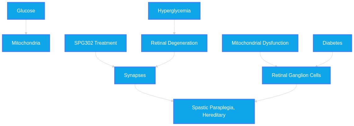
Keyword Analysis: diabetes; retinal diabetic neuropathy; ganglion cells; synapses; SPG302; tazbentetol; visual function; synaptic regeneration; neuroprotection; blindness; glaucoma
Plausibility Verdicts
SPG302 demonstrates potent neuroprotective and synaptic stabilizing effects in preclinical diabetic retinopathy and glaucoma models.
SPG302 is a highly promising synaptogenic candidate for diabetic retinal neuropathy, demonstrated to protect inner retinal layers in animal models.
SPG302 shows high potential in preclinical models for preserving synaptic integrity and preventing RGC loss in diabetic neuropathy.
Dataset Summary
Novel & Overlooked Insights
- Neurodegeneration in glaucoma often involves transsynaptic degeneration extending into secondary and higher-order visual brain regions.
- In diabetic retinopathy, mitochondrial fission acts as a pathological initiator, suppressing the Hippo pathway and promoting Müller cell activation.
- GPR75 knockdown provides a therapeutic strategy for alleviating mitochondrial dysfunction in retinal ganglion cells via the AMPK pathway.
- Sigma1 receptor (Sig1R) activation provides durable neuroprotection by coordinating redox, mitochondrial, and cell-survival pathways.
- Synaptic proteins such as Syntaxin-4 regulate membrane trafficking essential for maintaining neuronal homeostasis in the retina.
- Short-chain fatty acids like propionic acid show promise in reducing serum neurofilament light chain levels, indicating attenuation of neuroaxonal injury.
- The interaction between microglia and Müller cells is modulated by fibroblast growth factor 1 (FGF1), which is downregulated in glaucomatous retinas.
- Intranasal delivery of neuroprotective agents offers a potential non-invasive strategy for posterior segment ocular disease, bypassing the blood-retinal barrier.
- Panoptosis, an integrated programmed cell death modality, serves as a dynamic framework for interpreting inflammatory neurovascular degeneration in diabetic retinopathy.
- Targeting the liver-brain axis via Licochalcone A or other agents may provide systemic protection against metabolic neurodegeneration.
- Neurodegeneration, particularly RGC loss and synaptic impairment, often precedes clinical microvascular symptoms in diabetic retinopathy.
- SPG302 acts as a synaptogenic agent, capable of mitigating retinal injury across different disease etiologies, including glaucoma.
- The synaptic dysfunction in diabetes and glaucoma involves common molecular pathways, such as the modulation of postsynaptic density (PSD) proteins.
- Exosomes derived from specific physiological states (like hibernation) have been identified as potential mediators of intrinsic neuroprotection, suggesting novel intercellular signaling pathways.
- The use of GLP-1 receptor agonists and traditional Chinese medicines (e.g., Danshen, Ginsenoside Rg1) provides alternative, multi-target strategies for mitigating neuroinflammation in the retina.
- Calcium dysregulation acts as a "unifying pathogenic hub" for neurovascular unit dysfunction across multiple neurodegenerative diseases.
- Targeting the autophagy-lysosomal pathway (e.g., via the SNAI1-LAMP3 axis) represents an emerging therapeutic direction to preserve RPE and retinal neurons.
- Metabolic variability (e.g., glucose flux and uric acid levels) significantly influences the rate of ganglion cell thinning in diabetic patients without retinopathy.
- Advanced multimodal imaging (e.g., SS-OCTA) allows for the early detection of neurovascular uncoupling, which serves as a biomarker for disease progression.
- DRN often presents as a neurodegenerative disease manifesting before clinical microvascular damage is visible.
- SPG302 promotes glutamatergic synaptogenesis, offering a potential mechanism to restore synaptic connections that are lost early in the disease process.
- Mitochondrial transplantation and mitophagy regulation represent emerging frontiers in preserving RGC viability.
- Norrin, a protein secreted by Müller cells, is crucial for Wnt signaling and retinal capillary formation, and its downregulation is a key pathological event in diabetes.
- Neuroprotective effects of therapeutics such as fenofibrate, pelargonidin, and UAB126-MP occur via diverse signaling pathways (e.g., RXR agonism) distinct from conventional pressure-lowering.
- The integrity of the neurovascular unit is fundamentally tied to synaptic communication, which remains dysregulated following RGC injury.
- Emerging gene therapies, such as WFS1 delivery, show promise for genetic-based optic neuropathies.
- Advanced imaging and machine learning (e.g., 2.5D CFF module) are improving the precision of diagnostic markers like the Ganglion Cell Complex (GCC).
- Dietary and natural compounds, including eucalyptol and L-serine, show evidence for mitigating metabolic features of diabetic neuropathy.
- The relationship between systemic metabolic health and retinal neurodegeneration suggests that retinal assessment could serve as a systemic prognostic tool.
Extracted Discoveries
- Investigate the long-term visual outcomes of combining SPG302 with anti-VEGF therapies in human clinical trials.
- Perform single-nucleus RNA sequencing on human retinal samples treated with SPG302 to define cell-type specific molecular shifts.
- Assess SPG302 efficacy in modulating the SNAI1-LAMP3 axis to determine if enhanced autophagic flux contributes to its synaptogenic effect.
- Evaluate the impact of long-term SPG302 administration on pupillometry-derived autonomic indices to confirm systemic-retinal neuro-correlations.
- Clinical trial evaluating the impact of SPG302 on visual field stability in patients with early-stage diabetic retinopathy.
- Investigation of synaptic marker expression patterns in human retinal biopsy samples following neuroprotective treatment.
- A multicenter longitudinal observational study comparing the effectiveness of early versus late initiation of neuroprotective agents in patients with non-proliferative diabetic retinopathy.
- A phase 1 safety and pharmacokinetic study of intranasal delivery systems for neuroprotective agents in glaucoma patients.
- Conduct a longitudinal human clinical study using OCTA to correlate GCIPLT reduction rates with circulating SPG302-like small molecule levels in diabetic patients.
- Systematic comparison of GABAergic therapy versus SPG302 in reversing early-stage synaptopathy.
- Longitudinal observational study of synaptic marker dynamics in patients with diabetic retinopathy vs. controls.
- Comparative analysis of various neuroprotective agents (SPG302, Norrin, UAB126) on retinal neuro-glial vascular unit homeostasis.
- Discovered Hypothesis (A to C): PTP1B inhibition in microglia (Bridge B) could potentially improve the efficacy of Sig1R-mediated neuroprotection (Target C) following ischemic retinal injury (Origin A).
Literature A (Origin): Sig1R activation provides durable neuroprotection following neonatal ischemic retinal injury (ID: 42396530).
Literature C (Target): Microglia-specific deletion of Ptp1b prevents synaptic loss and cognitive impairment in neurodegeneration (ID: 42409182).
The Intersecting Bridge B: Modulation of the NF-κB pathway.
Biological Rationale: Sig1R activation modulates cell stress and mitochondrial function, while PTP1B is a known activator of NF-κB-dependent inflammation. Synergistic targeting could reduce the chronic inflammatory state that limits neuroprotective recovery. - SNAI1-mediated lysosomal dysfunction in RPE cells can be mitigated by SPG302-induced synaptic protein stabilization, potentially preventing retinal degeneration.
- SNAI1-LAMP3 axis in RPE cell autophagy (ID: 42410910)
- SPG302-mediated synaptic stability in RGCs (ID: 42456876)
- PSD95/Synaptic protein turnover regulation and lysosomal homeostasis.
- Since both synaptic density and RPE autophagic flux require rigid protein quality control, the stabilization of PSD proteins by SPG302 may indirectly reduce the lysosomal load and protect RPE cells from stress-induced SNAI1 upregulation.
- Enhancing autophagy via pharmacological mTOR activation could specifically mitigate microglial-induced neuroinflammation in diabetic retinopathy.
- Autophagy impairment in RGCs under hypoglycemia (Source 41294828)
- Microglia-driven neuroinflammation in diabetic retinopathy (Source 41497475)
- mTOR signaling pathway modulation
- mTOR signaling is a key regulator of autophagy and microglial phenotype. Restoring autophagic flux in RGCs while modulating mTOR may suppress pro-inflammatory microglia, thus providing a dual-action neuroprotective strategy.
- There is a notable discrepancy between the robust success of preclinical neuroprotective models (e.g., SPG302 in db/db mice) and the limited clinical translation of similar neuroprotective therapies, such as the landmark trial failure of memantine, as mentioned in ID: 42333387.
- There is a slight conflict regarding whether systemic therapies (like GAs) can act independently of vasodegeneration; one study (ID 42461929) suggests RXR agonism is neuroprotective without affecting acellular capillaries, while others (ID 41237937) suggest SRR inhibition affects both neural and vascular compartments.
- There is a minor contradiction regarding the impact of JNK inhibition on diabetic neuropathy (Source 42092483), where suppression of NLRP3 improves inflammatory markers but unexpectedly worsens pain, highlighting that anti-inflammatory success does not always correlate with symptomatic relief.
- The use of Mg2+ as an adjunct to opioid analgesia (ID: 42460019) could be repurposed for the management of chronic neuropathic pain in diabetic patients, potentially reducing the neuroinflammatory markers associated with diabetic retinal disease.
- SPG302, originally designed for glaucoma, serves as a high-potential repurposable candidate for early-stage diabetic retinal neuropathy due to its ability to prevent synaptic loss in both pathologies.
- Repurposing GLP-1 receptor agonists (often used for glucose-lowering) as secondary neuroprotective agents by leveraging their potential to modulate systemic and retinal neuroinflammation (Source 42059115).
PathMap is funded by sales of datasets and coversheets to researchers of any kind who wish to discover the most viable routes and paths to accelerate cures. We do not make theoretical molecules, we expose the truth in current PubMed literature. Commission a trace today.
PathMap Scores
How are these metrics evaluated?
Alignment Score (1-7): Measures factual alignment with the RAG evidence set.
[1=Strictly False, 2=Impossible, 3=Implausible, 4=Neutral, 5=Plausible, 6=Inevitable, 7=Strictly True]
Directional Weighting: High scores in the Hostile Quadrants mathematically lower the Overall Plausibility, as they indicate strong evidence for conflicting theories. Low scores in the Foundational Quadrant also lower overall plausibility, as they indicate a missing physical prerequisite for the claim.
Veridicality Audit Report
All Extracted Datapoints
Evaluated Perspectives & Quadrants
CLAIM EVALUATED AND ANSWER TO USER
The claim evaluated concerns the therapeutic potential of SPG302 and related neuroprotective mechanisms in mitigating diabetic retinal neuropathy (DRN), characterized by early ganglion cell loss and synaptic degeneration, and the potential for synaptic regeneration and preservation of visual function in conditions such as diabetes and glaucoma.ABSTRACT & REWRITTEN CLAIM
Diabetic retinopathy manifests early as diabetic retinal neuropathy (DRN), involving neurodegenerative processes that precede microvascular injury. Recent literature supports a shift toward neurovascular unit (NVU) protection, where pharmacological agents like SPG302 target synaptic and mitochondrial integrity. Evidence suggests that preserving retinal ganglion cells (RGCs) and synaptic markers through targeting metabolic pathways (such as Nrf2, AMPK, and nuclear receptors) may prevent progressive visual loss in diabetes and glaucoma.INTRODUCTION & JUSTIFICATION
Diabetic retinopathy is no longer classified solely as a microvascular pathology; it is increasingly recognized as a neurovascular degenerative disease involving coordinated injury to the retinal neurovascular unit. Diabetic retinal neuropathy (DRN), an early feature of DR that can precede the microvasculopathy, is characterized by inner retinal degeneration with loss of ganglion cells (GCs) and impaired GC function, decreased inner retina synaptic markers, and increased reactive gliosis and cell death. The molecular mechanisms driving this include mitochondrial dysfunction, where high glucose flux promotes mitochondrial reactive oxygen species overproduction and oxidative stress. SPG302, a novel pegylated benzothiazole derivative, promotes glutamatergic synaptogenesis and protects the inner retina in a mouse model of glaucoma, another leading cause of blindness associated with early synapse loss in the retina. Db/db mice demonstrated significant loss of GC ERG signal and GC cell number, decreased PSD95 and synaptophysin expression indicative of synapse loss, as well as increased reactive gliosis and expression of cell death markers such as pBAD and BAX. SPG302 treatment effectively preserved retinal integrity by reversing these changes. Such findings underscore the necessity of shifting clinical thinking from late vascular rescue toward mechanism-based neurovascular protection.Novel & Overlooked
EVIDENCE, METHODOLOGY & CITATIONS
1. ID: 42456876 - Application: Characterizes DRN as an early feature of DR. - *"Diabetic retinal neuropathy (DRN), an early feature of DR that can precede the microvasculopathy, is characterized by inner retinal degeneration with loss of ganglion cells (GCs) and impaired GC function, decreased inner retina synaptic markers, and increased reactive gliosis and cell death."* 2. ID: 42456876 - Application: Introduces SPG302 as a therapeutic agent. - *"SPG302, a novel pegylated benzothiazole derivative, promotes glutamatergic synaptogenesis and protects the inner retina in a mouse model of glaucoma, another leading cause of blindness associated with early synapse loss in the retina."* 3. ID: 42456876 - Application: Describes the protective effects of SPG302 in db/db mice. - *"Db/db mice demonstrated significant loss of GC ERG signal and GC cell number, decreased PSD95 and synaptophysin expression indicative of synapse loss, as well as increased reactive gliosis and expression of cell death markers such as pBAD and BAX. SPG302 treatment effectively preserved retinal integrity by reversing these changes."* 4. ID: 42398881 - Application: Explains metabolic overload in DR. - *"Under persistent hyperglycemic conditions, excessive glucose flux and metabolic overload promote mitochondrial reactive oxygen species (ROS) overproduction, mitochondrial DNA (mtDNA) damage, impaired oxidative phosphorylation, mitochondrial fusion-fission imbalance, defective mitochondrial biogenesis, dysregulated mitophagy, metabolic reprogramming, and epigenetic alterations."* 5. ID: 42460327 - Application: Discusses the shift in clinical thinking. - *"Reframing DR as a diabetes-driven disorder of early NVU uncoupling may help shift clinical thinking from late vascular rescue toward mechanism-based neurovascular protection, precision phenotyping, and stage-adapted intervention."* 6. ID: 42461929 - Application: Discusses long-acting microparticles. - *"Sustained release of UAB126-loaded microparticles enables intravitreal drug delivery for up to 6 months. Intravitreal UAB126 microparticles injection in diabetic mice confers long-term neuroprotection and anti-inflammatory benefits."* 7. ID: 42404883 - Application: Discusses PANoptosis. - *"This review systematically summarizes the latest advances in PANoptosis-associated mechanisms underlying DR pathogenesis, focusing on PANoptosome signaling networks, non-coding RNA-mediated regulation, and immune-metabolic crosstalk."* 8. ID: 42409182 - Application: Discusses microglial PTP1B deletion. - *"Crucially, microglia-specific deletion of Ptp1b prevented synaptic loss and cognitive impairment."* 9. ID: 42409919 - Application: Discusses the eGFR and DR association. - *"This cross-sectional analysis demonstrates a clear dose-response pattern: each incremental increase in estimated glomerular filtration rate (eGFR) is associated with a proportional reduction in the likelihood of diabetic retinopathy among adults with type 2 diabetes mellitus."* 10. ID: 42435652 - Application: Discusses Melatonin effects in I/R injury. - *"Mel improved retinal structural and visual function, visual-guidance behavior, and electrophysiological responses following I/R while concurrently reducing ferroptotic and inflammatory injury markers."* 11. ID: 42401762 - Application: CST outcomes for aflibercept. - *"Mean CST decreased by 105.9 µm, 90.4 µm, and 97.1 µm, respectively."* 12. ID: 42396532 - Application: Discusses DCP metrics. - *"Adding DCP VD to the clinical model significantly improved discrimination."* 13. ID: 42379865 - Application: Discusses SARM1 pathology. - *"SARM1 protein in the axons of the rat glaucoma model can promote retinal ganglion cell axonal pathology by localizing to axonal mitochondria and regulating SNPH expression."* 14. ID: 42458952 - Application: Defines Stx4 protein family. - *"Syntaxin-4 (Stx4), a member of the Qa-SNARE protein family, is a pivotal regulator of membrane trafficking."* 15. ID: 42352347 - Application: Discusses L-serine metabolic effects. - *"L-serine supplementation significantly improved metabolic status: S1 reduced HOMA-IR by 77.4% and S2 by 87.5% relative to diabetic controls (p < 0.001)."* 16. ID: 42346597 - Application: Discusses age and OCTA associations. - *"The strength of the association between aging and OCTA parameters differed significantly between the controls and those with early retinopathy, pointing to a potentially altered retinal vascular homeostasis secondary to diabetic pathophysiology."* 17. ID: 42460019 - Application: Discusses Mg2+ and morphine usage. - *"Overall, our findings support the concurrent use of Mg2+ and morphine in the treatment of neuropathic pain as a mechanism-informed adjunct strategy."* 18. ID: 42323468 - Application: Defines OL Piezo2. - *"OL-specific Piezo2 deficiency results in age-dependent motor impairment and selective disruption of myelin compaction in small-caliber optic nerve axons, a fiber population known to be particularly vulnerable in demyelinating disease."* 19. ID: 42367386 - Application: Discusses evidence architecture in glaucoma. - *"Glaucoma-related biomaterials are moving beyond passive delivery platforms, but their transition toward smart materials, additive manufacturing, and functional tissue engineering remains uneven."* 20. ID: 42371604 - Application: Discusses BBR and retinal morphology. - *"BBR improved retinal morphology, reduced edema, and restored the arrangement of retinal ganglion cells in DR rats."*CLAIM EVALUATED AND ANSWER TO USER
"The efficacy of SPG302 as a therapeutic candidate for diabetic retinal neuropathy (DRN), mediated through synaptic regeneration and neuroprotection, in the context of visual function preservation in diabetes and glaucoma."ABSTRACT & REWRITTEN CLAIM
Diabetic retinal neuropathy (DRN), an early neurodegenerative component of diabetic retinopathy, is characterized by synaptopathy and retinal ganglion cell (RGC) loss prior to microvasculopathy. SPG302, a pegylated benzothiazole derivative, promotes glutamatergic synaptogenesis and demonstrates neuroprotective potential in both diabetic and glaucomatous models by mitigating inner retinal damage and preserving visual function.INTRODUCTION & JUSTIFICATION
The paradigm of diabetic retinopathy has shifted from a primarily microvascular perspective to a neurovascular degenerative disorder where retinal ganglion cell (RGC) integrity and synaptic stability serve as critical therapeutic targets. The provided literature establishes that DRN manifests as inner retinal degeneration with a loss of ganglion cells and a reduction in synaptic markers, such as PSD95 and synaptophysin. SPG302 stands out as a "synaptogenic small molecule" that demonstrates broad applicability across neurodegenerative conditions, including glaucoma and diabetes, by reversing synaptic loss. By promoting synaptogenesis, SPG302 preserves retinal structural integrity and functional output as measured by electroretinography. This approach is aligned with the broader understanding that restoring synaptic architecture and metabolic homeostasis is requisite for preventing the irreversible vision loss associated with diabetic and glaucomatous neurodegeneration.Novel & Overlooked
EVIDENCE, METHODOLOGY & CITATIONS
1. ID: 42456876 - Application: Confirms SPG302's synaptogenic and neuroprotective role. - "SPG302, a novel pegylated benzothiazole derivative, promotes glutamatergic synaptogenesis and protects the inner retina in a mouse model of glaucoma" 2. ID: 42456876 - Application: Confirms SPG302 efficacy in diabetic models. - "SPG302 treatment effectively preserved retinal integrity by reversing these changes." 3. ID: 42352232 - Application: Highlights calcium as a central hub. - "Within the RNVU, calcium ions coordinate neurotransmission, glial activation, vascular tone, and blood-retinal barrier maintenance, and calcium dysregulation is emerging as a unifying pathogenic hub across these conditions." 4. ID: 42041557 - Application: Neurodegeneration vs. microvasculopathy timing. - "We found that retinal neurodegeneration may precede microvascular leakage in diabetic retinas." 5. ID: 41998758 - Application: Efficacy of WFS1 in preserving ganglion cells. - "Our results provide the first evidence that intravitreal injection of human WFS1 has significant neuroprotective effects on retinal ganglion cells and their axons and slows the loss of visual acuity." 6. ID: 41539543 - Application: Protective effect of betanin on retina. - "The rat experimental study validated this assumption and indicated a protective effect for betanin on rat retinas as shown by routine hematoxylin and eosin staining (retinal thickness and ganglion cell count)" 7. ID: 41024545 - Application: GCC utility in glaucoma diagnosis. - "GCC parameter is useful tool for diagnosis of Glaucoma. It has same ability to discriminate eyes with glaucoma and non glaucomatous eyes compared to RNFL." 8. ID: 40976316 - Application: Neurodegeneration precedes microvascular disease. - "Our findings suggest that neurodegenerative changes may precede microvascular alterations in non-DR patients, indicating that monitoring of GCIPLT is crucial" 9. ID: 40794319 - Application: HuD and CRYAA role in RGC survival. - "Silencing HuD and CRYAA enhanced high glucose-induced R-28 cell death, whereas their overexpression alleviated this effect." 10. ID: 40639562 - Application: Empagliflozin protective mechanism. - "Simultaneous intravitreal injection of EMPA (50 and 100 nmol) and NMDA reduced NMDA-induced cell loss in a dose-dependent manner." 11. ID: 40464812 - Application: RBM15 role in RGC pyroptosis. - "RBM15 silence restored cell viability and inhibited cell apoptosis and cell death in high glucose-triggered RGCs." 12. ID: 40211015 - Application: Autonomic and neurodegenerative overlap. - "There is a relationship between RNFL thickness and pupillometry parameters in diabetic patients, indicating simultaneous neurodegeneration and autonomic neuropathy." 13. ID: 38934389 - Application: GLP-1 eyedrops and RGC survival. - "Topical administration of glucagon-like peptide-1 eyedrops over a period of 2 weeks effectively countered the hyperglycemia-induced downregulation of GABAergic mIPSC frequency, subsequently enhancing the survival of retinal ganglion cells." 14. ID: 38318138 - Application: Compound Danshen Dripping Pills benefits. - "Moreover, CDDP reduced the expression levels of inflammatory factors in both the retina and serum. Conclusion: These findings strongly suggest that CDDP prevents early DR through vascular and neuroprotection." 15. ID: 37298544 - Application: Need for early neuroprotection. - "Neuronal cell death and axonal degeneration are irreversible, and their development can result in permanent visual loss." 16. ID: 40131295 - Application: DM and retinal degeneration in humans. - "The degeneration of neurons and synaptic connectivity within the macula in individuals with DM, even in the absence of clinical vascular signs, is associated with impaired visual function." 17. ID: 40215758 - Application: PERG/PVEP for early detection. - "All of the components of the PERG responses were significantly altered in the diabetic group, with a P value<0.05." 18. ID: 42410910 - Application: SNAI1-LAMP3 axis in autophagy. - "SNAI1 knockdown increased LAMP3 expression, thereby facilitating the recovery of lysosomal function and the alleviation of autophagic impairment." 19. ID: 42427680 - Application: Synaptic structure and gene therapy. - "Together, our results demonstrate that synaptic structure and function can be restored in the mature mouse retina in a model of complete synaptic disorganization." 20. ID: 42461929 - Application: RXR agonism for neuroprotection. - "These findings indicate that RXR agonism supports neuronal protection and immune modulation but does not prevent vasodegeneration, suggesting primarily neuroprotective and anti-inflammatory actions."CLAIM EVALUATED AND ANSWER TO USER
The therapeutic potential of SPG302 as a synaptogenic and neuroprotective agent in diabetic retinal neuropathy and glaucoma, and the underlying mechanistic role of synaptic integrity in preserving vision.ABSTRACT & REWRITTEN CLAIM
Diabetic retinopathy (DR) and glaucoma are significant neurodegenerative conditions characterized by early synaptic dysfunction and retinal ganglion cell (RGC) loss. SPG302, a pegylated benzothiazole derivative, demonstrates efficacy in promoting glutamatergic synaptogenesis and preserving retinal integrity. This evaluation synthesizes current literature on the impact of diabetes on RGCs, the role of synaptic loss in disease progression, and the therapeutic potential of SPG302 and similar neuroprotective agents.INTRODUCTION & JUSTIFICATION
Diabetic retinal neuropathy (DRN) is an early hallmark of diabetic retinopathy that frequently occurs prior to visible microvasculopathy. The pathology involves the loss of RGCs, impaired RGC function, and a significant decrease in inner retinal synaptic markers. Diabetic retinal neuropathy (DRN), an early feature of DR that can precede the microvasculopathy, is characterized by inner retinal degeneration with loss of ganglion cells (GCs) and impaired GC function. Because loss of glutamatergic synapses contributes to neuronal atrophy, therapeutic interventions targeting synaptic restoration offer a new point for disease mitigation. SPG302, a novel pegylated benzothiazole derivative, promotes glutamatergic synaptogenesis and protects the inner retina in a mouse model of glaucoma. Studies demonstrate that SPG302 treatment effectively preserved retinal integrity by reversing these changes. In both glaucoma and diabetes, RGC vulnerability is driven by metabolic and neurodegenerative processes. Accumulating evidence indicates that DR is not merely a microvascular disorder, but a complex neurovascular disease driven by long-standing hyperglycemia, metabolic dysregulation, oxidative stress, chronic inflammation, neurodegeneration, and impaired neurovascular coupling. In glaucomatous neurodegeneration, synaptic abnormalities are key, and SPG302 treatment effectively preserved synaptic integrity by reversing these changes. Further neuroprotective strategies include the use of norrin to restore PEDF levels, where Norrin, which is expressed in the inner retina, was significantly downregulated under diabetic conditions. Additionally, mitochondrial homeostasis is critical, as evidenced by studies showing sustained release of UAB126-loaded microparticles enables intravitreal drug delivery for up to 6 months.Novel & Overlooked
EVIDENCE, METHODOLOGY & CITATIONS
1. ID: 42456876 - Application: Demonstrates the role of SPG302 in mitigating diabetic retinal neuropathy. - "Diabetic retinal neuropathy (DRN), an early feature of DR that can precede the microvasculopathy, is characterized by inner retinal degeneration with loss of ganglion cells (GCs) and impaired GC function" 2. ID: 42456876 - Application: Discusses the mechanism of SPG302 in synaptogenesis. - "SPG302, a novel pegylated benzothiazole derivative, promotes glutamatergic synaptogenesis and protects the inner retina in a mouse model of glaucoma" 3. ID: 42456876 - Application: Shows efficacy of SPG302 in preserving retinal health. - "SPG302 treatment effectively preserved retinal integrity by reversing these changes." 4. ID: 42398881 - Application: Contextualizes DR as a neurovascular disease. - "Accumulating evidence indicates that DR is not merely a microvascular disorder, but a complex neurovascular disease driven by long-standing hyperglycemia, metabolic dysregulation, oxidative stress, chronic inflammation, neurodegeneration, and impaired neurovascular coupling." 5. ID: 42352232 - Application: Notes the pathogenic role of calcium. - "calcium dysregulation is emerging as a unifying pathogenic hub across these conditions." 6. ID: 42069589 - Application: Mentions ACA as an immunometabolic modulator. - "Acarbose (ACA), an α-glucosidase inhibitor widely used for diabetes, has recently been recognized for its dual regulatory potential on immune metabolism and aging-associated neurodegeneration." 7. ID: 42041557 - Application: Details the downregulation of norrin in diabetes. - "Norrin, which is expressed in the inner retina, was significantly downregulated under diabetic conditions." 8. ID: 41963265 - Application: Pelargonidin protective effects in RGCs. - "STZ-induced RGC loss was significantly reversed by pelargonidin, with the highest dose restoring RGC density to near-control or higher levels" 9. ID: 41548740 - Application: Discusses signaling at the synapse. - "Several key signaling pathways, including agrin-MuSK-LRP4, Wnt/β-catenin, and neuregulin/ErbB, govern NMJ maintenance with fiber-type-specific nuances." 10. ID: 41101191 - Application: GCC thickness importance. - "GCC (1 mm) may be interesting to explore in predicting visual outcomes after treatment" 11. ID: 41237937 - Application: SRR inhibition in diabetes. - "This is the first demonstration that pharmacological blockade of SRR improves retinopathy and normalizes glucose levels in a type 2 diabetes model." 12. ID: 40976316 - Application: Neurodegeneration precedes microvascular damage. - "Our findings suggest that neurodegenerative changes may precede microvascular alterations in non-DR patients" 13. ID: 40967391 - Application: SPG302 effect on synapses in glaucoma. - "SPG302 treatment effectively preserved synaptic integrity by reversing these changes." 14. ID: 40833325 - Application: NRXN role in RGCs. - "Our findings suggest that NRXNs may play cell-type-specific roles in the inner retina and associate decreased Nrxn3β expression in RGCs with inner retinal dysfunction in early DRD." 15. ID: 40794319 - Application: HuD/CRYAA axis role. - "Silencing HuD and CRYAA enhanced high glucose-induced R-28 cell death, whereas their overexpression alleviated this effect." 16. ID: 40759398 - Application: miRNA-122-5p as a therapeutic target. - "Targeted modulation of miRNA-122-5p may offer a novel therapeutic approach to preserve retinal neurons and counteract neurodegeneration in DR." 17. ID: 42461929 - Application: UAB126 microparticles delivery. - "Sustained release of UAB126-loaded microparticles enables intravitreal drug delivery for up to 6 months." 18. ID: 42461929 - Application: RXR agonism efficacy. - "Retinal explants exposed to high glucose showed downregulation of RXR, peroxisome proliferator-activated receptor (PPAR), and liver X receptor-β and their lipid metabolism targets (Abca1, Scd1, and Acox1)." 19. ID: 42352347 - Application: L-serine metabolic benefits. - "L-serine supplementation significantly improved metabolic status: S1 reduced HOMA-IR by 77.4% and S2 by 87.5% relative to diabetic controls (p < 0.001)." 20. ID: 42127585 - Application: Vitamin E impact on nerve conduction. - "Tocotrienol-rich vitamin E increased sural sensory nerve-conduction velocity (NCV) by 1.77 m s⁻¹ (0.80-2.74) and median sensory NCV by 1.53 m s⁻¹ (0.44-2.63)"Verbatim Quote Audit Console
Mapped Reference Directory (APA)
- [1] ID: 42456876 - Vanderklish PW, Bastola T, Secreast P, Brush M, Poudel M et al. (2026). SPG302 Mitigates Diabetic Retinal Neuropathy and Inner Retinal Damage in a Genetic Mouse Model of Diabetes.. Experimental eye research. ID: 42456876.
- [2] ID: 42398881 - Zhu X, Jin T, Zhang Y, Lian L, Du W (2026). Mitochondrial Dysfunction and Diabetic Retinopathy: Research Progress from Pathogenic Mechanisms to Therapeutic Targets.. Experimental eye research. ID: 42398881.
- [3] ID: 42460327 - Chen J, Zhang L (2026). Neurovascular unit uncoupling in diabetic retinopathy: molecular mechanisms and stage-adapted therapeutic strategies.. Frontiers in endocrinology. ID: 42460327.
- [4] ID: 42461929 - Johnson E, Kaffash E, Sheini F, Rennhack A, Sielski MS et al. (2026). Long-Acting RXR Agonism Preserves Retinal Function and Modulates Inflammation in Diabetic Retinopathy.. Diabetes. ID: 42461929.
- [5] ID: 42404883 - Ma L, Hou N, Zhao X, Li Z, Liu Q et al. (2026). PANoptosis in diabetic retinopathy: immunological insights into mechanisms and translational therapies.. Frontiers in immunology. ID: 42404883.
- [6] ID: 42409182 - Xu D, He C, Lv H, Weedor JG, Xing Y et al. (2026). Microglial PTP1B promotes synaptic pathology and cognitive deficits in chronic Toxoplasma gondii infection.. Brain, behavior, and immunity. ID: 42409182.
- [7] ID: 42409919 - Ling J, Xie Z, Zhang D, Gao Y, Hu Y et al. (2026). The linear association between estimated glomerular filtration rate and diabetic retinopathy: a cross-sectional study.. Scientific reports. ID: 42409919.
- [8] ID: 42435652 - Dou YN, Wen Y, Huang Y, Wu X, Zhang Z et al. (2026). Melatonin reprograms antioxidant defenses to suppress ferroptosis via Homer1a/mGluR1-Nrf2/xCT signaling after retinal ischemia-reperfusion.. Redox biology. ID: 42435652.
- [9] ID: 42401762 - Timoceanu L, Steinmann S, Gillies MC, Barthelmes D (2026). Aflibercept 24-Month Outcomes in Diabetic Macular Edema With and Without Prior Macular Laser Therapy: Data from the Fight Retinal Blindness! Registry.. Ophthalmology and therapy. ID: 42401762.
- [10] ID: 42396532 - Greenwood J, Kakihara S, Busza A, Fawzi A (2026). Validation of Deep Capillary Plexus OCTA Metrics as Predictors of Diabetic Retinopathy Complications: A One-Year Longitudinal Study.. Research square. ID: 42396532.
- [11] ID: 42379865 - Zhang XJ, Wu JH (2026). [Regulation of SARM1 on SNPH expression and its participation in glaucomatous optic neuropathy].. [Zhonghua yan ke za zhi] Chinese journal of ophthalmology. ID: 42379865.
- [12] ID: 42458952 - Li Y, Wei Y, Zhao J, Quan P, Wang C et al. (2026). Syntaxin-4 at the Crossroads of Synaptic Plasticity and Neurodegeneration: From Molecular Mechanisms to Biomarker and Therapeutic Potential.. CNS & neurological disorders drug targets. ID: 42458952.
- [13] ID: 42352347 - Hamdy M, Khodeer DM, Elsakka ME, Alaseem AM, Mostafa YM et al. (2026). L-Serine Attenuates Metabolic and Behavioural Features of Diabetic Neuropathy with Dose-Dependent Central Proteomic Correlates in a Rat Model.. Biomolecules. ID: 42352347.
- [14] ID: 42346597 - Taha A, Zhang YS, Ma CJ, Stewart JM (2026). Diabetes May Modulate the Association Between Age and Optical Coherence Tomography Angiography Parameters: A Serial, Cross-Sectional Study.. Journal of personalized medicine. ID: 42346597.
- [15] ID: 42460019 - Kurowski P, Kulik K, Kowalczyk A, Wróblewska N, Kondrat J et al. (2026). The influence of magnesium ions on the electrophysiological, analgesic, and BDNF-induced neuromodulation of morphine effects in diabetic rats.. Frontiers in pharmacology. ID: 42460019.
- [16] ID: 42323468 - Dyckow-Schubart J, Rabitsch AM, Geywitz C, Mayer C, Lerma-Martin C et al. (2026). Oligodendrocyte Piezo2 is a regulator of age-dependent myelin integrity and dysregulated in multiple sclerosis.. Communications biology. ID: 42323468.
- [17] ID: 42367386 - Wang J, Sui T, Ma Y, Sui Y, Qin Z et al. (2026). Evidence architecture of glaucoma-related biomaterials reveals an uneven transition toward smart materials, additive manufacturing, and functional tissue engineering.. Frontiers in bioengineering and biotechnology. ID: 42367386.
- [18] ID: 42371604 - Li N, Chen JL, Sun YJ, Sun JF, Pauzi FA et al. (2026). Berberine alleviates pyroptosis of retinal ganglion cells in diabetic retinopathy by regulating AKT1, JUN, and STAT3.. Ibrain. ID: 42371604.
- [19] ID: 42352232 - Ding S, Li J, Chen Z, Bai W, Li K (2026). Calcium at the Helm: Mechanisms and Therapeutic Targets in the Retinal Neurovascular Unit.. Biomolecules. ID: 42352232.
- [20] ID: 42041557 - Moon CH, Koh TY, Yoon JS, Kim M, Ha KS (2026). Norrin Ameliorates Retinal Ganglion Cell Apoptosis by Normalizing VEGF and PEDF Dysregulation in Diabetic Retinopathy.. Cells. ID: 42041557.
- [21] ID: 41998758 - Jagodzinska J, Péquignot M, Sarzi E, Quiles M, Cazevieille C et al. (2026). WFS1 gene delivery rescues visual function in a mouse model of Wolfram syndrome.. Acta neuropathologica communications. ID: 41998758.
- [22] ID: 41539543 - Zaitone S, Soliman N, Shalaby AM, Abdelgbar AA, Saleh MAK et al. (2026). Betanin protects against diabetic retinal damage via the inhibition of NF-κB/NLRP3/VEGF axis: Insights from network pharmacology and experimental studies.. Experimental eye research. ID: 41539543.
- [23] ID: 41024545 - Shuvo TR, Sayeed A, Alam M, Raju SMR, Das B et al. (2025). Role of Macular Ganglion Cell Complex Analysis for Diagnosis of Glaucoma Using Spectral Domain Optical Coherence Tomography.. Mymensingh medical journal : MMJ. ID: 41024545.
- [24] ID: 40976316 - Fan Y, Li L, Wu X, Yan Y, Li P et al. (2026). Longitudinal Neural and Microvascular Changes in Type 2 Diabetic Patients Without Retinopathy: A 2-Year Prospective Cohort Study.. American journal of ophthalmology. ID: 40976316.
- [25] ID: 40794319 - Kim C, Oh S, Park YH (2025). HuD and alpha-crystallin A axis protects neuro-retinal cells in early diabetes.. Molecular and cellular biochemistry. ID: 40794319.
- [26] ID: 40639562 - Ota M, Morita A, Kashihara T, Nakahara T (2025). Protective effects of empagliflozin against NMDA-induced excitotoxicity in the rat retina.. Neuroscience letters. ID: 40639562.
- [27] ID: 40464812 - Zhou L, Zhang C, Cheng Q, Ma M, Fan X et al. (2025). RBM15 promotes m6A methylation and stability of KLF6 mRNA to accelerate pyroptosis of retinal ganglion cells in early-stage diabetic retinopathy.. Journal of molecular histology. ID: 40464812.
- [28] ID: 40211015 - Thakar M, Tripathy SP, Dutta P, Bhattacharya S, Dhaka U (2025). Quantitative automated pupillometry in diabetic patients and correlation with retinal nerve fibre layer thickness.. Eye (London, England). ID: 40211015.
- [29] ID: 38934389 - Shao YQ, Wang YC, Wang L, Ruan HZ, Liu YF et al. (2026). Topical administration of GLP-1 eyedrops improves retinal ganglion cell function by facilitating presynaptic GABA release in early experimental diabetes.. Neural regeneration research. ID: 38934389.
- [30] ID: 38318138 - Xu X, Wang M, Zhang S, Wang J, Li X et al. (2024). Compound Danshen dripping pills prevent early diabetic retinopathy: roles of vascular protection and neuroprotection.. Frontiers in pharmacology. ID: 38318138.
- [31] ID: 37298544 - Tatsumi T (2023). Current Treatments for Diabetic Macular Edema.. International journal of molecular sciences. ID: 37298544.
- [32] ID: 40131295 - Albertos-Arranz H, Martínez-Gil N, Sánchez-Sáez X, Molina-Martín JC, Lax P et al. (2025). Neuronal Degeneration and Glial Activation in the Absence of Vascular Changes in Human Retinas of Patients With Diabetes.. Investigative ophthalmology & visual science. ID: 40131295.
- [33] ID: 40215758 - Nehme J, Raad P, Jalkh E, Karkouh R, Tamer Z et al. (2025). Pattern ERG, pattern VEP, and GCL thickness in diabetic patients with no diabetic retinopathy.. Journal francais d'ophtalmologie. ID: 40215758.
- [34] ID: 42410910 - Ji Y, Sun Y, Huang X, Liang J, Fang D et al. (2026). Targeting the SNAI1-LAMP3 axis to restore lysosomal function and alleviate autophagic flux impairment to delay retinal degeneration.. Autophagy. ID: 42410910.
- [35] ID: 42427680 - Hasan N, Paolo MD, McCall MA, Gregg RG (2026). Therapeutic restoration of synaptic architecture, retinal and visual function, and prevention of retinal degeneration in a mouse model of retinal dystrophy.. bioRxiv : the preprint server for biology. ID: 42427680.
- [36] ID: 42069589 - Wen Y, Dou YN, Chen X, Liu X, Yang Z et al. (2026). Acarbose modulates microglial Pkm2 acetylation to reshape immunometabolism and preserve retinal neurons after ischemia-reperfusion.. Journal of neuroinflammation. ID: 42069589.
- [37] ID: 41963265 - Yu H, Albrakati A, Wani EA, Li Y (2026). Pelargonidin protects retinal ganglion cells in a streptozotocin-induced diabetic rat model by reducing intraocular pressure, suppressing TGF-β and activating JAK2/STAT3 signalling pathway.. Acta pharmaceutica (Zagreb, Croatia). ID: 41963265.
- [38] ID: 41548740 - Qaisar R (2026). Fiber-type-specific architecture and pathophysiology of the neuromuscular junction.. Neuroscience. ID: 41548740.
- [39] ID: 41101191 - Condelipes A, Correia D, Fernandes I, Silva T, Correia E et al. (2025). Thickness profile of the ganglion cell complex and choroid in patients with persistent diabetic macular edema.. Computers in biology and medicine. ID: 41101191.
- [40] ID: 41237937 - Jiang H, Zhou P, Jiang X, Pei K, Liang W et al. (2026). Inhibition of serine racemase prevents retinopathy in diabetic mice.. Experimental eye research. ID: 41237937.
- [41] ID: 40967391 - Bastola T, Choi S, Shen Z, Kim KY, Vanderklish PW et al. (2025). SPG302 protects retinal ganglion cells and preserves visual function by preserving synaptic activity in a mouse model of glaucoma.. Experimental eye research. ID: 40967391.
- [42] ID: 40833325 - Unlu EK, Marx-Rattner R, Klein KA, Dawson VL, Dawson TM et al. (2025). Differential Expression of the Synapse Regulatory Proteins Neurexins in Early Diabetic Retinal Disease.. Investigative ophthalmology & visual science. ID: 40833325.
- [43] ID: 40759398 - Peng H, Li H, Liu S, Sun X, Zhang L et al. (2025). MiRNA-122-5p promotes retinal ganglion cell oxidative damage by targeting DJ-1 in hyperglycemic retina.. Free radical biology & medicine. ID: 40759398.
- [44] ID: 41528693 - Alhajaji R, Hassan AA, Al-Harahsheh MA, Saber RR, Soliman MA et al. (2026). Effectiveness of vitamin E in the treatment of diabetic neuropathy: systematic review and meta-analysis.. Hormones (Athens, Greece). ID: 41528693.
Abstract Repository (Raw Full-Texts) Show Database Collapse Database
ID: 37298544
Title: Current Treatments for Diabetic Macular Edema.
Abstract: Diabetic retinopathy is a major retinal disorder and a leading cause of blindness. Diabetic macular edema (DME) is an ocular complication in patients with diabetes, and it can impair vision significantly. DME is a disorder of the neurovascular system, and it causes obstructions of the retinal capillaries, damage of the blood vessels, and hyperpermeability due to the expression and action of vascular endothelial growth factor (VEGF). These changes result in hemorrhages and leakages of the serous components of blood that result in failures of the neurovascular units (NVUs). Persistent edema of the retina around the macula causes damage to the neural cells that constitute the NVUs resulting in diabetic neuropathy of the retina and a reduction in vision quality. The macular edema and NVU disorders can be monitored by optical coherence tomography (OCT). Neuronal cell death and axonal degeneration are irreversible, and their development can result in permanent visual loss. Treating the edema before these changes are detected in the OCT images is necessary for neuroprotection and maintenance of good vision. This review describes the effective treatments for the macular edema that are therefore neuroprotective.
View on PubMed
ID: 38318138
Title: Compound Danshen dripping pills prevent early diabetic retinopathy: roles of vascular protection and neuroprotection.
Abstract: Introduction: Diabetic retinopathy (DR) represents a major cause of adult blindness, and early discovery has led to significant increase in the number of patients with DR. The drugs currently used for treatment, such as ranibizumab, mainly focus on the middle and late periods of DR, and thus do not meet the clinical need. Here, the potential mechanisms by which compound Danshen Dripping Pills (CDDP) might protect against early DR were investigated. Methods: Db/db mice were used to establish a DR model. The initial weights and HbA1c levels of the mice were monitored, and retinal pathology was assessed by hematoxylin-eosin (HE) staining. The vascular permeability of the retina and thickness of each retinal layer were measured, and electroretinogram were performed together with fundus fluorescein angiography and optical coherence tomography. The levels of inflammatory factors were examined in retinal tissue, as well as those of intercellular adhesion molecule 1 (ICAM-1), IL-6, and monocyte chemoattractant protein 1 (MCP-1) in the serum using ELISA. Immunohistochemistry was used to evaluate levels of vascular endothelial growth factor (VEGF), B-cell lymphoma 2 (Bcl-2), and Bclassociated X protein (Bax). Retinal cell injury and apoptosis were examined by TdT-mediated dUTP Nick End Labeling (TUNEL) assays. Results: The data showed that CDDP significantly improved cellular disarrangement. Imaging data indicated that CDDP could reduce vascular permeability and the amplitude of oscillatory potentials (OPs), and restore the thickness of the ganglion cell layer. Moreover, CDDP reduced the expression levels of inflammatory factors in both the retina and serum. Conclusion: These findings strongly suggest that CDDP prevents early DR through vascular and neuroprotection.
View on PubMed
ID: 38934389
Title: Topical administration of GLP-1 eyedrops improves retinal ganglion cell function by facilitating presynaptic GABA release in early experimental diabetes.
Abstract: JOURNAL/nrgr/04.03/01300535-202602000-00048/figure1/v/2025-05-05T160104Z/r/image-tiff Diabetic retinopathy is a prominent cause of blindness in adults, with early retinal ganglion cell loss contributing to visual dysfunction or blindness. In the brain, defects in γ-aminobutyric acid synaptic transmission are associated with pathophysiological and neurodegenerative disorders, whereas glucagon-like peptide-1 has demonstrated neuroprotective effects. However, it is not yet clear whether diabetes causes alterations in inhibitory input to retinal ganglion cells and whether and how glucagon-like peptide-1 protects against neurodegeneration in the diabetic retina through regulating inhibitory synaptic transmission to retinal ganglion cells. In the present study, we used the patch-clamp technique to record γ-aminobutyric acid subtype A receptor-mediated miniature inhibitory postsynaptic currents in retinal ganglion cells from streptozotocin-induced diabetes model rats. We found that early diabetes (4 weeks of hyperglycemia) decreased the frequency of GABAergic miniature inhibitory postsynaptic currents in retinal ganglion cells without altering their amplitude, suggesting a reduction in the spontaneous release of γ-aminobutyric acid to retinal ganglion cells. Topical administration of glucagon-like peptide-1 eyedrops over a period of 2 weeks effectively countered the hyperglycemia-induced downregulation of GABAergic mIPSC frequency, subsequently enhancing the survival of retinal ganglion cells. Concurrently, the protective effects of glucagon-like peptide-1 on retinal ganglion cells in diabetic rats were eliminated by topical administration of exendin-9-39, a specific glucagon-like peptide-1 receptor antagonist, or SR95531, a specific antagonist of the γ-aminobutyric acid subtype A receptor. Furthermore, extracellular perfusion of glucagon-like peptide-1 was found to elevate the frequencies of GABAergic miniature inhibitory postsynaptic currents in both ON- and OFF-type retinal ganglion cells. This elevation was shown to be mediated by activation of the phosphatidylinositol-phospholipase C/inositol 1,4,5-trisphosphate receptor/Ca 2+ /protein kinase C signaling pathway downstream of glucagon-like peptide-1 receptor activation. Moreover, multielectrode array recordings revealed that glucagon-like peptide-1 functionally augmented the photoresponses of ON-type retinal ganglion cells. Optomotor response tests demonstrated that diabetic rats exhibited reductions in visual acuity and contrast sensitivity that were significantly ameliorated by topical administration of glucagon-like peptide-1. These results suggest that glucagon-like peptide-1 facilitates the release of γ-aminobutyric acid onto retinal ganglion cells through the activation of glucagon-like peptide-1 receptor, leading to the de-excitation of retinal ganglion cell circuits and the inhibition of excitotoxic processes associated with diabetic retinopathy. Collectively, our findings indicate that the γ-aminobutyric acid system has potential as a therapeutic target for mitigating early-stage diabetic retinopathy. Furthermore, the topical administration of glucagon-like peptide-1 eyedrops represents a non-invasive and effective treatment approach for managing early-stage diabetic retinopathy.
View on PubMed
ID: 40131295
Title: Neuronal Degeneration and Glial Activation in the Absence of Vascular Changes in Human Retinas of Patients With Diabetes.
Abstract: This study assessed retinal cells in the macula of human donors with diabetes with or without retinopathy. Seventeen human donor retinas were classified as diabetes mellitus (DM, n = 7), diabetes with diabetic retinopathy (DR, n = 3), or control (n = 8). Macular transversal sections were analyzed for photoreceptors, bipolar cells, horizontal cells, ganglion cells, their synaptic connections, and Müller cells using immunohistochemistry and confocal microscopy. The densities of bipolar cells, horizontal cells, and ganglion cells and the thickness of the inner plexiform layer (IPL) were quantified around the fovea. In the macula, cone photoreceptors elongated their axons to establish synapses with bipolar and horizontal cells in intraretinal cysts. Bipolar cells were reduced in the DM group compared to the control (P < 0.001), and rod bipolar cells showed morphological alterations in the cell body and synaptic terminals in both diabetic groups. Morphological changes were observed in both plexiform layers, with a decrease in the IPL thickness in DR. Horizontal cell terminals sprouted into the outer and inner retina in DR, despite no density differences existing between DM and control (P = 0.498). Ganglion cell density was reduced in the DM retinas compared to control (P < 0.001). Müller cells exhibited thickening of their cell bodies and end feet in all diabetic retinas. The degeneration of neurons and synaptic connectivity within the macula in individuals with DM, even in the absence of clinical vascular signs, is associated with impaired visual function. These early changes suggest potential new biomarkers for imaging techniques and emphasize the need for therapies for diabetic patients without clinical signs.
View on PubMed
ID: 40211015
Title: Quantitative automated pupillometry in diabetic patients and correlation with retinal nerve fibre layer thickness.
Abstract: To evaluate pupillary function in diabetic patients by automated pupillometry, and to study the correlation between retinal nerve fibre layer (RNFL) thickness and pupillary parameters. Diabetic patients underwent detailed systemic and ophthalmic examination including automated pupillometry. The pupillometer used a white stimulus and was equipped with a high-resolution infrared (880 nm) camera. Static pupillary diameters were captured at different levels of background intensity-photopic high (100 cd/m2), photopic low (10 cd/m2), mesopic high (1 cd/m2), and mesopic low (0.1 cd/m2). Dynamic pupillary responses were elicited with white-light flashes (total luminance 100 cd/m2, stimulus on time 200 ms, off time 3300 ms). RNFL thickness was measured using spectral domain optical coherence tomography (OCT) RESULTS: The study had 38 diabetic patients with retinopathy (DWR), 27 diabetic patients without retinopathy (DWOR), and 25 healthy controls. Static pupillometry showed significant differences between the three groups. Diabetic patients, both with and without retinopathy had significantly smaller pupillary diameters compared to controls, (p < 0.001). The amplitude of contraction and velocity of contraction was significantly lower in diabetic patients compared to controls (p < 0.001), and between DWR compared to DWOR (p < 0.001). Percent pupillary contraction differed between DWR and controls (p = 0.001) There was a significant difference in superior RNFL thickness between DWR and DWOR (p = 0.032). The superior quadrant RNFL correlated with the maximum number of pupillometry parameters. The amplitude and velocity of contraction are affected early in diabetic autonomic dysfunction. There is a relationship between RNFL thickness and pupillometry parameters in diabetic patients, indicating simultaneous neurodegeneration and autonomic neuropathy.
View on PubMed
ID: 40215758
Title: Pattern ERG, pattern VEP, and GCL thickness in diabetic patients with no diabetic retinopathy.
Abstract: The main objective of this study was to compare the retinal ganglion cell (RGC) function of diabetic patients without diabetic retinopathy with the RGC function of a control group, using functional tests and anatomical assessments. A cross-sectional prospective pilot study was conducted on two groups. We compared the results of functional tests (Pattern ERG and Pattern VEP - PERG and PVEP) and anatomical tests (macular and RNFL OCT) in a diabetic group without diabetic retinopathy to a control group. The χ2 test was used to study qualitative data, and the t test was used for quantitative data. The significance threshold was a P value less than 0.05. A total of 37 eyes were included in the study. None of the demographic variables showed any significant association or effect on any of the two groups. GCL thickness was significantly reduced in the diabetic group in the superior, inferior, and nasal outer circles, with a P value <0.001. The amplitude of the P100 wave was significantly reduced in the diabetic group, with a P value<0.05 for the pattern sizes of 60' and 30', and the diabetic group had a longer latency for the 15' VEPs. All of the components of the PERG responses were significantly altered in the diabetic group, with a P value<0.05. Our study indicates that combining different tests may be used as an early means of detection of compromised retinal neuron function in diabetic eyes during the course of early diabetic retinopathy.
View on PubMed
ID: 40464812
Title: RBM15 promotes m6A methylation and stability of KLF6 mRNA to accelerate pyroptosis of retinal ganglion cells in early-stage diabetic retinopathy.
Abstract: Neurodegeneration in early-stage diabetes retinopathy (DR) is mainly caused by the loss of retinal ganglion cells (RGCs), and high glucose-treated cell pyroptosis contributes to an important cause. However, the detailed molecular regulatory mechanism has not yet been thoroughly examined. In this study, primary mouse RGCs were stimulated with different concentrations of glucose, and mouse was intraperitoneally injected with streptozotocin (STZ) to construct DR model in vitro and in vivo. We found that compared to normal controls, RNA binding motif protein 15 (RBM15) was significantly upregulated in high glucose-treated RGCs and STZ-induced mice. RBM15 silence restored cell viability and inhibited cell apoptosis and cell death in high glucose-triggered RGCs. In parallel, RBM15 knockdown distinctly improved pathological damage such as thinning of retinal tissue thickness and loss of RGCs in STZ-modeling mice. Interestingly, the production of inflammatory cytokines and the expression of Cleaved caspase-1, NLRP3 and GSDMD-N were significantly reduced by RBM15 silence in vivo and in vitro. Mechanistically, RBM15 bound to kruppel like factor 6 (KLF6) mRNA to promote m6A modification and stabilize KLF6 mRNA, upregulating KLF6 expression in model cells and model mice retinal tissues. KLF6 overexpression increased the production of inflammatory cytokines and the expression of proteins related to pyroptosis, reversing the protective effects of RBM15 silence in high glucose-treated RGCs and diabetic retina. In conclusion, RBM15 is upregulated by high glucose, and stabilizes KLF6 mRNA to activate NLRP3-mediated pyroptosis pathway, exacerbating inflammation and apoptosis of RGCs and accelerating the progression of DR.
View on PubMed
ID: 40639562
Title: Protective effects of empagliflozin against NMDA-induced excitotoxicity in the rat retina.
Abstract: Empagliflozin (EMPA), a sodium-glucose cotransporter 2 inhibitor used in patients with type 2 diabetes, exerts various beneficial effects, including anti-inflammatory and antioxidant properties, in addition to its glucose-lowering effect. In this study, we examined whether EMPA protects against N-methyl-d-aspartic acid (NMDA)-induced retinal excitotoxicity and the mechanisms underlying its protective effects. Male Sprague-Dawley rats (7-8 weeks old) were used in this study. The number of cells in the ganglion cell layer (GCL) decreased 7 days after intravitreal injection of NMDA (50 nmol). Simultaneous intravitreal injection of EMPA (50 and 100 nmol) and NMDA reduced NMDA-induced cell loss in a dose-dependent manner. The protective effect of EMPA was significantly attenuated by the AMP-activated protein kinase (AMPK) inhibitor, compound C (10 nmol). NMDA increased the number of terminal deoxynucleotidyl transferase-mediated dUTP nick-end labeling (TUNEL)-positive cells in the GCL 6 h after injection, and the response was significantly, but not completely, attenuated by EMPA. These results suggest that EMPA protects against NMDA-induced retinal excitotoxicity in rats. The protective effect of EMPA may be partly attributed to the activation of the AMPK pathway and inhibition of neuronal cell apoptosis.
View on PubMed
ID: 40759398
Title: MiRNA-122-5p promotes retinal ganglion cell oxidative damage by targeting DJ-1 in hyperglycemic retina.
Abstract: Mitochondrial dysfunction, induced by prolonged hyperglycemia, is widely regarded as a central factor in oxidative stress and retinal ganglion cell (RGC) degeneration in diabetic retinopathy (DR). DJ-1 (PARK7) acts as a crucial antioxidant defense mechanism, stabilizing mitochondrial structure and redox balance. However, its expression is dramatically suppressed under diabetic conditions, and the upstream regulatory mechanisms remain incompletely characterized. Growing evidence from epigenetic research implicates microRNAs (miRNAs) as important players in the molecular pathways underlying DR progression. Among these, miRNA-122-5p has drawn increasing attention due to its aberrant activity under diabetic stress and its putative interaction with PARK7. In this study, we employed a streptozotocin-induced type 1 diabetes mouse model and glucose-stimulated R28 cells to explore the contribution of miRNA-122-5p to mitochondrial damage, oxidative stress, and RGC injury. In diabetic mice, we observed significant oxidative imbalance, increased apoptotic activity, RGC loss, and diminished retinal function. MiRNA profiling identified miRNA-122-5p as the most upregulated among candidate miRNAs. Inhibition of miRNA-122-5p attenuated these pathological changes and preserved both cellular integrity and visual function. In vitro, high glucose triggered mitochondrial fragmentation, membrane potential collapse, and excessive reactive oxygen species generation in R28 cells. Suppressing miRNA-122-5p alleviated these injuries. Dual-luciferase assays confirmed that miRNA-122-5p directly targets PARK7, thereby post-transcriptionally repressing DJ-1 expression and compromising mitochondrial resilience. These findings establish miRNA-122-5p as a critical upstream regulator of DJ-1-mediated antioxidant defense. By aggravating mitochondrial oxidative stress, it contributes to RGC vulnerability in DR. Targeted modulation of miRNA-122-5p may offer a novel therapeutic approach to preserve retinal neurons and counteract neurodegeneration in DR.
View on PubMed
ID: 40794319
Title: HuD and alpha-crystallin A axis protects neuro-retinal cells in early diabetes.
Abstract: Diabetic retinopathy (DR) is a prevalent microvascular complication of diabetes; however, neuro-retinal degeneration is also observed in patients with diabetes without signs of DR. The mechanisms leading to neuro-retinal cell loss before vascular complications manifest in diabetes remain poorly understood. In this study, we investigated the neuronal RNA-binding protein HuD as a novel regulator of neuro-retinal degeneration in the early stage of diabetes. We determined the expression of HuD and alpha-crystallin A (CRYAA) in the retinal ganglion cell layer. HuD and CRYAA were down-regulated in the retinas of streptozotocin-induced diabetic rats and in neuro-retinal cells (R-28) treated with high glucose. Cryaa mRNA was identified as a novel target transcript of HuD, and we demonstrated that HuD post-transcriptionally regulates the expression of Cryaa mRNA by binding to its 3'-untranslated region. Silencing and overexpression of HuD positively regulated the expressions of Cryaa mRNA and protein. We demonstrated that the increase in inflammatory cytokines such as TNFα, IL-1β, and IL-6 in R-28 cells under hyperglycemic conditions was a result of both CRYAA and HuD levels. Silencing HuD and CRYAA enhanced high glucose-induced R-28 cell death, whereas their overexpression alleviated this effect. HuD post-transcriptionally regulates CRYAA expression, influencing the function and viability of neuro-retinal cells under diabetic conditions. Our results suggest that the HuD/CRYAA axis plays a crucial role in neuro-retinal cells and has the potential to serve as a prognostic factor and therapeutic target for diabetic neuro-retinal degeneration.
View on PubMed
ID: 40833325
Title: Differential Expression of the Synapse Regulatory Proteins Neurexins in Early Diabetic Retinal Disease.
Abstract: Neurexins (NRXNs), a complex family of synapse regulatory proteins, represent attractive candidate molecular mediators of retinal neuronal dysfunction in early diabetic retinal disease (DRD) given their roles in the central nervous system and recent work suggesting a potential link with diabetes mellitus (DM). As antibodies are unable to distinguish NRXN family members and isoforms, the purpose of this study was to investigate differential expression of individual NRXN transcripts in relevant retinal cell types in early DRD. RNAscope multiplexed fluorescent in situ hybridization was used to quantify transcripts of NRXNs in mouse and human retina. DM was induced in C57BL/6J mice using streptozotocin (STZ) and the retinal phenotype characterized by electroretinogram (ERG), optical coherence tomography (OCT), and optokinetic tracking (OKT) after 6 weeks. Retinal ganglion cells (RGCs) were generated in vitro by directed differentiation from human embryonic stem cells (hESCs), cultured in normal or high glucose, and NRXN expression assessed by qPCR and Western blot. All NRXN family members (NRXN1, NRXN2, and NRXN3) were enriched in inner retinal neurons in both human and mouse tissue. We identified decreased Nrxn3 transcripts, specifically the Nrxn3β isoform, in RGCs of diabetic mice at 6 weeks after STZ treatment. Interestingly, Nrxn3α and Nrxn3β were differentially expressed in inner retinal layers. Finally, we confirmed decreased Nrxn3β expression in hESC-RGCs cultured in high glucose in vitro. Our findings suggest that NRXNs may play cell-type-specific roles in the inner retina and associate decreased Nrxn3β expression in RGCs with inner retinal dysfunction in early DRD.
View on PubMed
ID: 40967391
Title: SPG302 protects retinal ganglion cells and preserves visual function by preserving synaptic activity in a mouse model of glaucoma.
Abstract: Glaucoma, a leading cause of irreversible vision loss worldwide, is an optic neuropathy characterized by optic nerve degeneration and retinal ganglion cell (RGC) death. Early glaucomatous damage is often associated with dendritic and synaptic abnormalities in RGCs, yet the mechanisms linking these synaptic alterations to RGC death remain unclear. In a mouse model of glaucoma, treatment with the clinical-stage, synaptogenic small molecule SPG302, a pegylated benzothiazole derivative, demonstrated neuroprotective effects, protecting RGCs and their axons in the glaucomatous retina and also improving retinal function as assessed by pattern electroretinogram testing. Elevated intraocular pressure disrupted synapses, as evidenced by reduced synaptophysin expression and homeostatic increases in Bassoon and PSD95 levels in the inner plexiform layer. SPG302 treatment effectively preserved synaptic integrity by reversing these changes. These findings highlight the therapeutic potential of SPG302 for protecting RGCs and preserving vision by modulating synaptic activity in glaucomatous neurodegeneration.
View on PubMed
ID: 40976316
Title: Longitudinal Neural and Microvascular Changes in Type 2 Diabetic Patients Without Retinopathy: A 2-Year Prospective Cohort Study.
Abstract: To compare the 2-year longitudinal changes in macular ganglion cell-inner plexiform layer thickness (GCIPLT) and superficial capillary plexus (SCP) vessel density between patients with type 2 diabetes mellitus (T2DM) without retinopathy (non-DR) and healthy controls. Prospective observational cohort study. T2DM patients without clinical retinopathy at baseline and after a 2-year follow-up, along with age- and sex-matched healthy controls, were recruited from the community in Guangzhou, China. Measurements of macular GCIPLT, retinal thickness (RT), and SCP vessel density were conducted at baseline and at the 2-year follow-up. Linear mixed-effects models were used to estimate absolute and relative rates of changes and to compare absolute rates between the 2 groups. A total of 282 eyes (141 in the control group and 141 in the non-DR group) were included in the analysis. Significant reductions in GCIPLT and GCIPLT/RT occurred only in the non-DR group, with GCIPLT decreasing by -0.229 µm/y (95% CI = -0.313 to -0.144; P < .001) and 0.324 % (95% CI = 0.444 to 0.204; P < .001), approximately 5-fold faster than in the control group. After adjusting for confounding factors, the longitudinal rates of RT, GCIPLT, and GCIPLT/RT were significantly accelerated in the non-DR group compared to the control group by -0.603 µm/y (95% CI = -0.939 to -0.268; P < .001), -0.189 µm/y (95% CI = -0.306 to -0.073; P = .001), and -0.073 % (95% CI = -0.118 to -0.028; P = .001), respectively. Although longitudinal changes in SCP vessel density did not show significant differences between the 2 groups (P = .861). Our findings suggest that neurodegenerative changes may precede microvascular alterations in non-DR patients, indicating that monitoring of GCIPLT is crucial for patients with long-standing diabetes, even in the absence of retinopathy.
View on PubMed
ID: 41024545
Title: Role of Macular Ganglion Cell Complex Analysis for Diagnosis of Glaucoma Using Spectral Domain Optical Coherence Tomography.
Abstract: Glaucoma is chronic progressive irreversible optic neuropathy characterized by significant visual field (VF) loss. So, early diagnosis and proper treatment can preserve the useful vision in lifetime. The objective of the study was to determine the importance of ganglion cell complex (GCC) analysis as a parameter for diagnosis of glaucoma. This case control study was conducted in the Department of Ophthalmology, Bangladesh Institute of Research and Rehabilitation in Diabetes, Endocrine and Metabolic Disorders (BIRDEM) General Hospital, Bangladesh over a period of one year from July 2017 to June 2018. Twenty five (25) Type-2 Diabetes control subjects and 50 subjects with Type-2 diabetes were divided into two groups, 25 diabetic with pre perimetric and 25 diabetic with perimetric glaucoma. In this study, there was no significant difference in age (50.92±5.53 years vs. 52.44±4.75 years vs. 52.64±7.80 years), gender. Regarding Optical coherence tomography (OCT) Retinal Nerve Fiber Layer (RNFL) of right eyes of the study subjects, superior and inferior RNFL were significantly thinner in both pre-perimetric right eye (109.18±8.95 and 111.21±10.53) and perimetric right eye (90.28±8.94 and 91.51±7.87) comparing normal eyes (129.12±2.68 and 132.17±3.22). Superior and inferior RNFL were significantly lower in both pre-perimetric (110.13±11.53 and 113.75±9.61) and perimetric (95.93±15.08 and 93.29±12.68) left eyes comparing normal left eyes (129.71±5.50 and 132.57±5.22). Regarding OCT GCC layer of right eyes of the study subjects, superior and inferior GCC layer were significantly lower in both pre-perimetric (87.66±3.81 and 89.70±4.98) and perimetric (77.48±6.97 and 79.21±6.06) right eyes comparing normal eyes (104.53±2.73 and 106.88±3.29). Regarding OCT GCC layer of left eyes of the study subjects, superior and inferior GCC layer were significantly lower in both pre-perimetric (84.88±3.82 and 87.21±3.77) and perimetric (81.08±9.51 and 80.01±10.02) left eyes comparing normal eyes (102.64±2.29 and 105.20±1.27). GCC parameter is useful tool for diagnosis of Glaucoma. It has same ability to discriminate eyes with glaucoma and non glaucomatous eyes compared to RNFL. In glaucoma, GCC layer is significantly thin which correlates strongly with glaucomatous damage. According to our result GCC layer analysis is important investigation to diagnosis of Glaucoma.
View on PubMed
ID: 41101191
Title: Thickness profile of the ganglion cell complex and choroid in patients with persistent diabetic macular edema.
Abstract: About 40 % of patients with diabetic macular edema (DME) do not respond optimally to first-line treatment with intravitreal injection of anti-vascular endothelial growth factor (AVEGF). Evidence suggests that additional vascular and neurodegenerative mechanisms may be involved. This study aimed to characterise the thickness of the Ganglion Cell Complex (GCC) and investigate the Choroidal Vascularity Index (CVI) in patients with different patterns of therapeutic response to AVEGF DME. This cross-sectional study included 27 diabetic patients into 3 different groups based on their response to AVEGF therapy: control group, responder DME group, and persistent DME group. The study's approach to vascular and neurodegenerative imaging biomarkers involved three steps: (1) Automatic quantification of GCC thickness, with manual correction when necessary; (2) Semi-automatic measurement of choroidal thickness; and (3) Analysis of choroidal area and choroidal luminal area using ImageJ software to calculate the CVI. In the overall characterization of the sample, a significant difference was observed only in the Best Corrected Visual Acuity (BCVA). There was a significant difference in Average Retinal Thickness (1 mm, 3 mm, and 6 mm) between the 3 groups and in GCC thickness at 1 mm. BCVA was negatively correlated with mean retinal thickness, while CVI showed a potential positive correlation with BCVA. While demographic and general clinical characteristics showed minimal differences across the groups, important differences in GCC and choroidal characteristics were observed. GCC (1 mm) may be interesting to explore in predicting visual outcomes after treatment, and CVI may impact visual gain.
View on PubMed
ID: 41237937
Title: Inhibition of serine racemase prevents retinopathy in diabetic mice.
Abstract: A single-nucleotide polymorphism in the serine racemase (SRR)-encoding gene, Srr, is associated with an increased risk of type 2 diabetes. Studies, including our own, demonstrate that SRR is linked to a complication of diabetes, diabetic retinopathy (DR), and deletion of Srr protects diabetic animals from developing retinopathy. We investigate the feasibility and mechanisms of SRR inhibition as a strategy to prevent DR. SRR proteins in the retinas of db/db mice were approximately 50 % higher than C57BLKS/J wild-type (WT) mice, while D-serine levels in the aqueous humor were more than two-fold higher. Oral gavage of l-aspartic acid β-hydroxamate (L-ABH) improved b-wave amplitudes in electroretinogram recordings under both photopic and scotopic conditions. It also protected the inner retina in db/db mice by reducing the loss of retinal ganglion cells, endothelial cells, and pericytes, as well as decreasing the activation of Müller cells. In high glucose and hypoxia, interleukin-1 and interleukin-18 mRNA levels were lower in Srr-deleted Müller cell cultures than in WT cultures. Intravitreal injection of L-ABH mitigated glutamate-induced neurotoxicity in the retina. Systemically, L-ABH maintained euglycemia and improved glucose tolerance in db/db mice by inhibiting liver gluconeogenesis through restricting the expansion of pancreatic islet α-cells. Altogether, our results demonstrate that SRR inhibition prevents retinopathy in diabetic animals by blocking excitotoxicity and Müller cell-mediated inflammation in the retina, while also maintaining glucose homeostasis. This is the first demonstration that pharmacological blockade of SRR improves retinopathy and normalizes glucose levels in a type 2 diabetes model.
View on PubMed
ID: 41528693
Title: Effectiveness of vitamin E in the treatment of diabetic neuropathy: systematic review and meta-analysis.
Abstract: To evaluate whether tocotrienol-rich vitamin E improves nerve-conduction parameters and symptoms in diabetic peripheral neuropathy (DPN). Following Preferred Reporting Items for Systematic Reviews and Meta-Analyses PRISMA 2020 (PROSPERO CRD420250653145), we searched the PubMed, Web of Science, Scopus, and Embase databases up until 20 March 2025, for parallel-group randomized controlled trials (RCTs) of oral vitamin E in adults (≥ 18 years) with electrophysiologically confirmed DPN. Two independent reviewers performed screening, extraction, and the revised Cochrane Risk of Bias tool version 2.0 (RoB 2.0). Random-effects meta-analyses were conducted using the mean differences (MD) with 95% confidence intervals (CI). Five RCTs (n = 660) met the criteria. Tocotrienol-rich vitamin E increased sural sensory nerve-conduction velocity (NCV) by 1.77 m s⁻¹ (0.80-2.74) and median sensory NCV by 1.53 m s⁻¹ (0.44-2.63); tibial motor NCV rose 1.47 m s⁻¹ (0.36-2.58) versus placebo. Nerve-action-potential amplitudes and glycated hemoglobin A1c (HbA₁c) were unchanged (MD - 0.06%, - 0.18-0.06). Adverse-event rates were similar between groups. Two trials had a low risk of bias; one presented some concerns. Vitamin E yielded selective NCV gains without amplitude change, suggesting preservation or remyelination of sensory fibers rather than axonal regeneration. The absence of glycemic effects indicates neuroprotection independent of glucose control, positioning vitamin E as a potential adjunct-not substitute-to antidiabetic therapy. Heterogeneity in isoform, dose, and treatment duration (≤ 12 months) and modest sample sizes limit certainty. Larger, longer trials incorporating functional outcomes (pain, gait, and quality of life) are required.
View on PubMed
ID: 41539543
Title: Betanin protects against diabetic retinal damage via the inhibition of NF-κB/NLRP3/VEGF axis: Insights from network pharmacology and experimental studies.
Abstract: Diabetic retinopathy (DIR) is a predominant diabetic microvascular complication that may cause vision loss. Retinal inflammation and angiogenesis contribute largely to the neuronal degeneration in DIR. The current study is aiming to test the effect of oral betanin doses in protection from DIR in rats along with a network pharmacology study to investigate an assumption that betanin may inhibit nuclear factor-κ B (NF-κB). Three rat groups were assigned as vehicle, DIR, and DIR + Betanin 100 mg/kg. Molecular docking indicated the possible binding between betanin and NF-κB while the bioinformatic study highlighted a relation between this possible inhibition and suppression of NOD-like receptor pyrin domain-containing protein 3/vascular endothelial growth factor (NLRP3/VEGF) axis. The rat experimental study validated this assumption and indicated a protective effect for betanin on rat retinas as shown by routine hematoxylin and eosin staining (retinal thickness and ganglion cell count) and periodic acid-Schiff staining that was mediated through mitigating expression/protein level for of NF-κB, NLRP3, TNF-α, IL-6 and VEGF proteins. Immunohistochemistry showed that betanin was able to suppress retinal content of the glial fibrillary acidic protein (GFAP). In conclusion, the current study indicated that betanin was a good candidate for DIR in rats through suppression of the inflammatory cascade and may be suggested for diabetic patients if appropriated clinical studies will be available.
View on PubMed
ID: 41548740
Title: Fiber-type-specific architecture and pathophysiology of the neuromuscular junction.
Abstract: The neuromuscular junction (NMJ) is a specialized synapse essential for translating neuronal signals into muscle contraction. This review examines the complex structural, functional, and molecular differences in NMJs that innervate fast- and slow-twitch skeletal muscle fibers. Fast-twitch fibers, optimized for rapid and powerful contractions, possess elaborate NMJs with deep folds, high neurotransmitter turnover, and greater vulnerability to synaptic fatigue and degeneration. In contrast, slow-twitch fiber NMJs exhibit simpler but more stable architectures that support sustained, fatigue-resistant activity. These differences are not fixed but subject to activity-dependent plasticity and pathological remodeling. Chronic stimulation, injury, and aging influence NMJ morphology, with fast-twitch junctions more prone to degeneration in conditions such as ALS, myasthenia gravis, and diabetic neuropathy. Slow-twitch NMJs often resist early deterioration due to superior trophic support, metabolic stability, and more robust expression of synaptic organizers, such as agrin and PGC-1α. Several key signaling pathways, including agrin-MuSK-LRP4, Wnt/β-catenin, and neuregulin/ErbB, govern NMJ maintenance with fiber-type-specific nuances. These insights underscore the importance of tailoring therapeutic strategies to the muscle fiber phenotype. Gene therapies, neuromuscular electrical stimulation, and biomaterial scaffolds are emerging as promising modalities for preserving or restoring NMJ integrity, especially in fast-twitch fibers at higher risk of degeneration. Understanding fiber-type-specific NMJ biology enhances our understanding of motor control, muscle aging, and neuromuscular disease progression, and it opens pathways for precision therapeutics that target vulnerable synapses with structural and functional specificity. This review introduces a novel perspective by emphasizing fiber-type-specific NMJ differences and their implications for targeted therapies.
View on PubMed
ID: 41963265
Title: Pelargonidin protects retinal ganglion cells in a streptozotocin-induced diabetic rat model by reducing intraocular pressure, suppressing TGF-β and activating JAK2/STAT3 signalling pathway.
Abstract: Diabetic retinopathy (DR) is one of the primary causes of vision impairment, affecting individuals with diabetes, and is marked by the neurodegeneration of the retina along with increased intraocular pressure (IOP). This study sought to determine the effects of pelargonidin on extracellular matrix (ECM) modulation and the inhibition of transforming growth factor-β (TGF-β) and Janus Kinase 2/Signal Transducer and Activator of Transcription 3 (JAK2/STAT3) pathway in retinal ganglion cells of streptozotocin-induced diabetic rats. Male Sprague-Dawley rats (180-200 g) were rendered diabetic by intraperitoneal administration of streptozotocin (STZ). The rats were divided into 5 groups: control, diabetic model (STZ), STZ + low dose pelargonidin (12.5 mg kg-1 per day), STZ + medium dose pelargonidin (25 mg kg-1 per day) and STZ + high dose pelargonidin (50 mg kg-1 per day). IOP was monitored using a tonometer. Whole-mount retinal immunofluorescence staining using RNA-binding protein with multiple splicing (RBPMS) was performed to assess retinal ganglion cell (RGC) density. Protein expression levels of apoptotic markers, ECM components, and TGF-β and JAK2/STAT3 signalling pathways were evaluated by Western blotting. Pelargonidin treatment dose-dependently reduced the elevated IOP. Importantly, immunofluorescence analysis revealed a marked dose-dependent preservation of retinal ganglion cell (RGC) density: STZ-induced RGC loss was significantly reversed by pelargonidin, with the highest dose restoring RGC density to near-control or higher levels in both the central and peripheral retina. This was achieved via modulation of apoptosis-related proteins through the upregulation of Bcl-xL, Bcl-2, and downregulation of Bad, Bax and cleaved caspase-3. Furthermore, pelargonidin modulated ECM remodelling protein expression in the RGC layer. In particular, TGF-β2/Smad2/3 signalling was downregulated, and the JAK2/STAT3 pathway was upregulated. By reducing IOP, preserving RGC density, modulating ECM deposition, inhibiting TGF-β and upregulating the JAK2/STAT3 pathway, pelargonidin exerts protective effects against diabetic retinal injury. The results of this study further confirm the pharmacological potential of pelargonidin as a therapeutic agent for diabetic retinopathy.
View on PubMed
ID: 41998758
Title: WFS1 gene delivery rescues visual function in a mouse model of Wolfram syndrome.
Abstract: Wolfram syndrome is a rare childhood neurodegenerative disease characterized by diabetes followed by severe and rapid optic atrophy leading to blindness before the age of 20. Patients often develop other symptoms, such as deafness and neurological dysfunction. Wolfram syndrome is caused by mutations in the WFS1 gene, which encodes wolframin protein. Despite decades of intensive research, the complex mechanisms of optic neuropathy are not fully understood, and there are currently no therapies to prevent vision loss in Wolfram patients. Here, we showed that the Wfs1 knockout mice produced by the Estonian group, in which exon 8 of the Wfs1 gene was disrupted, exhibit a progressive loss of visual acuity, optic disc pallor and severe optic nerve damage. We tested the efficiency of gene therapy using AAV2 to deliver human WFS1 to retinal ganglion cells in Wfs1 knockout mice. Our results provide the first evidence that intravitreal injection of human WFS1 has significant neuroprotective effects on retinal ganglion cells and their axons and slows the loss of visual acuity. These results demonstrate that WFS1 is able to provide both functional and structural protection to retinal ganglion cells in Wfs1 knockout mice and provide important evidence for the efficacy of WFS1 as a neuroprotective treatment for Wolfram syndrome. These results demonstrate the promising effects of gene therapy for Wolfram syndrome and encourage future research aimed at conducting clinical trials in patients.
View on PubMed
ID: 42041557
Title: Norrin Ameliorates Retinal Ganglion Cell Apoptosis by Normalizing VEGF and PEDF Dysregulation in Diabetic Retinopathy.
Abstract: Diabetic retinopathy is increasingly recognized as a neurovascular disorder rather than a purely vascular disease; however, therapeutic strategies targeting retinal neurodegeneration remain limited. In this study, we investigated the protective effects of norrin against hyperglycemia-induced retinal neurodegeneration and elucidated its underlying molecular mechanisms in diabetic mice. We found that retinal neurodegeneration may precede microvascular leakage in diabetic retinas. Norrin, which is expressed in the inner retina, was significantly downregulated under diabetic conditions. Intravitreal supplementation of norrin markedly attenuated hyperglycemia-induced neurodegenerative processes, leading to retinal ganglion cell (RGC) apoptosis, including oxidative stress, inflammation, and neuropathological alterations such as reactive gliosis, glutamate excitotoxicity, and synaptic dysfunction. Norrin also reduced hyperglycemia-induced microvascular leakage and RGC apoptosis by normalizing vascular endothelial growth factor (VEGF) overexpression and restoring pigment epithelium-derived factor (PEDF) levels. Notably, PEDF upregulated by norrin effectively suppressed neurodegenerative processes induced by hyperglycemia or VEGF, thereby preserving RGC function. These findings identify norrin as a critical modulator of hyperglycemia-induced retinal neurodegeneration through restoration of the VEGF-PEDF balance. Our results highlight norrin as a potential therapeutic target for early neurodegenerative changes in diabetic retinopathy.
View on PubMed
ID: 42069589
Title: Acarbose modulates microglial Pkm2 acetylation to reshape immunometabolism and preserve retinal neurons after ischemia-reperfusion.
Abstract: Retinal ischemia-reperfusion (IR) elicits microglia-driven neuroinflammation and mitochondrial failure that led to retinal ganglion cell (RGCs) loss, yet effective disease-modifying therapies remain limited. Acarbose (ACA), an α-glucosidase inhibitor widely used for diabetes, has recently been recognized for its dual regulatory potential on immune metabolism and aging-associated neurodegeneration. Here, we demonstrate that intravitreal ACA administration attenuates retinal inflammation and improves RGCs survival following IR injury. Single-cell RNA sequencing revealed extensive inflammatory activation and metabolic reprogramming across the retina, characterized by enhanced nicotinamide adenine dinucleotide (NAD) catabolism, particularly in microglia. ACA treatment was associated with reversal of these alterations, replenished NAD levels, and restored mitochondrial integrity. Integrative proteomic and biochemical analyses identified pyruvate kinase, muscle-type 2 (Pkm2) as a candidate regulatory node affected by ACA. Intravitreal delivery of siPkm2 partially protected against IR injury, and co-administration with ACA produced an additive trend in neuroprotection. Mechanistically, ACA upregulated sirtuin 1 (Sirt1) and reduced Pkm2 acetylation at lysine 270 (K270), which was linked to pro-inflammatory microglial activation. Structure-based virtual screening further identified HY-113082, a small molecule targeting Pkm2-K270, which synergized with ACA to suppress inflammation and enhance retinal protection. Moreover, Pkm2fl/flCx3cr1-Cre mice conferred partial resistance to IR injury, but blunted the additional benefit of HY-113082 when combined with ACA, consistent with on-target engagement. Our findings support that ACA exerts retinal protection through the Sirt1-Pkm2-NAD axis, suggesting a metabolic checkpoint that integrates immune and mitochondrial regulation. This study provides mechanistic insight into ACA's dual immunometabolic and neuroprotective actions, holding promise for therapeutic insights into neuroinflammation.
View on PubMed
ID: 42323468
Title: Oligodendrocyte Piezo2 is a regulator of age-dependent myelin integrity and dysregulated in multiple sclerosis.
Abstract: Studies on the mechanosensitive ion channel Piezo2 largely focus on its role in the peripheral nervous system, particularly in touch and pain sensation. Here, we investigate Piezo2 function in the anterior visual pathway of the central nervous system with a focus on oligodendrocyte (OL) biology and myelin integrity. Using single-nucleus RNA sequencing, we identify Piezo2 expression in late differentiated OLs of the murine optic nerve, with minor expression in retinal ganglion cells. OL-specific Piezo2 deficiency results in age-dependent motor impairment and selective disruption of myelin compaction in small-caliber optic nerve axons, a fiber population known to be particularly vulnerable in demyelinating disease. Differential gene expression analysis further indicates that Piezo2 regulates myelin compaction and white matter integrity in mature OLs. Consistent with these findings, OL-encoded PIEZO2 expression is reduced in optic nerve lesion areas from multiple sclerosis patients, highlighting a convergent mechanism of small-caliber fiber vulnerability. Together, these data identify Piezo2 as an age-related regulator of OL function and myelin integrity, with potential relevance for preserving white matter structure in multiple sclerosis.
View on PubMed
ID: 42346597
Title: Diabetes May Modulate the Association Between Age and Optical Coherence Tomography Angiography Parameters: A Serial, Cross-Sectional Study.
Abstract: Purpose: We investigated the association between age and retinal microvasculature parameters as measured by optical coherence tomography angiography (OCTA) and the modifying effect of diabetes status on this association. Methods: In this serial, cross-sectional study, 3 × 3 mm2 macular OCTA images were obtained from healthy adults and adults with diabetes mellitus (DM) with no diabetic retinopathy (DR) or with mild non-proliferative DR (NPDR). The parameters analyzed included foveal avascular zone (FAZ) area and perimeter, vessel density (VD), vessel length density (VLD), and flow index (FI) of the superficial capillary plexus (SCP) and deep capillary plexus (DCP). The associations between OCTA parameters and age were explored using multivariable linear regression models. Results: For the included 1855 patients (1855 eyes) (49% male; mean age: 55 years), the results were as follows: no diabetes (N = 217), DM no DR (N = 1352), and mild NPDR (N = 286). Increasing age was significantly associated with decreased SCP and DCP VD and VLD in the diabetic and non-diabetic groups. The slope of association between SCP and DCP FI and age in the diabetic patients was significantly different than that in the control patients. Conclusions: The strength of the association between aging and OCTA parameters differed significantly between the controls and those with early retinopathy, pointing to a potentially altered retinal vascular homeostasis secondary to diabetic pathophysiology. This finding offers insight into the early pathological biomarkers of DR and may guide early DR management for patients based on personalized risk scores.
View on PubMed
ID: 42352232
Title: Calcium at the Helm: Mechanisms and Therapeutic Targets in the Retinal Neurovascular Unit.
Abstract: Retinal neurovascular unit (RNVU) dysfunction underlies major blinding and neurodegenerative conditions including glaucoma, diabetic retinopathy (DR), age-related macular degeneration (AMD), retinal ischemia-reperfusion (RIR) injury, and Alzheimer's disease (AD)-associated retinopathy. Within the RNVU, calcium ions coordinate neurotransmission, glial activation, vascular tone, and blood-retinal barrier maintenance, and calcium dysregulation is emerging as a unifying pathogenic hub across these conditions. Although upstream triggers differ, including mechanical stress in glaucoma, hyperglycemia in DR, oxidative damage in AMD, ischemic energy failure in RIR, and amyloid-β-driven endoplasmic reticulum stress in AD, all converge on disruption of intracellular calcium homeostasis, producing shared downstream consequences including excitotoxic injury of retinal ganglion cells (RGCs), Müller cell reactive gliosis, and pericyte hypercontraction. Broad-spectrum calcium channel blockade has shown limited clinical success, underscoring the need for cell-type-specific and pathway-selective approaches. This review therefore catalogs key interventional nodes, including transient receptor potential (TRP) channel antagonists, T-type calcium channel inhibitors, calcium/calmodulin-dependent protein kinase II (CaMKII) suppressors, and mitochondrial permeability transition pore (mPTP) inhibitors, and discusses how precision targeting of these pathways may restore RNVU homeostasis and open a therapeutic window into central nervous system (CNS) degenerative disorders.
View on PubMed
ID: 42352347
Title: L-Serine Attenuates Metabolic and Behavioural Features of Diabetic Neuropathy with Dose-Dependent Central Proteomic Correlates in a Rat Model.
Abstract: Diabetic neuropathy (DN) is a multifactorial complication of diabetes mellitus driven by chronic hyperglycemia, insulin resistance, and disturbed metabolic homeostasis, leading to progressive injury of both the peripheral and central nervous systems. This study investigated whether L-serine supplementation could attenuate DN through dose-dependent metabolic and neuroprotective mechanisms in a high-fat diet (HFD) plus streptozotocin (STZ)-induced diabetic rat model. Male Wistar rats (n = 8 per group) were allocated to five groups: normal control (NC), diabetic control (DC), pioglitazone (PIO; 1.5 mg/kg/day), low-dose L-serine (S1; 200 mg/kg/day), and high-dose L-serine (S2; 400 mg/kg/day). After 60 days of oral gavage, behavioural testing, glucose and insulin profiling, HOMA-IR calculation, brain histopathology, nerve growth factor (NGF) immunohistochemistry, and LC-MS/MS-based proteomic analysis of cerebral tissue were performed. Diabetic rats exhibited marked hyperglycaemia (355.33 ± 4.72 mg/dL), hyperinsulinaemia, severe insulin resistance (HOMA-IR 16.8 ± 3.2; a 14-fold increase), impaired thermal nociception, motor dysfunction, and pronounced neuronal degeneration. L-serine supplementation significantly improved metabolic status: S1 reduced HOMA-IR by 77.4% and S2 by 87.5% relative to diabetic controls (p < 0.001). High-dose L-serine produced greater improvements in thermal sensitivity, motor coordination (rotarod latency 26.67 ± 1.52 s vs. 16.1 ± 0.85 s in DC; p < 0.05), and NGF expression (8.6-fold increase vs. DC). Histopathology confirmed attenuation of neuronal injury and gliosis in both treatment groups. Exploratory, group-level proteomic profiling identified dose-specific molecular signatures: S1 was predominantly associated with carbohydrate, lipid, and biosynthetic pathways, whereas S2 was associated with synaptic, neurotransmission-related, and proteostasis pathways. Within the constraints of an exploratory design-group-level pooled proteomics, analysis of cerebral rather than peripheral-nerve tissue, and only two doses-these findings indicate that L-serine attenuates the metabolic and behavioural features of experimental diabetic neuropathy and generates the testable hypothesis of dose-dependent neuro-metabolic remodelling. The proteomic signatures are hypothesis-generating and require orthogonal validation before any mechanistic or translational inference can be drawn.
View on PubMed
ID: 42367386
Title: Evidence architecture of glaucoma-related biomaterials reveals an uneven transition toward smart materials, additive manufacturing, and functional tissue engineering.
Abstract: Glaucoma-related biomaterial research has expanded from ocular drug delivery to responsive hydrogels, anti-fibrotic systems, glaucoma drainage device (GDD) and minimally invasive glaucoma surgery (MIGS)-related interfaces, retinal ganglion cell protection, trabecular meshwork models, and additive manufacturing. Whether this expansion represents a coherent transition toward smart, manufacturable, and function-oriented tissue-engineering systems remains unclear. We conducted an AI-assisted, rule-guided, and manually audited evidence architecture reconstruction of glaucoma-related biomaterial studies published from 2006 to 2025. Records from Web of Science Core Collection, Scopus, and PubMed were integrated, deduplicated, parsed from RIS files, screened, and quality controlled. Retained studies were classified by application scenario, evidence level, and translational features. Core evidence records were assigned to five operational levels, from material preparation and physicochemical characterization to disease-microenvironment intervention, long-term functional integration, and clinical or advanced translational evidence. Theme maturity and the convergence of smart material, additive manufacturing, and tissue-engineering relevance were further assessed. From 1,227 parsed records, 596 were retained, including 547 core evidence studies and 49 review or background records. In the core evidence set, Level 1 to Level 5 evidence included 93, 57, 140, 99, and 158 records, respectively. Level 1-3 evidence accounted for 53.0% of the core evidence set, whereas Level 4-5 evidence accounted for 47.0%, indicating a substantial but unevenly distributed translational component. In situ hydrogels and contact lens-based delivery systems represented the largest application categories, whereas retinal ganglion cell protection, trabecular meshwork modeling, anti-fibrosis after glaucoma surgery, GDD/MIGS-related interfaces, and 3D printing represented smaller but more disease-specific or integration-oriented domains. Glaucoma-related biomaterials are moving beyond passive delivery platforms, but their transition toward smart materials, additive manufacturing, and functional tissue engineering remains uneven. Future studies should emphasize reproducible material design, disease-relevant functional endpoints, outflow-pathway models, neuroprotection, and engineered surgical interfaces.
View on PubMed
ID: 42371604
Title: Berberine alleviates pyroptosis of retinal ganglion cells in diabetic retinopathy by regulating AKT1, JUN, and STAT3.
Abstract: Berberine (BBR) exerts an effective protection for diabetic retinopathy (DR), but the underlying key molecular mechanism remains unknown; this study investigated the protective mechanism of BBR on DR by alleviating cell pyroptosis. A rat DR model was established and treated with BBR, and histological analyses, including hematoxylin and eosin staining, Nissl staining, and immunofluorescence, were executed to evaluate tissue changes. Core target genes were identified using the GeneCards database, Venn diagram analysis, Gene Ontology (GO), Kyoto Encyclopedia of Genes and Genomes (KEGG) pathway enrichment, protein-protein interaction networks, and molecular docking. Validation of key genes was performed via reverse transcription-quantitative polymerase chain reaction (RT-qPCR), Western blot, and RNA interference. BBR improved retinal morphology, reduced edema, and restored the arrangement of retinal ganglion cells in DR rats. BBR significantly reduced the levels of pyroptosis markers such as IL-1β and IL-18, which were elevated in DR. Network pharmacology identified 10 hub genes, with six genes (JUN, STAT3, AKT1, TP53, IL-1B, EGFR) further analyzed. BBR reversed DR-induced upregulation of JUN, STAT3, and AKT1 at both the mRNA and protein levels, as confirmed by RT-qPCR and Western blot. Silencing these genes enhanced cell viability and amplified BBR's protective effects. Altogether, BBR alleviates retinal inflammation and pyroptosis in diabetic retinal ganglion cells by targeting JUN, STAT3, and AKT1, providing insights into its therapeutic potential for DR.
View on PubMed
ID: 42379865
Title: [Regulation of SARM1 on SNPH expression and its participation in glaucomatous optic neuropathy].
Abstract: Objective: To investigate the molecular mechanism of toll-like receptor adaptor protein SARM1 in glaucomatous optic neuropathy. Methods: The experimental study was conducted from February 2024 to October 2025. A chronic ocular hypertension glaucoma model was established by injecting micro-magnetic beads into the anterior chamber of 8- to 10-week-old male Wistar rats. At 3 days, 1 week, and 2 weeks post-modeling, retinal and optic nerve tissues from 6 eyes of 6 rats were collected as the chronic ocular hypertension glaucoma model group, and 6 eyes from 6 wild-type rats that received an equal volume of saline via anterior chamber injection served as the control group. Intraocular pressure was measured using a TonoLab tonometer. Retinal whole-mounts were prepared and POU domain class 4 transcription factor 1 (POU4F1 or Brn3A) immunofluorescence staining was used to detect retinal ganglion cell loss. Western blotting was performed to detect the expression levels of SARM1 and SNPH in the rat retina and optic nerve. Immunofluorescence staining was used to examine their distribution in these tissues. Furthermore, CRISPR/Cas9 technology was used to knock down the expression of SARM1 and SNPH in mouse 661W retinal ganglion cells, respectively. Cells were collected 48 hours after transfection, and Western blotting was performed to detect the expression levels of SARM1 and SNPH. Normally distributed continuous data are presented as mean±SEM. Comparisons between two groups were performed using the Student's t-test, while comparisons among multiple groups were assessed by the one-way analysis of variance followed by the Tukey's multiple comparisons test. Results: Western blot analysis revealed that in the glaucoma model group, the relative expression level of SARM1 protein in the optic nerve at one week post-modeling (1.22±0.06) was significantly higher than that in the control group (1.03±0.01; P=0.027, q=4.38). In contrast, the expression level of SARM1 in the retina at three days post-modeling (0.79±0.02) was significantly lower than that in the control group (1.04±0.03; P<0.001, q=6.86). Concurrently, the expression level of SNPH at three days post-modeling (0.74±0.01) was lower than that in the control group (1.03±0.04; P=0.040, q=0.58), and its expression at one week post-modeling (1.19±0.10; P=0.002, q=4.36) was significantly higher than that at three days (0.74±0.01). Consistent with the Western blot results, immunofluorescence staining results showed that in the optic nerve of the glaucoma model group, the expression of SARM1 was significantly higher than that in the control group at one week post-modeling, while the expression of SNPH was lower than that in the control group at three days post-modeling. Both proteins partially co-localized with the neuronal marker β3-tubulin. Immunofluorescence staining also revealed co-localization of these two proteins within axons. Additionally, SARM1 co-localized with the mitochondrial marker protein TOM20. Western blot results from 661W cells showed that the knockdown of SARM1 expression (0.54±0.04) significantly reduced SNPH expression (0.54±0.05; P=0.003, q=7.98), whereas the knockdown of SNPH expression (0.39±0.06) did not markedly affect SARM1 levels (0.75±0.05; P=0.010, q=6.39). Conclusion: The elevated expression of SARM1 protein in the axons of the rat glaucoma model can promote retinal ganglion cell axonal pathology by localizing to axonal mitochondria and regulating SNPH expression. 目的: 探讨Toll样受体适配蛋白含无菌α基序及Toll/白介素受体基序蛋白1(SARM1)调控轴突线粒体锚定蛋白(SNPH)表达参与青光眼视神经病变的机制。 方法: 实验研究,于2024年2月至2025年10月开展。用简单随机法将8~10周Wistar雄性大鼠分为对照组和青光眼模型组,每组6只动物,均取右眼纳入实验。模型组进行前房微粒磁珠注射,构建慢性高眼压青光眼模型,在造模后3 d、1周和2周取视网膜和视神经进行实验;对照组大鼠前房注射等体积生理盐水。使用TonoLab眼压计测量大鼠眼压。采用视网膜铺片Brn3A免疫荧光染色检测视网膜神经节细胞(RGC)丢失情况。采用Western印迹检测大鼠视网膜和视神经中SARM1和SNPH的表达情况。采用免疫荧光染色法检测大鼠SARM1和SNPH在视网膜和轴突中的表达分布情况。并利用成簇规律间隔短回文重复序列(CRISPR)/核酸内切酶9(Cas9)技术分别降低小鼠视网膜神经节细胞系661W中SARM1和SNPH的表达,转染48 h后收集细胞。采用Western印迹检测细胞中SARM1和SNPH的表达情况。采用独立样本t检验、单因素方差分析、Tukey多重比较进行统计学分析。 结果: Western印迹结果显示,模型组造模后1周,视神经中的SARM1蛋白的相对表达量(1.22±0.06)高于对照组(1.03±0.01,P=0.027,q=4.38)。而视网膜中SARM1蛋白表达量在造模后3 d(0.79±0.02)低于对照组(1.04±0.03,P<0.001,q=6.86)。SNPH的表达量在造模后3 d(0.74±0.01)低于对照组(1.03±0.04,P=0.040,q=0.58),并在造模后1周(1.19±0.10,P=0.002,q=4.36)表达量高于造模后3 d(0.74±0.01)。免疫荧光染色结果显示,模型组RGC轴突中SARM1的表达在造模后1周高于对照组,SNPH的表达在造模后3 d低于对照组与Western印迹结果一致。并且都与神经元标志物微管蛋白部分共定位。并且免疫荧光染色结果还显示这两种蛋白在视神经中共定位。SARM1与线粒体标志物蛋白TOM20共定位。661W细胞中Western印迹结果显示,SARM1表达降低(0.54±0.04)可降低SNPH(0.54±0.05,P=0.003,q=7.98)的表达,但SNPH表达降低(0.39±0.06)对SARM1表达影响较小(0.75±0.05,P=0.010,q=6.39)。 结论: SARM1蛋白在大鼠青光眼模型组中的视神经中表达升高,通过定位于线粒体调控SNPH表达参与RGCs视神经病变。.
View on PubMed
ID: 42396532
Title: Validation of Deep Capillary Plexus OCTA Metrics as Predictors of Diabetic Retinopathy Complications: A One-Year Longitudinal Study.
Abstract: Diabetic retinopathy (DR) can lead to vision-threatening complications, and tools that capture microvascular damage beyond standard clinical grading of disease severity may improve risk prediction. Optical coherence tomography angiography (OCTA) quantifies retinal perfusion, but its prognostic value in referable DR is not fully established. We aimed to validate baseline deep capillary plexus (DCP) OCTA metrics for predicting one-year complications in eyes with referable DR. In this prospective longitudinal study in 137 eyes of 96 participants, we assessed baseline predictors of DR complications, defined as best-corrected visual acuity (BCVA) loss (≥ 10 letters on the ETDRS chart), center-involving diabetic macular edema, anti-VEGF injections, pan-retinal photocoagulation, or vitreous hemorrhage. Baseline variables included BCVA, low-luminance visual acuity (LLVA), ocular parameters, demographic characteristics, systemic variables, and OCTA metrics (foveal avascular zone area, vessel density [VD] and geometric perfusion deficit in the superficial capillary plexuses [SCP] and [DCP]). Logistic regression prioritized variables with pathophysiologic relevance while minimizing risk of collinearity. Receiver operating characteristic (ROC) curves assessed whether DCP OCTA metrics improved discrimination beyond a clinical model with traditional risk factors. Over one year, 34 eyes (24.8%) experienced one or more complications. Multivariate analysis including DR severity, DCP VD, and LLVA identified higher baseline DR severity (OR, 5.77; 95% CI: 1.93 to 17.27; P = 0.002) and lower DCP VD (OR, 0.59; 95% CI: 0.36 to 0.95; P = 0.031) as significant predictors. Adding DCP VD to the clinical model significantly improved discrimination. These findings support DCP OCTA metrics as capillary-level biomarkers for risk stratification in referable DR and highlight the need for larger longitudinal studies to confirm clinical utility.
View on PubMed
ID: 42398881
Title: Mitochondrial Dysfunction and Diabetic Retinopathy: Research Progress from Pathogenic Mechanisms to Therapeutic Targets.
Abstract: Diabetic retinopathy (DR) is one of the most common microvascular complications of diabetes mellitus (DM) and remains a major cause of visual impairment and blindness in adults. Accumulating evidence indicates that DR is not merely a microvascular disorder, but a complex neurovascular disease driven by long-standing hyperglycemia, metabolic dysregulation, oxidative stress, chronic inflammation, neurodegeneration, and impaired neurovascular coupling. Mitochondria are central regulators of cellular energy metabolism and redox homeostasis, and mitochondrial dysfunction is increasingly recognized as a pivotal mechanism linking hyperglycemia-induced metabolic abnormalities to retinal neurovascular unit injury. Under persistent hyperglycemic conditions, excessive glucose flux and metabolic overload promote mitochondrial reactive oxygen species (ROS) overproduction, mitochondrial DNA (mtDNA) damage, impaired oxidative phosphorylation, mitochondrial fusion-fission imbalance, defective mitochondrial biogenesis, dysregulated mitophagy, metabolic reprogramming, and epigenetic alterations. These abnormalities lead to ATP depletion, inflammatory amplification, and activation of multiple forms of programmed cell death, including apoptosis, ferroptosis, pyroptosis, necroptosis, and poly(ADP-ribose) polymerase 1 (PARP1)-dependent cell death. Mitochondrial injury affects retinal endothelial cells, pericytes, Muller cells, microglia, retinal ganglion cells, photoreceptors, and retinal pigment epithelial cells in a cell-type-specific manner, ultimately contributing to blood-retinal barrier disruption, capillary occlusion, neurovascular coupling impairment, retinal neurodegeneration, and progression from non-proliferative to proliferative DR. This review summarizes recent advances in mitochondrial dysfunction in DR, focusing on oxidative stress, mtDNA injury, mitochondrial metabolic reprogramming, mitochondrial dynamics, mitochondrial biogenesis, mitophagy, epigenetic regulation, mitochondria-associated cell death, and neurovascular unit dysfunction. Emerging mitochondria-targeted therapeutic strategies, including mitochondrial antioxidants, modulation of mitochondrial biogenesis and dynamics, mitophagy regulation, mtDNA protection, ferroptosis and inflammasome inhibition, epigenetic intervention, are also discussed. A deeper understanding of mitochondrial mechanisms may provide new therapeutic targets and translational opportunities for DR prevention and treatment.
View on PubMed
ID: 42401762
Title: Aflibercept 24-Month Outcomes in Diabetic Macular Edema With and Without Prior Macular Laser Therapy: Data from the Fight Retinal Blindness! Registry.
Abstract: We evaluated 24-month real-world outcomes of intravitreal aflibercept 2 mg for diabetic macular edema (DME) in treatment-naive eyes and eyes previously treated with macular thermal laser photocoagulation. This retrospective observational cohort study analyzed data from a prospectively designed treatment outcomes registry. We described outcomes separately in treatment-naive eyes and eyes previously treated with macular thermal laser photocoagulation, all of which received intravitreal aflibercept 2 mg for DME in routine clinical practice. Given the small number of eyes with prior macular laser, findings in this subgroup were considered descriptive and exploratory. The main functional outcome was change in visual acuity (VA) from baseline at 6, 12, and 24 months. The main anatomical outcome was change in central subfield thickness (CST) from baseline at the same time points. Secondary outcomes were injection and visit burden, changes in center-involving clinically significant macular edema activity, time to additional macular laser treatment, and ocular adverse events. The cohort included 122 eyes from 78 patients: 110 treatment-naive eyes and 12 eyes with prior macular laser. In treatment-naive eyes, mean VA improved from baseline by 5.5, 5.0, and 5.5 letters at 6, 12, and 24 months, respectively. Mean CST decreased by 105.9 µm, 90.4 µm, and 97.1 µm, respectively. In eyes with prior macular laser, changes in VA were not statistically significant at any follow-up point. CST decreased significantly at 6 months (-36.5 µm) and 12 months (-37.5 µm), but not at 24 months (-26.1 µm). By month 24, the median number of injections was 12 in treatment-naive eyes and 11 in eyes with prior macular laser. Additional macular laser was rarely needed, and ocular adverse events were rare. In routine clinical care, intravitreal aflibercept 2 mg for DME was associated with sustained anatomical improvement and modest, largely stable visual gains over 24 months, particularly in treatment-naive eyes. These findings support favorable real-world outcomes with aflibercept, although subgroup findings in eyes with prior macular laser should be interpreted cautiously.
View on PubMed
ID: 42404883
Title: PANoptosis in diabetic retinopathy: immunological insights into mechanisms and translational therapies.
Abstract: Diabetic retinopathy (DR), a leading cause of blindness, is driven by hyperglycemia-induced neurovascular damage. Emerging evidence indicates that PANoptosis, an integrated inflammatory programmed cell death modality encompassing apoptosis, pyroptosis, and necroptosis, participates in the progression of diabetic retinal damage; however, the supporting evidence varies substantially across clinical specimens, diabetic animal models, high-glucose cultured retinal cells, and non-DR inflammatory disease models. This review systematically summarizes the latest advances in PANoptosis-associated mechanisms underlying DR pathogenesis, focusing on PANoptosome signaling networks, non-coding RNA-mediated regulation, and immune-metabolic crosstalk. We outline promising candidate biomarkers including PANoptosis-related gene signatures and inflammatory cell death molecules, and critically evaluate multiple translational therapeutic strategies covering small-molecule inhibitors, gene intervention, and nanomedicine delivery. Importantly, we further address context-dependent dual roles of PANoptosis, potential safety risks of non-selective PANoptosis blockade, challenges in blood-retinal barrier penetration, systemic immune side effects, and pathological heterogeneity among DR subtypes and disease stages. Since PANoptosis also fundamentally contributes to host defense and retinal innate immune homeostasis, DR therapeutic strategies should prioritize biomarker-guided, local ocular delivery, time-dependent stage intervention, and cell-type-specific fine tuning, rather than generalized systemic suppression. By reconciling mechanistic progress with unresolved translational bottlenecks, this review proposes that PANoptosis serves as a dynamic and evolving conceptual framework for interpreting inflammatory neurovascular degeneration in DR, instead of an entirely confirmed clinical therapeutic target. We emphasize the urgent need for DR-specific mechanistic verification, longitudinal biomarker cohort studies, and well-designed multicenter clinical trials to advance safe and precise targeted interventions.
View on PubMed
ID: 42409182
Title: Microglial PTP1B promotes synaptic pathology and cognitive deficits in chronic Toxoplasma gondii infection.
Abstract: Chronic infection with the neurotropic pathogen Toxoplasma gondii has been epidemiologically associated with a risk of neurodegeneration; however, the mechanisms driving infection-associated cognitive decline remain unclear. We investigated the role of microglial protein-tyrosine phosphatase 1B (PTP1B) as a potential driver of neuropathology in chronic toxoplasmosis. Using a murine model, we demonstrate that PTP1B expression is elevated in the hippocampus following infection. Global genetic ablation or pharmacological inhibition of PTP1B rescued infection-induced cognitive deficits and mitigated neuroinflammation. Crucially, microglia-specific deletion of Ptp1b prevented synaptic loss and cognitive impairment. Mechanistically, we show that microglial PTP1B potentiates the nuclear factor-kappa B (NF-κB) pathway, promoting complement component 1q (C1q)-mediated synaptic tagging and subsequent neuronal structural damage. Validating the clinical relevance of these findings, we observed significantly elevated PTP1B levels in peripheral blood mononuclear cells from T. gondii-seropositive individuals, which correlated with inflammatory markers. Overall, our findings identify microglial PTP1B as a pivotal mediator of T. gondii-associated neurodegeneration.
View on PubMed
ID: 42409919
Title: The linear association between estimated glomerular filtration rate and diabetic retinopathy: a cross-sectional study.
Abstract: Diabetic retinopathy (DR) is the predominant microvascular complication of diabetes and the main cause of preventable blindness in working-age adults. Because the retina and kidney share similar microvascular architecture, renal dysfunction-routinely expressed as the estimated glomerular filtration rate (eGFR)-is biologically plausible as a marker of DR risk. However, the relationship between eGFR and DR remains controversial. This study aimed to investigate the association between eGFR and DR prevalence in diabetic patients with type 2 diabetes mellitus. This study represents a secondary analysis of data derived from a cross-sectional study. We included 2001 adults with diabetes mellitus (858 men and 1143 women; mean age 64.0 ± 11.3 years) who attended the internal-medicine outpatient clinics of two hospitals in southern Taiwan between April 2002 and November 2004. Demographic and clinical variables were recorded, and eGFR was calculated using the simplified MDRD equation. The association between eGFR and DR was examined with multivariable logistic regression, adjusting for potential confounders. To explore potential non-linear relationships, we further applied a generalized additive model (GAM) with smooth-spline fitting. Higher eGFR was inversely associated with the odds of DR across progressively adjusted models. When modeled as a continuous variable, higher eGFR was linked to lower DR odds in Model 1 (OR 0.88; 95% CI 0.83-0.92; P < 0.0001), Model 2 (OR 0.90; 95% CI 0.85-0.95; P < 0.0001), and remained significant after full adjustment in Model 3 (OR 0.92; 95% CI 0.87-0.98; P = 0.0056), indicating a robust inverse association. Compared with the lowest eGFR tertile (15.36-60.50 mL/min/1.73 m2), the highest tertile (76.68-141.22 mL/min/1.73 m2) showed a 30% lower risk of DR (adjusted OR 0.70, 95% CI 0.50-0.90). Smooth-spline analysis confirmed a linear inverse relationship, and no significant effect modification was observed across subgroups of age, sex, BMI, blood pressure, glycaemic control or cardiovascular comorbidities. This cross-sectional analysis demonstrates a clear dose-response pattern: each incremental increase in estimated glomerular filtration rate (eGFR) is associated with a proportional reduction in the likelihood of diabetic retinopathy among adults with type 2 diabetes mellitus. Future work should dissect the shared microvascular pathways linking the kidney and retina and determine whether interventions that preserve renal function can translate into meaningful reductions in retinopathy risk.
View on PubMed
ID: 42410910
Title: Targeting the SNAI1-LAMP3 axis to restore lysosomal function and alleviate autophagic flux impairment to delay retinal degeneration.
Abstract: Retinal degenerative diseases are a leading cause of irreversible blindness. Their pathogenesis is intricately linked to oxidative stress-induced dysfunction of retinal pigment epithelial (RPE) cells and subsequent retinal degeneration. Macroautophagy/autophagy, a critical cellular degradation pathway, plays a vital role in maintaining RPE homeostasis, yet its dysregulation in retinal degenerative diseases remains poorly understood. In this study, we observed that sodium iodate (NaIO3), an oxidative stress inducer, triggered lysosomal dysfunction via lysosomal membrane permeabilization (LMP), thereby impairing autophagic flux in RPE cells and exacerbating retinal degeneration. RNA sequencing identified LAMP3 (lysosomal-associated membrane protein 3) as a downregulated gene following NaIO3 treatment. Functionally, LAMP3 overexpression alleviated NaIO3-induced LMP, improved lysosomal function, and alleviated autophagic impairment. Furthermore, upregulation of LAMP3 reduced oxidative stress and apoptosis in RPE cells, while alleviating retinal degeneration in a NaIO3-induced mouse model. Mechanistically, our data suggested that NaIO3 upregulated the transcription factor SNAI1, which acts as a transcriptional repressor of LAMP3. SNAI1 knockdown increased LAMP3 expression, thereby facilitating the recovery of lysosomal function and the alleviation of autophagic impairment. Collectively, our findings indicate that the SNAI1-LAMP3 axis contributes to the regulation of the autophagy-lysosomal pathway in retinal degeneration, highlighting a potential therapeutic target for delaying disease progression.Abbreviations: AMD: age-related macular degeneration; AO: acridine orange; Baf A1: bafilomycin A1; BAX: BCL2-associated X protein; BCL2: B cell leukemia/lymphoma 2; BSA: bovine serum albumin; CCK-8: cell counting kit-8; ChIP: chromatin immunoprecipitation; CM-H2DCFDA: chloromethyl-2',7'-dichlorodihydrofluorescein diacetate; CTSD: cathepsin D; DAPI: 4',6-diamidino-2-phenylindole; DEGs: differentially expressed genes; DHE: dihydroethidium; EdU: 5-ethynyl-2'-deoxyuridine; ERG: electroretinography; GSEA: gene set enrichment analysis; H&E: hematoxylin and eosin; HsRPE: human primary retinal pigment epithelial; JC-1: 5,5',6,6'-tetrachloro-1,1',3,3'-tetraethylbenzimidazolylcarbocyanine iodide; LAMP1: lysosomal-associated membrane protein 1; LAMP2: lysosomal-associated membrane protein 2; LAMP3: lysosomal-associated membrane protein 3; LGALS3: lectin, galactose binding, soluble 3; LLOMe: leu-leu methyl ester; LMP: lysosomal membrane permeabilization; MAP1LC3B/LC3B: microtubule-associated protein 1 light chain 3 beta; MMP: mitochondrial membrane potential; NAC: N-acetyl-L-cysteine; NaIO3: sodium iodte; NC: negative control; OCT: optical coherence tomography; PCA: principal component analysis; PI: propidium iodide; qRT-PCR: quantitative real-time polymerase chain reaction; Rapa: rapamycin; ROS: reactive oxygen species; RP: retinitis pigmentosa; RPE: retinal pigment epithelium; RPE65: retinal pigment epithelium 65; siRNA: small interfering RNA; SNAI1: snail family zinc finger 1; SQSTM1/p62: sequestosome 1; TJP1/ZO-1: tight junction protein 1; ZNF135: zinc finger protein 135.
View on PubMed
ID: 42427680
Title: Therapeutic restoration of synaptic architecture, retinal and visual function, and prevention of retinal degeneration in a mouse model of retinal dystrophy.
Abstract: Vision depends on the transfer of photoreceptor signals through the retina and then to many CNS visual nuclei. While the most common inherited retinal diseases (IRDs) involve defects in rod and/or cone function, another group (referred to as congenital stationary night blindness (CSNB)) results from defects in glutamate release from photoreceptors, or conversion of the glutamatergic signal in bipolar cells. One example results from mutations in the CACNA2D4 gene, which encodes a subunit of the voltage-gated calcium channel that is critical for glutamate release from both rod and cone photoreceptors. Mutations in CACNA2D4 result in a range of phenotypes in human patients, from incomplete CSNB to rod-cone dystrophy. In the CACNA2D4 knockout mouse ( α2δ4 -/- ), there is slow photoreceptor degeneration, the photoreceptor-to-bipolar cell synapse is disorganized, and the retina lacks scotopic and photopic full-field electroretinogram b-waves; this also results in low visual acuity. Using adult α2δ4 -/- mice, we show that recombinant adeno-associated virus (rAAV)-mediated gene therapy directed to rod photoreceptors prevents rod degeneration, restores synaptic organization, retinal function, and improves visual acuity under both light- and dark-adapted conditions. This rescue was maintained for up to 14 months post-treatment. Together, our results demonstrate that synaptic structure and function can be restored in the mature mouse retina in a model of complete synaptic disorganization. The results highlight the neuroprotective potential of targeting synaptic organizing proteins in retinal gene therapy.
View on PubMed
ID: 42435652
Title: Melatonin reprograms antioxidant defenses to suppress ferroptosis via Homer1a/mGluR1-Nrf2/xCT signaling after retinal ischemia-reperfusion.
Abstract: Melatonin (Mel) exerts antioxidant and anti-ferroptosis effects not only via the canonical receptors Mt1/Mt2 but also via metabotropic glutamate receptor 1 (mGluR1)/glutamate-mediated signaling. Nevertheless, the regulatory mechanism by which Mel modulates mGluR1/glutamate signaling in retinal ischemia-reperfusion (I/R) injury and its downstream effects on ferroptosis and oxidative stress remain poorly understood. Here, bulk RNA sequencing identified that the Mel receptors Mt1/Mt2 and ferroptosis/iron-redox regulators (Gpx4, Fth1, and xCT) were significantly downregulated after retinal I/R. Mel improved retinal structural and visual function, visual-guidance behavior, and electrophysiological responses following I/R while concurrently reducing ferroptotic and inflammatory injury markers. These benefits were largely abolished by Mel membrane receptor antagonists. Mechanistically, Mel upregulated Homer1a in vivo and in vitro in a receptor-dependent manner. Conditional deletion of Homer1a in transgenic mice, as well as Homer1a knockdown in retinal ganglion cells (RGCs), abolished the protective effects of Mel on visual function, along with its anti-ferroptotic and anti-inflammatory activities. Molecular docking and immunoprecipitation assays demonstrated that Mel restored the interaction between Homer1a and mGluR1 and activated the xCT/GSH/Gpx4 antioxidant axis. Pharmacologic inhibition of xCT with the buthionine sulfoximine (BSO) simulation pr genetic interference with xCT expression counteracted the protective effects of Mel. Mel also enhanced Nrf2-mediated transcriptional expression of cCT in a Homer1a/mGluR1-dependent manner. Collectively, our results define a Mel-Homer1a/mGluR1-Nrf2/xCT signaling cascade that promotes RGCs survival after I/R by suppressing ferroptosis/iron-redox reactions, thereby providing a potential translatable strategy for receptor-targeted intervention in I/R-related retinal damage.
View on PubMed
ID: 42456876
Title: SPG302 Mitigates Diabetic Retinal Neuropathy and Inner Retinal Damage in a Genetic Mouse Model of Diabetes.
Abstract: Diabetic retinopathy (DR) is a leading cause of vision loss stemming from vascular and neurodegenerative sequelae of impaired glucose metabolism. Diabetic retinal neuropathy (DRN), an early feature of DR that can precede the microvasculopathy, is characterized by inner retinal degeneration with loss of ganglion cells (GCs) and impaired GC function, decreased inner retina synaptic markers, and increased reactive gliosis and cell death. Loss of glutamatergic synapses in the retina precedes, and may contribute to, overt neuron loss in DR, thus offering a potential new point for therapeutic intervention. SPG302, a novel pegylated benzothiazole derivative, promotes glutamatergic synaptogenesis and protects the inner retina in a mouse model of glaucoma, another leading cause of blindness associated with early synapse loss in the retina. To evaluate potential protective effects of SPG302 in DR, db/db mice (a diabetic mouse model with an inactivation mutation in the leptin receptor) were treated with intraperitoneal SPG302 or vehicle for 8 weeks (16-24 weeks of age). Db/+ mice served as an additional control. Db/db mice demonstrated weight gain, hyperglycemia, insulin intolerance, and glucose intolerance, while db/+ mice did not. SPG302 treatment did not change these metabolic parameters. Following treatment, GC function was assessed using electroretinography (ERG). Mice were then euthanized for quantification of retinal GCs, synaptic markers, glial markers, and markers of cell death using immunofluorescence staining and Western blot methods. Db/db mice demonstrated significant loss of GC ERG signal and GC cell number, decreased PSD95 and synaptophysin expression indicative of synapse loss, as well as increased reactive gliosis and expression of cell death markers such as pBAD and BAX. SPG302 treatment effectively preserved retinal integrity by reversing these changes. Thus, SPG302 has therapeutic potential for protecting the inner retina and mitigating DRN in diabetes.
View on PubMed
ID: 42458952
Title: Syntaxin-4 at the Crossroads of Synaptic Plasticity and Neurodegeneration: From Molecular Mechanisms to Biomarker and Therapeutic Potential.
Abstract: Syntaxin-4 (Stx4), a member of the Qa-SNARE protein family, is a pivotal regulator of membrane trafficking. Stx4 is predominantly localized at the postsynaptic membrane of neurons and in glial cells, where it is essential for synaptic plasticity and the maintenance of neuronal homeostasis. Stx4 facilitates the activity-dependent exocytosis of glutamatergic receptors, thereby governing the balance between Long-Term Potentiation (LTP) and Long-Term Depression (LTD). Emerging evidence suggests that dysregulation of Stx4 is intricately linked to the pathogenesis of neurodegenerative diseases. In Alzheimer's disease, Stx4 is involved in synaptic dysfunction and cognitive impairment through its interactions with amyloid-β (Aβ) and tau pathologies, affecting receptor recruitment at dendritic spines. In Parkinson's Disease (PD), Stx4 contributes to α-synuclein proteostasis, dopaminergic signaling, neuroinflammation, and the maintenance of blood-brain barrier integrity. While its presence in cerebrospinal fluid highlights its potential as a candidate biomarker for these diseases, further validation is required to establish clinical utility. This review summarizes current mechanistic insights into Stx4's multifaceted roles in the neurodegenerative disorders and explores the therapeutic prospects of targeting Stx4-mediated pathways due to its translational potential.
View on PubMed
ID: 42460019
Title: The influence of magnesium ions on the electrophysiological, analgesic, and BDNF-induced neuromodulation of morphine effects in diabetic rats.
Abstract: Neuropathic pain is difficult to treat due to the involvement of multiple signaling pathways, including increased expression of sodium channels, decreased expression of potassium channels, heightened neuronal excitability from the activation of N-methyl-D-aspartate receptors (NMDARs), nerve damage leading to changes in neurotrophic factor levels, and reduced opioid efficacy. As a result, the treatment of neuropathic pain often requires a multifaceted approach. It has been shown that magnesium ions (Mg2+), which act as physiological antagonists of NMDARs, can augment opioid analgesia in chronic pain. The aim of this study was to assess the influence of Mg2+ on the electrophysiological, analgesic, and brain-derived neurotrophic factor (BDNF)-induced neuromodulation of the morphine profile in diabetic rats. Diabetes in male Wistar rats was induced by a single intramuscular injection of streptozotocin (STZ). The Plantar Test was used to assess the effect of Mg2+ and morphine co-treatment on STZ-induced hyperalgesia, expressed as increased warm sensitivity of the experimental rat hind paw. Ex vivo whole-cell recordings from ventrolateral periaqueductal gray (vlPAG) neurons were conducted to evaluate the frequency of action potentials evoked by incremental depolarizing current steps (current-clamp, 1-s steps). To assess glutamatergic signaling, neurons were voltage-clamped to measure NBQX-sensitive current at -70 mV and NMDA-evoked currents during depolarization in the presence of NMDA/glycine. Changes in BDNF concentrations in PAG structures were measured using an enzyme-linked immunosorbent assay. The results of our study suggest that although magnesium sulfate shows limited analgesic properties when administered alone, it can significantly enhance opioid efficacy when used as an adjunct. Moreover, the co-administration of morphine with magnesium sulfate resulted in a greater reduction in NMDA-evoked currents in diabetic rats compared to either agent alone. Overall, our findings support the concurrent use of Mg2+ and morphine in the treatment of neuropathic pain as a mechanism-informed adjunct strategy.
View on PubMed
ID: 42460327
Title: Neurovascular unit uncoupling in diabetic retinopathy: molecular mechanisms and stage-adapted therapeutic strategies.
Abstract: Diabetic retinopathy (DR) is a major neurovascular complication of diabetes and remains a leading cause of vision loss among working-age adults worldwide. Although DR has traditionally been classified as a microvascular complication, it is now increasingly recognized as a neurovascular degenerative disorder involving coordinated injury to neuronal, glial, vascular, and extracellular matrix components of the retinal neurovascular unit (NVU). The NVU provides the structural and functional basis for coupling neuronal activity to local blood flow and for maintaining retinal immune and barrier homeostasis. In diabetes, chronic hyperglycemia, oxidative stress, inflammation, metabolic dysregulation, impaired vascular endothelial growth factor (VEGF)/angiopoietin-Tie (Ang/Tie) signaling, abnormal intercellular communication, and epigenetic memory progressively disrupt the coordinated interactions among NVU components, leading to neurovascular uncoupling. This concept helps explain why retinal functional abnormalities and neurodegenerative changes may precede clinically visible vascular lesions. In this review, we summarize cell-specific NVU alterations and the molecular mechanisms that drive neurovascular uncoupling in DR. We also discuss how this framework may support earlier diagnosis, mechanism-based phenotyping, and stage-adapted treatment strategies. Established therapies, including anti-vascular endothelial growth factor (anti-VEGF) agents, corticosteroids, and angiopoietin-2 (Ang-2)/Tie-2-directed vascular stabilization, are considered together with investigational approaches targeting oxidative stress, inflammation, neuroprotection, metabolic reprogramming, epigenetic regulation, and drug delivery. Reframing DR as a diabetes-driven disorder of early NVU uncoupling may help shift clinical thinking from late vascular rescue toward mechanism-based neurovascular protection, precision phenotyping, and stage-adapted intervention.
View on PubMed
ID: 42461929
Title: Long-Acting RXR Agonism Preserves Retinal Function and Modulates Inflammation in Diabetic Retinopathy.
Abstract: Diabetic retinopathy (DR) is a leading cause of vision loss and is associated with reduced nuclear retinoid X receptor (RXR) activity. Retinal explants exposed to high glucose showed downregulation of RXR, peroxisome proliferator-activated receptor (PPAR), and liver X receptor-β and their lipid metabolism targets (Abca1, Scd1, and Acox1). Supplementation with the RXR agonist UAB126 restored nuclear receptor signaling and preferentially activated RXR/PPAR heterodimers. We evaluated UAB126 microparticles (UAB126-MP), a sustained-release RXR agonist formulation, for pharmacokinetic, molecular, and functional effects in db/db mouse retinas. A single intravitreal injection of UAB126-MP provided extended drug exposure for up to 6 months and increased retinal RXRα expression. Functionally, UAB126-MP preserved scotopic and photopic electroretinographic responses at 2 months and sustained cone photoreceptor function at 6 months. However, it did not reduce acellular capillaries. At 6 months, immunostaining and flow cytometry revealed decreased retinal macrophage/microglial infiltration and reduced CD45-positive myeloid cells and monocytes. These findings indicate that RXR agonism supports neuronal protection and immune modulation but does not prevent vasodegeneration, suggesting primarily neuroprotective and anti-inflammatory actions. Collectively, UAB126-MP confers durable retinal protection through nuclear receptor-mediated pathways, underscoring its potential as a long-acting therapeutic strategy for DR. High glucose exposure of retinal explants reduces expression of nuclear receptors retinoid X receptor (RXR), peroxisome proliferator-activated receptor, and liver X receptor-β, and treatment with UAB126 increases the formation of RXR-peroxisome proliferator-activated receptor heterodimers. Sustained release of UAB126-loaded microparticles enables intravitreal drug delivery for up to 6 months. Intravitreal UAB126 microparticles injection in diabetic mice confers long-term neuroprotection and anti-inflammatory benefits. RXR-targeted therapy represents a promising approach strategy for managing diabetic retinopathy.
View on PubMed
Investigator Profile