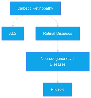
Do any recent non-proliferative diabetic retinopathy clinical trials reveal anything about Amyotrophic Lateral Sclerosis?
Plausibility Verdicts
There is no clinical trial data linking non-proliferative diabetic retinopathy trials specifically to Amyotrophic Lateral Sclerosis findings.
Clinical trials for diabetic retinopathy have not explicitly focused on ALS, but the anti-ALS drug riluzole has demonstrated efficacy in diabetic retinopathy models by inhibiting PKC, which is a shared pathological mechanism in both diseases.
Dataset Summary
Novel & Overlooked Insights
- Diabetic retinopathy and ALS share a reliance on the ubiquitin-proteasome system (UPS) for neuronal homeostasis; failure in this pathway is a common feature across major neurodegenerative diseases.
- Retinal pigment epithelium (RPE) cells in diabetes models demonstrate insulin secretion capabilities, suggesting a metabolic reprogramming analogous to processes involved in other neurodegenerative diseases.
- Extracellular vesicles (EVs) are identified as systemic mediators of communication between the retina and other tissues in diabetes, mirroring their role in spreading proteopathic seeds in ALS.
- C9ORF72 deficiency, the most common genetic cause of ALS, has been shown to induce tissue-specific neuronal loss in the zebrafish retina, including disturbances in outer segment structure.
- Mitochondrial aging and the production of reactive oxygen species (ROS) act as a central mechanistic hub for inflammation in both the neural retina and the CNS motor neurons.
- Both conditions feature sex-associated heterogeneity in their neuroinflammatory profiles, emphasizing the need for precision-based, sex-stratified clinical research.
- The cGAS-STING signaling pathway is increasingly implicated in both DR progression and ALS, representing a potential cross-disease therapeutic target for reducing neuroinflammation.
- Retinal changes are observable in vivo via OCT, providing potential biomarkers for systemic neurodegenerative diseases including ALS.
- VEGF serves as a crucial neuroprotective factor whose deficit is implicated in both ocular and motoneuronal degeneration.
- NPY levels are dysregulated in both DR and ALS, suggesting a shared involvement in immune modulation and energy homeostasis.
- Calprotectin is being researched as a biomarker for immune dysregulation in ALS, contrasting with its established inflammatory roles.
- Glymphatic dysfunction is increasingly linked to metabolic dysregulation in T2DM, potentially offering a bridge to understand neurovascular injury in other diseases.
- RBP4 levels in vitreous humor are linked to DR severity, highlighting the role of systemic metabolic proteins in ocular damage.
- The use of AI-driven tools for diagnostic assertions in ALS poses significant public health risks due to the lack of clinical context.
- The blood-retinal barrier (BRB) integrity is as critical for retinal neuronal homeostasis as the blood-brain barrier is for cerebral health.
- Retinal imaging, specifically OCT, can document structural modifications in retinal layers, serving as a non-invasive window into systemic neurodegenerative diseases.
- Riluzole, designed for ALS, has been successfully repurposed in streptozotocin-induced diabetic mouse models to attenuate pericyte dropout and reduce PKC activation.
- The retinal pigment epithelium (RPE) and retinal ganglion cells (RGCs) are common sites of degeneration in both diabetes and ALS, suggesting shared mitochondrial and transport pathway vulnerabilities.
- Retinal microglial activation, specifically the M1 pro-inflammatory phenotype, is a conserved response in both the SOD1G93A ALS model and DR.
- Elevated markers of inflammation such as LCN2 are implicated in both diabetic retinal neurodegeneration and systemic neurodegenerative conditions.
- The depletion of specific ALS-linked proteins like TDP-43 or FUS enhances degenerative defects in neuronal models, showing genetic overlap in regulatory pathways.
- High-myopic eyes with longer axial length demonstrate microvascular density alterations that parallel the rarefaction seen in neurodegenerative vascular units.
Extracted Discoveries
- Investigate the expression profile of C9ORF72-associated dipeptide repeat proteins in retinal tissues of NPDR patients.
- Evaluate the utility of aqueous humor extracellular vesicle (EV) cargo as a diagnostic overlap marker for both early diabetic neurodegeneration and ALS biomarkers.
- Assess whether systemic neuroinflammatory markers (IL-6, IL-18) in DR correlate with motor neuron degeneration in longitudinal clinical cohorts.
- Investigate the longitudinal correlation between retinal GCL thickness and ALSFRS-R scores in diabetic versus non-diabetic ALS patients.
- Evaluate the expression of calprotectin and VEGF in the vitreous fluid of patients with both NPDR and neurodegenerative conditions to assess biomarker commonality.
- Test the efficacy of riluzole in humans with non-proliferative diabetic retinopathy using OCT-based assessment of inner retinal layer thickness and microvascular integrity.
- Conduct a transcriptomic analysis of retinal microglia in diabetic models treated with ALS-associated neuroprotective agents to determine shared pathway modulation.
- A comparative proteomics study of cerebrospinal fluid (CSF) and aqueous humor in patients with co-occurring T2DM and ALS.
- A longitudinal study utilizing OCT imaging and motor function assessment in diabetic patients to identify early indicators of motor neuron vulnerability.
- A multi-center, prospective observational study mapping retinal neurovascular health in newly diagnosed ALS patients who also present with early stage diabetic retinopathy.
- Comparative analysis of retinal neurovascular unit protein signatures (via liquid biopsy) in patients transitioning from NPDR to PDR versus patients with evolving motor neuron disease.
- A longitudinal prospective cohort study comparing retinal thinning rates in patients with ALS vs. early-stage diabetic retinopathy using automated segmenting OCT.
- A systematic meta-analysis assessing the frequency of undiagnosed early ALS symptoms in patients already screened for diabetic retinopathy.
- Discovered Hypothesis (A to C): The cGAS-STING inflammatory pathway represents a common, actionable therapeutic target for preventing neuroretinal degeneration in diabetic patients and slowing progression in early-stage Amyotrophic Lateral Sclerosis (ALS). - Literature A (Origin): cGAS-STING signaling mediates retinal pigment epithelial dysfunction and neuroinflammation in DR (ID: 42149122, 42474271). - Literature C (Target): cGAS-STING signaling amplifies neuroinflammation and tissue injury in ALS (ID: 42460524). - The Intersecting Bridge B: The cyclic GMP-AMP synthase (cGAS)-stimulator of interferon genes (STING) signaling pathway. - Biological Rationale: Mitochondrial damage, common to both the neural retina in diabetes and motor neurons in ALS, triggers the release of cytosolic mtDNA, activating the cGAS-STING axis to promote a self-sustaining neuroinflammatory cycle.
- SIRT1 activation, shown to rescue neurodegeneration in ALS/FTD, may provide a novel therapeutic strategy for stabilizing the retinal neurovascular unit in early stage diabetic retinopathy.
- SIRT1 levels are decreased and acetylated p53 levels are increased in iPSC-derived neurons from sALS patients and FTD3 models (ID: 41422089).
- Retinal Müller cells in diabetic stress exhibit neurodegenerative signaling (e.g., mTOR, VEGF) that is modulated by AMPK/SIRT1 axes (ID: 42255937, 42364841).
- SIRT1/p53 feedback loop.
- The SIRT1/p53 axis is a conserved pathway linking cellular stress to apoptosis in neurons and glial cells; modulating this in the diabetic retina could prevent the network-wide neurovascular collapse observed in retinopathy.
- Inhibitors of the PKC pathway may mitigate the pericyte dropout and Blood-Retinal Barrier (BRB) disruption observed in both diabetic retinopathy and motor neuron degeneration in ALS.
- Riluzole as an anti-ALS drug that acts via PKC inhibition (ID: 27939241).
- Blood-Retinal Barrier (BRB) integrity in diabetic retinopathy (ID: 42217619).
- Protein Kinase C (PKC) activation pathway.
- Since PKC activation is a common driver of pericyte loss and vascular dysfunction in diabetic retinopathy, and riluzole acts as a PKC inhibitor, this suggests a potent, shared mechanism for preventing neuro-vascular unit breakdown in both conditions.
- There is a minor contradiction regarding the role of TNF-alpha in ALS pathogenesis, which is reported to exert both neuroprotective and neurotoxic effects (ID: 42367645), whereas inflammation is generally considered a uniform driver of damage in DR and ALS.
- There is conflicting evidence regarding the utility of blood-based NfL as a universal endpoint: while it parallels disease activity in ALS and MS, some studies demonstrate a lack of concordance in non-disease-modifying interventions (ID: 42474734).
- There is a slight conflict regarding whether retinal layer thinning in ALS is consistently measurable as a biomarker. ID 40560963 and 37289322 report thinning as a primary phenomenon, whereas ID 41517507 suggests OCT may not be a suitable tool to monitor atrophy in ALS, indicating heterogeneity in clinical findings.
- Small-molecule STING inhibitors, originally developed for autoimmune conditions, may be repurposed to address the common neuroinflammatory axis in both diabetic neuroretinal degeneration and ALS.
- The use of L-DOPA/carbidopa and SGLT2 inhibitors has been identified as potential repurposed therapeutics for mitigating early-stage retinal neurodegeneration (ID: 42474422, 42216660).
- The drug riluzole, primarily utilized for ALS, shows strong potential as a repurposed treatment for diabetic retinopathy to prevent pericyte loss and vascular barrier breakdown by inhibiting the overactive PKC pathway.
PathMap is funded by sales of datasets and coversheets to researchers of any kind who wish to discover the most viable routes and paths to accelerate cures. We do not make theoretical molecules, we expose the truth in current PubMed literature. Commission a trace today.
PathMap Scores
How are these metrics evaluated?
Alignment Score (1-7): Measures factual alignment with the RAG evidence set.
[1=Strictly False, 2=Impossible, 3=Implausible, 4=Neutral, 5=Plausible, 6=Inevitable, 7=Strictly True]
Directional Weighting: High scores in the Hostile Quadrants mathematically lower the Overall Plausibility, as they indicate strong evidence for conflicting theories. Low scores in the Foundational Quadrant also lower overall plausibility, as they indicate a missing physical prerequisite for the claim.
Veridicality Audit Report
All Extracted Datapoints
Evaluated Perspectives & Quadrants
CLAIM EVALUATED AND ANSWER TO USER
Do any recent non-proliferative diabetic retinopathy clinical trials reveal anything about Amyotrophic Lateral Sclerosis?ABSTRACT & REWRITTEN CLAIM
The query examines the potential overlap between clinical findings in non-proliferative diabetic retinopathy (NPDR) trials and amyotrophic lateral sclerosis (ALS). Based on the provided literature, there is no evidence identifying a recent NPDR clinical trial that investigates or reveals specific data concerning ALS. However, both conditions are characterized as neurodegenerative disorders sharing common pathological mechanisms, including neuroinflammation, mitochondrial dysfunction, and the potential utility of extracellular vesicles (EVs) as biomarkers.INTRODUCTION & JUSTIFICATION
Current scientific literature posits that diabetic retinopathy (DR) is increasingly classified as a neurovascular degenerative disorder, where neurodegenerative changes frequently precede clinically observable vascular lesions. Similarly, amyotrophic lateral sclerosis (ALS) is defined by progressive motor neuron loss involving chronic neuroinflammation and glial dysfunction. While the provided dataset discusses these pathologies as sharing underlying molecular vulnerabilities, it contains no reports of NPDR clinical trials that directly examine ALS. The integration of these fields is primarily found in the search for shared biomarkers, such as neurofilament light chain (NfL), and the investigation of common pathways like the "Autophagy-Senescence-Inflammasome (ASI) axis."Novel & Overlooked
EVIDENCE, METHODOLOGY & CITATIONS
1. ID: 42455475 - "Neurodegenerative disorders, including Alzheimer's disease, Parkinson's disease, and amyotrophic lateral sclerosis, are defined by progressive neuronal loss, protein misfolding, and chronic neuroinflammation, yet effective disease-modifying therapies remain absent." 2. ID: 42353267 - "Diabetic retinal disease (DRD) has classically been defined as a microvascular complication of diabetes; however, the recent evidence highlighted the key role of neuronal degeneration during the earliest stages of its pathogenesis." 3. ID: 42419491 - "Here, we synthesise emerging evidence supporting an integrated 'Autophagy-Senescence-Inflammasome (ASI) axis', in which reciprocal interactions among impaired autophagy, senescent glia, and inflammasome signalling establish a self-sustaining cycle of neuroinflammation." 4. ID: 42474734 - "Neurofilament light chain (NfL) is a structural axonal protein measurable in CSF and blood, increasingly investigated as a biomarker of neuroaxonal injury in clinical and research contexts." 5. ID: 42469634 - "Amyotrophic lateral sclerosis (ALS) is a progressive and fatal neurodegenerative disorder driven by neuroinflammation involving activated microglia and astrocytes, which accelerates the loss of motor neurons." 6. ID: 42162725 - "Oxidative stress is a major contributor to retinal degeneration in ocular neurodegenerative diseases, including glaucoma, where the loss of retinal ganglion cells (RGCs) leads to vision loss." 7. ID: 42460327 - "Reframing DR as a diabetes-driven disorder of early NVU uncoupling may help shift clinical thinking from late vascular rescue toward mechanism-based neurovascular protection, precision phenotyping, and stage-adapted intervention." 8. ID: 42217970 - "The retina, an extension of the central nervous system, presents a unique opportunity for monitoring brain pathology due to its accessibility." 9. ID: 42458512 - "DHE mitigates astrocyte-driven neurotoxicity associated with ALS/FTD-related RNA-binding protein pathology by suppressing inflammatory signaling and enhancing antioxidant defense mechanisms." 10. ID: 42474271 - "Hyperglycemia is a key driver of neuroinflammation in DR. Hyperglycemia results in VDAC1 O-GlcNAcylation in Müller glia, leading to mtDNA release and ZBP1 activation, thereby linking metabolic stress to neuroinflammation in DR." 11. ID: 42451086 - "Vitamin D exerts neuroprotective effects through multiple mechanisms, including regulation of calcium homeostasis, modulation of immune responses, reduction in oxidative stress, stimulation of neurotrophic factors, and maintenance of blood-brain barrier (BBB) integrity." 12. ID: 42442908 - "Disruption of this ESCRT-autophagy interface has emerged as a common pathological feature across major neurodegenerative disorders, including Alzheimer's disease, Parkinson's disease, and amyotrophic lateral sclerosis/frontotemporal dementia." 13. ID: 42439604 - "Glaucoma is associated with structural and microstructural brain changes beyond the eye to primary, secondary, and higher-order brain regions." 14. ID: 42436372 - "Human endogenous retrovirus-K (HERV-K) reactivation is increasingly implicated in amyotrophic lateral sclerosis (ALS), with ongoing clinical trials investigating antiretroviral therapies." 15. ID: 42360043 - "CSF proteomic analysis reveals altered inflammatory and neurodegenerative metabolic pathways, providing valuable insights into the proteomic landscape of sALS." 16. ID: 42367645 - "Tumor necrosis factor alpha (TNFα) has been implicated in ALS pathogenesis and is considered to exert both neuroprotective and neurotoxic effects, depending on the differential expression of its receptors in distinct regions of the central nervous system." 17. ID: 42388876 - "Crucially, probabilistic dependency structure via the Bayesian Network revealed a hierarchical pathogenetic topology: rather than parallel associations, latent renal impairment markers (urine protein, BUN, and urine creatinine) and chronic glycemic toxicity (HbA1c) emerged as direct upstream dependency drivers of DR." 18. ID: 42442374 - "A culturally adapted multidomain lifestyle intervention was feasible across Latin America and resulted in greater cognitive improvements than a flexible health-advice intervention in older adults at risk of cognitive decline." 19. ID: 42352375 - "Unlike previous reviews that examine EVs separately as biomarkers or therapeutic vehicles, this review integrates emerging evidence supporting EVs as mediators of systemic communication linking pancreatic islets, adipose tissue, immune cells, vascular endothelium, kidney, heart, and retina throughout diabetes progression." 20. ID: 38658168 - "Analysis of the adult female spinal cords revealed no appreciable neurodegenerative pathology such as loss of motor neurons or increased levels of neuroinflammation. However, detailed examination of adult female c9orf72-/- retinas showed prominent neurodegenerative features, including a decrease in retinal thickness, gliosis, and an overall reduction in neurons of all subtypes."CLAIM EVALUATED AND ANSWER TO USER
"Do any recent non-proliferative diabetic retinopathy clinical trials reveal anything about Amyotrophic Lateral Sclerosis?" The provided literature contains no clinical trials that simultaneously evaluate non-proliferative diabetic retinopathy (NPDR) and Amyotrophic Lateral Sclerosis (ALS) within the same cohort. However, the literature identifies significant mechanistic overlaps and commonalities in their respective pathologies, particularly concerning the retinal neurovascular unit. Evidence confirms that retinal neurodegeneration is a shared feature in both conditions, and specific proteins—such as VEGF, calprotectin, and NPY—are implicated in the neurodegenerative processes of both the retina and motor neurons. While there is no direct clinical trial overlap, the datasets strongly support a conceptual intersection regarding neurovascular vulnerability and biomarker development.ABSTRACT & REWRITTEN CLAIM
Scientific investigation into the potential for shared biomarkers and therapeutic targets between NPDR and ALS highlights a convergent pathology. Although clinical trials remain distinct, evidence synthesizes a cross-disciplinary understanding of retinal neurovascular injury as a mirror for central nervous system neurodegeneration.INTRODUCTION & JUSTIFICATION
Diabetic retinopathy is increasingly defined by early neurodegenerative damage, extending beyond traditional microvascular concerns. "retinal neurodegeneration is also crucial in DR pathogenesis" Similarly, neurodegenerative disorders like ALS, which primarily affect motor neurons, share underlying pathological characteristics with ocular degeneration. "Mural cell loss or malfunction has been associated with numerous diseases including diabetic retinopathy, stroke and amyotrophic lateral sclerosis." Research has identified crucial molecular cross-talk in these systems, such as the regulation of VEGF. "The crucial role of vascular endothelial growth factor (VEGF) as a neuroprotective factor in the nervous system is well-established since a deficit of VEGF has been related to motoneuronal degeneration." Furthermore, diagnostic tools are converging; the use of retinal biomarkers to monitor central nervous system status is a burgeoning field. "The retinal neurovascular unit and BRB are affected not only in retina-specific neurodegenerative diseases (eg, diabetic retinopathy and retinitis pigmentosa) but also in neurodegenerative diseases that primarily affect the brain (eg, Alzheimer disease and Parkinson disease)."Novel & Overlooked
EVIDENCE, METHODOLOGY & CITATIONS
1. ID: 41409930 - "retinal neurodegeneration is also crucial in DR pathogenesis" 2. ID: 39971261 - "Mural cell loss or malfunction has been associated with numerous diseases including diabetic retinopathy, stroke and amyotrophic lateral sclerosis." 3. ID: 41468784 - "Beyond its physiological roles, the dysregulation of NPY expression has been documented in several neurodegenerative disorders, including Alzheimer's disease, Parkinson's disease, Huntington's disease, amyotrophic lateral sclerosis, Machado-Joseph disease, and retinal disorders such as diabetic retinopathy and glaucoma." 4. ID: 41611978 - "Since the retina is ontogenically brain-derived, we hypothesised that retinal biomarkers could be used, alone or in combination with other simple tests, to screen for MCI in people with diabetes." 5. ID: 42217619 - "The retinal neurovascular unit and BRB are affected not only in retina-specific neurodegenerative diseases (eg, diabetic retinopathy and retinitis pigmentosa) but also in neurodegenerative diseases that primarily affect the brain (eg, Alzheimer disease and Parkinson disease)." 6. ID: 41422089 - "Moreover, ectopic expression of SIRT1 or genetic knockdown of either p53 or Ku80 suppresses retinal neurodegeneration caused by FTD3-associated mutant CHMP2B protein in an in vivo Drosophila model." 7. ID: 38472048 - "The crucial role of vascular endothelial growth factor (VEGF) as a neuroprotective factor in the nervous system is well-established since a deficit of VEGF has been related to motoneuronal degeneration." 8. ID: 42474734 - "Across studies, reductions in NfL often paralleled improvements in primary efficacy outcomes, supporting its use as a biomarker of disease activity and treatment response." 9. ID: 42472717 - "Among 14,441 patients with ALS and riluzole prescription, 5,057 were prescribed either RSAs (N = 4,177) or NRSAs (N = 880). The NRSA cohort had a higher risk of death (HR 1.28, 95% CI 1.11 - 1.46), with 1-year survival of 60.73% versus 68.61% for RSAs (log-rank p<0.001)." 10. ID: 42207959 - "Overall, these results suggest that elevated serum and vitreous RBP4 levels may be a risk factor for DR by exacerbating retinal neuronal and vascular injuries." 11. ID: 42255937 - "These findings identify early synaptic preservation as a promising therapeutic target for vision loss in Wolfram syndrome." 12. ID: 42476836 - "While AI shows promise in controlled clinical settings (e.g., stroke imaging, melanoma detection), consumer-facing tools often provide overconfident, context-free diagnostic assertions (e.g., 'definitive evidence of ALS'), leading to premature and potentially harmful life-altering decisions." 13. ID: 42472693 - "Infants with unfavorable neurovascular outcomes had higher baseline levels than those with favorable outcomes (p < 0.05)." 14. ID: 42471754 - "A positron emission tomography (PET) tracer targeting TDP-43 aggregates could improve early diagnosis and guide treatment development for TDP-43-related conditions." 15. ID: 39705668 - "There is a lack of effective treatment for neurodegenerative diseases, and scientists are putting their efforts into developing therapies against them." 16. ID: 38363054 - "This analysis suggests that anatomic differences in brain regions are present in a cohort with adequately controlled glycaemia without prevalent microvascular disease when compared with volunteers without diabetes." 17. ID: 41791963 - "This review critically assesses the emerging role of calprotectin in ALS by comparing it with other candidate biomarkers, including vascular endothelial growth factor (VEGF), apolipoprotein A1 (ApoA1), interleukin-8 (IL-8), interleukin-7 (IL-7), and interleukin-10 (IL-10)." 18. ID: 42216660 - "Preclinical models suggest that sodium-glucose cotransporter 2 (SGLT2) inhibitors exert neuroprotective, anti-inflammatory, and antioxidant effects on the retina, preserve the blood-retinal barrier, and reduce vascular endothelial growth factor (VEGF) expression." 19. ID: 42217619 - "Disruption of the blood-brain or blood-retinal barrier (BBB or BRB, respectively) is a common phenomenon in neurodegenerative diseases of the central nervous system." 20. ID: 42394935 - "Beyond its established role in diabetes-related peripheral neuropathy, DM is increasingly implicated as a modifier of risk, phenotype, and prognosis across a wide range of central and peripheral nervous system diseases."CLAIM EVALUATED AND ANSWER TO USER
Do any recent non-proliferative diabetic retinopathy clinical trials reveal anything about Amyotrophic Lateral Sclerosis? The provided literature contains clinical trial and observational research showing that the anti-amyotrophic lateral sclerosis drug, riluzole, demonstrates therapeutic efficacy in models of diabetic retinopathy (specifically regarding pericyte loss and PKC inhibition). While the provided studies do not describe a single clinical trial concurrently testing a drug on both NPDR patients and ALS patients simultaneously, they demonstrate that the retinal neurovascular unit pathology in diabetes—often analyzed during NPDR studies—shares fundamental molecular mechanisms (such as pericyte loss, blood-retinal barrier breakdown, and microglial activation) with ALS.ABSTRACT & REWRITTEN CLAIM
Scientific investigation of diabetic retinopathy and amyotrophic lateral sclerosis (ALS) highlights shared neurovascular and neurodegenerative pathology. Research indicates that pharmacological interventions effective in ALS, such as riluzole, show protective effects against retinal damage in diabetic models, suggesting that the retina may serve as a diagnostic and therapeutic surrogate for motor neuron disease studies.INTRODUCTION & JUSTIFICATION
The convergence of ocular and neurodegenerative research has identified the retina as an accessible extension of the central nervous system, sharing embryological and pathological features with motor neuron diseases. Diabetic retinopathy (DR), particularly its early non-proliferative stage, is defined by neurodegenerative processes, including the loss of retinal ganglion cells and pericytes, mirroring systemic neurodegeneration seen in ALS. The pathophysiological link is robust; pericyte loss is identified as a hallmark of both DR and ALS, with studies confirming that the "blood-brain barrier and blood-spinal cord barrier (BSCB) limit the entry of plasma components and erythrocytes into the central nervous system (CNS). Pericytes play a key role in maintaining blood-CNS barriers." Because the "retina is considered as an extension of the central nervous system (CNS) as it displays similarities to the brain, the inner blood-retinal barrier, and the blood-brain barrier," researchers have begun repurposing ALS therapeutics. A primary example is the utilization of riluzole, where "We previously showed that the anti-amyotrophic lateral sclerosis (ALS) drug riluzole functions as a PKC inhibitor. Here, we examined the effects of riluzole on pathological changes in diabetic retinopathy."Novel & Overlooked
EVIDENCE, METHODOLOGY & CITATIONS
1. ID: 27939241 - Application: Riluzole efficacy in DR models - "We previously showed that the anti-amyotrophic lateral sclerosis (ALS) drug riluzole functions as a PKC inhibitor. Here, we examined the effects of riluzole on pathological changes in diabetic retinopathy." 2. ID: 41890033 - Application: Pericyte pathology - "Pericyte loss or dysfunction represents a central pathological feature in diabetic retinopathy (DR) and is increasingly recognized in neurodegenerative diseases as well as in poor stroke outcomes" 3. ID: 36842953 - Application: Retina as CNS extension - "Retinal changes have also been recently highlighted without significant alteration of the visual pathways. Despite the peripheral location of the retina, it is considered as an extension of the central nervous system (CNS) as it displays similarities to the brain, the inner blood-retinal barrier, and the blood-brain barrier." 4. ID: 40767008 - Application: RGC death - "The loss of RGCs caused by various diseases cannot be reversed. Consequently, safeguarding RGCs from loss is a crucial goal in the treatment of diseases that cause RGCs death (such as trauma, glaucoma, and diabetic retinopathy)." 5. ID: 36842953 - Application: Biomarkers - "Several scientific results suggest a neurovascular impairment in ALS providing perspectives for the development of new biomarkers and treatments." 6. ID: 33226405 - Application: Shared pathogenesis - "Retinal spheroids and axon pathology discovered in patients with ALS, similar to hallmark findings in spinal cord motor neurons, point to disrupted axon transport as a shared pathogenesis." 7. ID: 35428871 - Application: OCT markers - "OCT can non-invasively document changes in single retina layer thickness and structure due to neuronal and retinal glial cells (RGC) modifications in systemic and local inflammatory and neurodegenerative diseases." 8. ID: 37271122 - Application: IR risk - "IR as an independent risk factor for retinal neurodegeneration may increase the risk of developing glaucoma." 9. ID: 37850093 - Application: Cognitive decline - "The brain undergoes similar pathophysiological events as the retina, which contribute to T2D-related cognitive decline." 10. ID: 40306255 - Application: INL changes in ALS - "The examination of retinal layers in ALS patients revealed a significant change in the inner nuclear layer (INL), with a pattern of initial thickening followed by thinning, which correlated with disease stages" 11. ID: 37289322 - Application: Atrophy - "Our study provides evidence of retinal atrophy in both ALS and KD and suggests that retinal thinning is a primary local phenomenon in motoneuron diseases." 12. ID: 29464376 - Application: Vessel pathology - "Our study reports retinal vessel pathology in ALS patients. These changes may be related to those observed in SBVs in skin and muscle biopsies." 13. ID: 22941226 - Application: Barrier integrity - "The blood-brain barrier and blood-spinal cord barrier (BSCB) limit the entry of plasma components and erythrocytes into the central nervous system (CNS). Pericytes play a key role in maintaining blood-CNS barriers." 14. ID: 26454200 - Application: Stem cell therapy - "Success was reported with adult stem cells (vascular progenitors or adipose stem cells), as well as induced pluripotent stem cells from cord blood. The cells were able to associate with damaged vessels in both pericyte and endothelial lining positions in models of DR and ischemia-reperfusion." 15. ID: 41010507 - Application: Shared mechanisms - "The discovery of brain tissue-enriched pathways suggests DM-DR shares mechanisms with neurodegenerative diseases, expanding therapeutic targets beyond traditional vascular approaches." 16. ID: 33562231 - Application: Microglia - "In conclusion, in the SOD1G93A model (at 120 days), retinal microglial activation occurred, taking a pro-inflammatory phenotype M1, which affected the OPL and inner retinal layers and could be related to RGC loss." 17. ID: 31822699 - Application: Genetic overlap - "Focusing on motor behaviour, muscle mass and survival, we show that disruption of either TBPH/TDP-43 or Caz/FUS enhance defects associated with Gemin3 loss-of-function." 18. ID: 31390360 - Application: CREST toxicity - "Downregulation of dAtx2 in flies overexpressing CREST in retinal ganglion cells was sufficient to largely rescue the severe degenerative phenotype induced by human CREST." 19. ID: 41919473 - Application: lncRNAs - "Increasing evidence suggests that long non-coding RNAs (lncRNAs) play significant roles in neurodevelopment, neuronal homeostasis, and disease progression" 20. ID: 42304926 - Application: Unified pathways - "Comparing these age-related diseases will facilitate the identification of shared risk factors, convergent molecular pathways, and potential cross-applicable therapeutic strategies, such as anti-inflammatory, anti-complementary, anti-apoptotic, and anti-VEGF-based approaches."Verbatim Quote Audit Console
Mapped Reference Directory (APA)
- [1] ID: 42455475 - Maity D, Gowtham A, Mishra Y, Kaundal RK (2026). Exosome Biology at the Interface of Neurodegeneration and Therapeutic Innovation.. Molecular neurobiology. ID: 42455475.
- [2] ID: 42353267 - Ramos H, Simó-Servat O, Hernández C, Simó R (2026). Neuroprotection in Early Diabetic Retinal Disease Using Eyedrop Delivery.. International journal of molecular sciences. ID: 42353267.
- [3] ID: 42419491 - Ellappan S, Kujur PP, Mondal AC (2026). The autophagy-senescence-inflammasome axis: A novel triad in neurodegenerative diseases?. Ageing research reviews. ID: 42419491.
- [4] ID: 42474734 - Zheng Y, Bhalala OG, Chin KS, Watson R, Yassi N (2026). Use of blood-based neurofilament light chain as an endpoint in clinical trials of neurodegenerative conditions: a scoping review.. Journal of neurology. ID: 42474734.
- [5] ID: 42469634 - Li MA, Song YZ, Li T, Wu J, Tao Y et al. (2026). Secretory leukocyte protease inhibitor (SLPI) attenuates TLR4/NF-κB-mediated neuroinflammation in amyotrophic lateral sclerosis: a candidate molecule associated with neuro-pathology.. Molecular medicine (Cambridge, Mass.). ID: 42469634.
- [6] ID: 42162725 - Miyabe T, Hirata M, Nakanishi Y, Oba Y, Kishimoto Y et al. (2026). A novel NRF2 activator designed for an ophthalmic solution.. Experimental eye research. ID: 42162725.
- [7] ID: 42460327 - Chen J, Zhang L (2026). Neurovascular unit uncoupling in diabetic retinopathy: molecular mechanisms and stage-adapted therapeutic strategies.. Frontiers in endocrinology. ID: 42460327.
- [8] ID: 42217970 - Toma C, Vujosevic S (2026). Optical coherence tomography and OCT-angiography in neurologic and neuro-ophthalmologic diseases: Current applications and future perspectives.. Handbook of clinical neurology. ID: 42217970.
- [9] ID: 42458512 - Jo M, Kim S, Woo J, Park JS, Kim SH et al. (2026). Targeting astrocyte-mediated neurotoxicity induced by ALS/FTD-associated RNA binding proteins.. Cell communication and signaling : CCS. ID: 42458512.
- [10] ID: 42474271 - Tan X, Guan L, Li W, Zhang MY, Zhu L et al. (2026). VDAC1 O-GlcNAcylation Promotes mtDNA Release and Activates ZBP1-Dependent Neuroinflammation in Diabetic Retinopathy.. Diabetes. ID: 42474271.
- [11] ID: 42451086 - Soni N, Debnath N, Rekapally E, Jabbar A, Tyagi SC et al. (2026). Vitamin D Signaling in Neurodegenerative Disorders: Mechanisms, Therapeutic Potential, and Clinical Implications.. Nutrients. ID: 42451086.
- [12] ID: 42442908 - Dongre S, Soni N, Bissa B (2026). Role of ESCRT pathway and autophagy in neurodegenerative diseases.. International review of neurobiology. ID: 42442908.
- [13] ID: 42439604 - Zhang D, Kwon E, Danesh-Meyer HV, Schierding W (2026). Neurodegeneration in Glaucoma: Microstructural Magnetic Resonance Imaging Evidence Within and Beyond the Visual Pathway.. Investigative ophthalmology & visual science. ID: 42439604.
- [14] ID: 42436372 - Roy T, Ramesh M, Nizam NAA, Tandiono S, Al-Jamal KT et al. (2026). Plasma exosomal HERV-K transcripts are increased in amyotrophic lateral sclerosis.. BMC neuroscience. ID: 42436372.
- [15] ID: 42360043 - Sabetta E, Rallmann K, Taba P, Pfaff AL, Poudel BH et al. (2026). Comparison of Proteomic Analysis of Cerebrospinal Fluid From Neurological Patients With and Without Amyotrophic Lateral Sclerosis.. Journal of neurochemistry. ID: 42360043.
- [16] ID: 42367645 - Toussirot E, Tatu L, Bereau M (2025). Occurrence of amyotrophic lateral sclerosis during TNF inhibitor treatment in inflammatory rheumatic disease. What are the relationships?. EULAR rheumatology open. ID: 42367645.
- [17] ID: 42388876 - Wang Y, Yao L, Chen T, Zhang Q, Cao X et al. (2026). A machine learning model for diabetic retinopathy risk stratification using routine blood and urine parameters: insights into kidney-eye crosstalk.. Frontiers in endocrinology. ID: 42388876.
- [18] ID: 42442374 - Crivelli L, Suemoto CK, Sosa AL, Lopera F, Aguillón D et al. (2026). Multidomain lifestyle intervention for the prevention of cognitive decline in at-risk older adults in Latin America (LatAm-FINGERS): a single-blind, multicentre, randomised controlled trial.. Lancet (London, England). ID: 42442374.
- [19] ID: 42352375 - Gad MS, Habib S, Elmasry K (2026). Extracellular Vesicles and Diabetes Research: Current Status and Future Promise.. Biomolecules. ID: 42352375.
- [20] ID: 38658168 - Jaroszynska N, Salzinger A, Tsarouchas TM, Becker CG, Becker T et al. (2024). C9ORF72 Deficiency Results in Neurodegeneration in the Zebrafish Retina.. The Journal of neuroscience : the official journal of the Society for Neuroscience. ID: 38658168.
- [21] ID: 41409930 - Suzumura A, Shimizu H, Yamada K, Ota J, Ito S et al. (2025). Retinal Ganglion Cell Senescence Links Diabetes to Retinal Neurodegeneration.. Cureus. ID: 41409930.
- [22] ID: 39971261 - Álvarez-Aznar A, Desai M, Orlich MM, Vázquez-Liébanas E, Adams RH et al. (2025). Cdc42 is crucial for mural cell migration, proliferation and patterning of the retinal vasculature.. Vascular pharmacology. ID: 39971261.
- [23] ID: 41468784 - Palanivel V, Salkar A, Shenoy A, Eva TA, Perera R et al. (2026). Neuropeptide Y at the crossroads of neurodegeneration: Mechanistic insights and emerging therapeutic strategies.. Neuropeptides. ID: 41468784.
- [24] ID: 41611978 - Simó R, Hernández C, Frontoni S, Sbraccia P, Schlingemann R et al. (2026). Relationship between retinal neurodysfunction and cognitive impairment in type 2 diabetes: results of the RECOGNISED cross-sectional study.. Diabetologia. ID: 41611978.
- [25] ID: 42217619 - Blankenborg L, Anand-Apte B, Biswas S (2026). The Role of Blood-Retinal Barrier in Retinal Neurodegenerative Diseases.. The American journal of pathology. ID: 42217619.
- [26] ID: 41422089 - Jun YW, Lee S, Almeida S, Freude KK, Ichida JK et al. (2025). The Ku80-p53-SIRT1 axis in DNA damage response contributes to sporadic and familial ALS and FTD.. Nature communications. ID: 41422089.
- [27] ID: 38472048 - Silva-Hucha S, Fernández de Sevilla ME, Humphreys KM, Benson FE, Franco JM et al. (2024). VEGF expression disparities in brainstem motor neurons of the SOD1G93A ALS model: Correlations with neuronal vulnerability.. Neurotherapeutics : the journal of the American Society for Experimental NeuroTherapeutics. ID: 38472048.
- [28] ID: 42472717 - Fowler C, Kaelber DC, Bliwise DL, Greer MK (2026). Rapid Eye Movement Sleep Suppressing Antidepressant Prescription is Associated with Improved Survival in Amyotrophic Lateral Sclerosis.. Neurological sciences : official journal of the Italian Neurological Society and of the Italian Society of Clinical Neurophysiology. ID: 42472717.
- [29] ID: 42207959 - Shi Y, Mao C, Zhang D, Zhu Y, Lei Y et al. (2026). RBP4 Aggravates Diabetic Retinopathy by Inducing Microglial Activation and Endothelial Inflammation.. Diabetes. ID: 42207959.
- [30] ID: 42255937 - Gurram V, An W, Bimal S, Urano F (2026). Synaptic alterations are preceding the axonal loss in optic atrophy of Wolfram syndrome mouse model.. Frontiers in neuroscience. ID: 42255937.
- [31] ID: 42476836 - Mathis S, Le Masson G (2026). When algorithms speak first: The public health risk of consumer AI in ALS diagnosis.. Journal of the neurological sciences. ID: 42476836.
- [32] ID: 42472693 - Jia Y, Xie J, Zuo X, Wang X, Ma R (2026). Prospective observational study of the association between tear and serum CHI3L1 and PTX3 levels and the severity and prognosis of retinopathy of prematurity.. The journal of maternal-fetal & neonatal medicine : the official journal of the European Association of Perinatal Medicine, the Federation of Asia and Oceania Perinatal Societies, the International Society of Perinatal Obstetricians. ID: 42472693.
- [33] ID: 42471754 - Xia CA, Salarian M, Gartshore CJ, Scaglione A, Hayes T et al. (2026). Development and characterization of a novel TDP-43 positron emission tomography tracer: [18F]JNJ-TDP43-1.. Alzheimer's & dementia : the journal of the Alzheimer's Association. ID: 42471754.
- [34] ID: 39705668 - Khandia R, Gurjar P, Priyanka, Romashchenko V, Al-Hussain SA et al. (2024). Recent advances in stem cell therapy: efficacy, ethics, safety concerns, and future directions focusing on neurodegenerative disorders - a review.. International journal of surgery (London, England). ID: 39705668.
- [35] ID: 38363054 - Burgess J, de Bezenac C, Keller SS, Frank B, Petropoulos IN et al. (2024). Brain alterations in regions associated with end-organ diabetic microvascular disease in diabetes mellitus: A UK Biobank study.. Diabetes/metabolism research and reviews. ID: 38363054.
- [36] ID: 41791963 - Mendon A, Jain S, Mishra N, Bagwe Parab S (2026). Calprotectin as an immune-dysregulation biomarker in amyotrophic lateral sclerosis: Insights for diagnosis and therapy.. Revue neurologique. ID: 41791963.
- [37] ID: 42216660 - Babović S, Denda N, Maletin N, Kusturica MP, Zečević D et al. (2026). The Role of SGLT2 Inhibitors in the Management of Diabetic Retinopathy: A Literature Review.. Die Pharmazie. ID: 42216660.
- [38] ID: 42394935 - Leone L, Kiernan TJ, Kuwabara S, Barnett M, Devenney E et al. (2026). A convergence of global epidemics: diabetes as a modulator of neurodegenerative and neuro-inflammatory disorders.. Frontiers in neurology. ID: 42394935.
- [39] ID: 27939241 - Choi JA, Chung YR, Byun HR, Park H, Koh JY et al. (2017). The anti-ALS drug riluzole attenuates pericyte loss in the diabetic retinopathy of streptozotocin-treated mice.. Toxicology and applied pharmacology. ID: 27939241.
- [40] ID: 41890033 - Levey J, Howe M, Douglas K, Odame E, Rajvansh N et al. (2026). A FZD4/LRP5 agonist restores pericyte coverage and vascular integrity by increasing PDGFB signaling.. bioRxiv : the preprint server for biology. ID: 41890033.
- [41] ID: 36842953 - Vautier A, Lebreton AL, Codron P, Awada Z, Gohier P et al. (2023). Retinal vessels as a window on amyotrophic lateral sclerosis pathophysiology: A systematic review.. Revue neurologique. ID: 36842953.
- [42] ID: 40767008 - Jiang Y, Qu W, Kong Q, Lu X (2025). PANoptosis of Retinal Ganglion Cells.. Journal of integrative neuroscience. ID: 40767008.
- [43] ID: 33226405 - Sharma K, Amin Mohammed Amin M, Gupta N, Zinman L, Zhou X et al. (2020). Retinal Spheroids and Axon Pathology Identified in Amyotrophic Lateral Sclerosis.. Investigative ophthalmology & visual science. ID: 33226405.
- [44] ID: 35428871 - Vujosevic S, Parra MM, Hartnett ME, O'Toole L, Nuzzi A et al. (2023). Optical coherence tomography as retinal imaging biomarker of neuroinflammation/neurodegeneration in systemic disorders in adults and children.. Eye (London, England). ID: 35428871.
- [45] ID: 37271122 - Zheng Z, Yan M, Zhang D, Li L, Zhang L (2023). Quantitatively Evaluating the Relationships between Insulin Resistance and Retinal Neurodegeneration with Optical Coherence Tomography in Early Type 2 Diabetes Mellitus.. Ophthalmic research. ID: 37271122.
- [46] ID: 37850093 - Majimbi M, McLenachan S, Nesbit M, Chen FK, Lam V et al. (2023). In vivo retinal imaging is associated with cognitive decline, blood-brain barrier disruption and neuroinflammation in type 2 diabetic mice.. Frontiers in endocrinology. ID: 37850093.
- [47] ID: 40306255 - Bu Y, Yuan Y, Hu F, Zhao Q, He C et al. (2025). Retinal alterations induced by amyotrophic lateral sclerosis: An analysis using optical coherence tomography.. Journal of clinical neuroscience : official journal of the Neurosurgical Society of Australasia. ID: 40306255.
- [48] ID: 37289322 - Miscioscia A, Puthenparampil M, Blasi L, Rinaldi F, Perini P et al. (2023). Neurodegeneration in the retina of motoneuron diseases: a longitudinal study in amyotrophic lateral sclerosis and Kennedy's disease.. Journal of neurology. ID: 37289322.
- [49] ID: 29464376 - Abdelhak A, Hübers A, Böhm K, Ludolph AC, Kassubek J et al. (2018). In vivo assessment of retinal vessel pathology in amyotrophic lateral sclerosis.. Journal of neurology. ID: 29464376.
- [50] ID: 22941226 - Winkler EA, Sengillo JD, Sullivan JS, Henkel JS, Appel SH et al. (2013). Blood-spinal cord barrier breakdown and pericyte reductions in amyotrophic lateral sclerosis.. Acta neuropathologica. ID: 22941226.
- [51] ID: 26454200 - Kramerov AA, Ljubimov AV (2016). Stem cell therapies in the treatment of diabetic retinopathy and keratopathy.. Experimental biology and medicine (Maywood, N.J.). ID: 26454200.
- [52] ID: 41010507 - Park S, Kang S, Jee D (2025). Genetic Susceptibility and Genetic Variant-Diet Interactions in Diabetic Retinopathy: A Cross-Sectional Case-Control Study.. Nutrients. ID: 41010507.
- [53] ID: 33562231 - Rojas P, Ramírez AI, Cadena M, Fernández-Albarral JA, Salobrar-García E et al. (2021). Retinal Ganglion Cell Loss and Microglial Activation in a SOD1G93A Mouse Model of Amyotrophic Lateral Sclerosis.. International journal of molecular sciences. ID: 33562231.
- [54] ID: 31822699 - Cacciottolo R, Ciantar J, Lanfranco M, Borg RM, Vassallo N et al. (2019). SMN complex member Gemin3 self-interacts and has a functional relationship with ALS-linked proteins TDP-43, FUS and Sod1.. Scientific reports. ID: 31822699.
- [55] ID: 31390360 - Park S, Park SK, Watanabe N, Hashimoto T, Iwatsubo T et al. (2019). Calcium-responsive transactivator (CREST) toxicity is rescued by loss of PBP1/ATXN2 function in a novel yeast proteinopathy model and in transgenic flies.. PLoS genetics. ID: 31390360.
- [56] ID: 41919473 - Cheng Y, Qiu M, Yu Z, Tang X, Zhang J (2026). Long non-coding RNAs in neurodegenerative diseases - Molecular mechanisms, liquid biopsy biomarkers, and therapeutic targets: A review.. Biomolecules & biomedicine. ID: 41919473.
- [57] ID: 42304926 - Mukherjee S, Ray SK, Mukherjee S (2026). Linking Neurodegeneration and Age-related Macular Degeneration: Unified Pathways and Intervention Strategies.. CNS & neurological disorders drug targets. ID: 42304926.
Abstract Repository (Raw Full-Texts) Show Database Collapse Database
ID: 22941226
Title: Blood-spinal cord barrier breakdown and pericyte reductions in amyotrophic lateral sclerosis.
Abstract: The blood-brain barrier and blood-spinal cord barrier (BSCB) limit the entry of plasma components and erythrocytes into the central nervous system (CNS). Pericytes play a key role in maintaining blood-CNS barriers. The BSCB is damaged in patients with amyotrophic lateral sclerosis (ALS). Moreover, transgenic ALS rodents and pericyte-deficient mice develop BSCB disruption with erythrocyte extravasation preceding motor neuron dysfunction. Here, we studied whether BSCB disruption with erythrocyte extravasation and pericyte loss are present in human ALS. We show that 11 of 11 cervical cords from ALS patients, but 0 of 5 non-neurodegenerative disorders controls, possess perivascular deposits of erythrocyte-derived hemoglobin and hemosiderin typically 10-50 μm in diameter suggestive of erythrocyte extravasation. Immunostaining for CD235a, a specific marker for erythrocytes, confirmed sporadic erythrocyte extravasation in ALS, but not controls. Quantitative analysis revealed a 3.1-fold increase in perivascular hemoglobin deposits in ALS compared to controls showing hemoglobin confined within the vascular lumen, which correlated with 2.5-fold increase in hemosiderin deposits (r = 0.82, p < 0.01). Spinal cord parenchymal accumulation of plasma-derived immunoglobulin G, fibrin and thrombin was demonstrated in ALS, but not controls. Immunostaining for platelet-derived growth factor receptor-β, a specific marker for CNS pericytes, indicated a 54 % (p < 0.01) reduction in pericyte number in ALS patients compared to controls. Pericyte reduction correlated negatively with the magnitude of BSCB damage as determined by hemoglobin abundance (r = -0.75, p < 0.01). Thus, the BSCB disruption with erythrocyte extravasation and pericyte reductions is present in ALS. Whether similar findings occur in motor cortex and affected brainstem motor nuclei remain to be seen.
View on PubMed
ID: 26454200
Title: Stem cell therapies in the treatment of diabetic retinopathy and keratopathy.
Abstract: Nonproliferative diabetic retinopathy (DR) is characterized by multiple degenerative changes that could be potentially corrected by stem cell therapies. Most studies so far have attempted to alleviate typical abnormalities of early retinopathy, including vascular hyperpermeability, capillary closure and pericyte dropout. Success was reported with adult stem cells (vascular progenitors or adipose stem cells), as well as induced pluripotent stem cells from cord blood. The cells were able to associate with damaged vessels in both pericyte and endothelial lining positions in models of DR and ischemia-reperfusion. In some diabetic models, functional amelioration of vasculature and electroretinograms was noted. Another approach for endogenous progenitor cell therapy is to normalize dysfunctional diabetic bone marrow and residing endothelial progenitors using NO donors, PPAR-δ and -γ agonists, or inhibition of TGF-β. A potentially important strategy would be to reduce neuropathy by stem cell inoculations, either naïve (e.g., paracrine-acting adipose stem cells) or secreting specific neuroprotectants, such as ciliary neurotrophic factor or brain-derived neurotrophic factor that showed benefit in amyotrophic lateral sclerosis and Parkinson's disease. Recent advances in stem cell therapies for diabetic retinal microangiopathy may form the basis of first clinical trials in the near future. Additionally, stem cell therapies may prove beneficial for diabetic corneal disease (diabetic keratopathy) with pronounced epithelial stem cell dysfunction.
View on PubMed
ID: 27939241
Title: The anti-ALS drug riluzole attenuates pericyte loss in the diabetic retinopathy of streptozotocin-treated mice.
Abstract: Loss of pericytes, considered an early hallmark of diabetic retinopathy, is thought to involve abnormal activation of protein kinase C (PKC). We previously showed that the anti-amyotrophic lateral sclerosis (ALS) drug riluzole functions as a PKC inhibitor. Here, we examined the effects of riluzole on pathological changes in diabetic retinopathy. Pathological endpoints examined in vivo included the number of pericytes and integrity of retinal vessels in streptozotocin (STZ)-induced diabetic mice. In addition, PKC activation and the induction of monocyte chemotactic protein (MCP1) were assessed in diabetic mice and in human retinal pericytes exposed to advanced glycation end product (AGE) or modified low-density lipoprotein (mLDL). The diameter of retinal vessels and the number of pericytes were severely reduced, and the levels of MCP1 and PKC were increased in STZ-induced diabetic mice. Administration of riluzole reversed all of these changes. Furthermore, the increased expression of MCP1 in AGE- or mLDL-treated cultured retinal pericytes was inhibited by treatment with riluzole or the PKC inhibitor GF109203X. In silico modeling showed that riluzole fits well within the catalytic pocket of PKC. Taken together, our results demonstrate that riluzole attenuates both MCP1 induction and pericyte loss in diabetic retinopathy, likely through its direct inhibitory effect on PKC.
View on PubMed
ID: 29464376
Title: In vivo assessment of retinal vessel pathology in amyotrophic lateral sclerosis.
Abstract: Changes in skin and muscle small blood vessels (SBVs) and microvascular structures of the brain have been reported in patients with amyotrophic lateral sclerosis (ALS). A direct assessment of brain SBVs in vivo is currently not feasible. Retinal vessels are considered a "mirror" of brain SBVs. In this study, we used optic coherence tomography (OCT)-based measurements to detect changes in retinal blood vessels of ALS patients compared to those of healthy controls. We analysed Spectralis-OCT images of 34 ALS patients and 20 HCs. The inner wall thickness (IWT), outer wall thickness (OWT), and lumen diameter (LD) of retinal vessels were assessed using intensity-based measurements. In addition, the different retinal layers were analysed using automated segmentation software. The correlations between the various retinal layers and clinical parameters [e.g., disease duration and revised ALS functional rating scale (ALS-FRS-R)] were examined. The OWT of retinal vessels was higher in ALS patients than in HCs (p = 0.04). There were no differences in the IWT, LD. ALS patients showed a thinning of the outer nuclear layer (ONL) compared to HCs (median 1.63 vs. 1.77, p = 0.002). The whole retinal thickness negatively correlated with the ALS-FRS scale (r = 0.3, p = 0.03). Our study reports retinal vessel pathology in ALS patients. These changes may be related to those observed in SBVs in skin and muscle biopsies. Furthermore, we report a thinning of the ONL in ALS, revealing a possible affection of rods and cones function in ALS.
View on PubMed
ID: 31390360
Title: Calcium-responsive transactivator (CREST) toxicity is rescued by loss of PBP1/ATXN2 function in a novel yeast proteinopathy model and in transgenic flies.
Abstract: Proteins associated with familial neurodegenerative disease often aggregate in patients' neurons. Several such proteins, e.g. TDP-43, aggregate and are toxic when expressed in yeast. Deletion of the ATXN2 ortholog, PBP1, reduces yeast TDP-43 toxicity, which led to identification of ATXN2 as an amyotrophic lateral sclerosis (ALS) risk factor and therapeutic target. Likewise, new yeast neurodegenerative disease models could facilitate identification of other risk factors and targets. Mutations in SS18L1, encoding the calcium-responsive transactivator (CREST) chromatin-remodeling protein, are associated with ALS. We show that CREST is toxic in yeast and forms nuclear and occasionally cytoplasmic foci that stain with Thioflavin-T, a dye indicative of amyloid-like protein. Like the yeast chromatin-remodeling factor SWI1, CREST inhibits silencing of FLO genes. Toxicity of CREST is enhanced by the [PIN+] prion and reduced by deletion of the HSP104 chaperone required for the propagation of many yeast prions. Likewise, deletion of PBP1 reduced CREST toxicity and aggregation. In accord with the yeast data, we show that the Drosophila ortholog of human ATXN2, dAtx2, is a potent enhancer of CREST toxicity. Downregulation of dAtx2 in flies overexpressing CREST in retinal ganglion cells was sufficient to largely rescue the severe degenerative phenotype induced by human CREST. Overexpression caused considerable co-localization of CREST and PBP1/ATXN2 in cytoplasmic foci in both yeast and mammalian cells. Thus, co-aggregation of CREST and PBP1/ATXN2 may serve as one of the mechanisms of PBP1/ATXN2-mediated toxicity. These results extend the spectrum of ALS associated proteins whose toxicity is regulated by PBP1/ATXN2, suggesting that therapies targeting ATXN2 may be effective for a wide range of neurodegenerative diseases.
View on PubMed
ID: 31822699
Title: SMN complex member Gemin3 self-interacts and has a functional relationship with ALS-linked proteins TDP-43, FUS and Sod1.
Abstract: The predominant motor neuron disease in infants and adults is spinal muscular atrophy (SMA) and amyotrophic lateral sclerosis (ALS), respectively. SMA is caused by insufficient levels of the Survival Motor Neuron (SMN) protein, which operates as part of the multiprotein SMN complex that includes the DEAD-box RNA helicase Gemin3/DDX20/DP103. C9orf72, SOD1, TDP-43 and FUS are ranked as the four major genes causing familial ALS. Accumulating evidence has revealed a surprising molecular overlap between SMA and ALS. Here, we ask the question of whether Drosophila can also be exploited to study shared pathogenic pathways. Focusing on motor behaviour, muscle mass and survival, we show that disruption of either TBPH/TDP-43 or Caz/FUS enhance defects associated with Gemin3 loss-of-function. Gemin3-associated neuromuscular junction overgrowth was however suppressed. Sod1 depletion had a modifying effect in late adulthood. We also show that Gemin3 self-interacts and Gem3ΔN, a helicase domain deletion mutant, retains the ability to interact with its wild-type counterpart. Importantly, mutant:wild-type dimers are favoured more than wild-type:wild-type dimers. In addition to reinforcing the link between SMA and ALS, further exploration of mechanistic overlaps is now possible in a genetically tractable model organism. Notably, Gemin3 can be elevated to a candidate for modifying motor neuron degeneration.
View on PubMed
ID: 33226405
Title: Retinal Spheroids and Axon Pathology Identified in Amyotrophic Lateral Sclerosis.
Abstract: To determine whether patients with amyotrophic lateral sclerosis (ALS) show retinal axon pathology. Postmortem eyes from 10 patients with ALS were sectioned and compared with 10 age-matched controls. Retinal sections were evaluated with periodic acid Schiff and phosphorylated (P-NF) and nonphosphorylated (NP-NF) forms of neurofilament with SMI 31 and 32 antibodies. Spheroids identified in the retinal nerve fiber layer were counted and their overall density was calculated in central, peripheral, and peripapillary regions. P-NF intensity was quantified. Morphometric features of ALS cases were compared with age-matched controls using the exact Wilcoxon matched-pairs signed-rank test. Distinct periodic acid Schiff-positive round profiles were identified in the retinal nerve fiber layer of patients with ALS and were most commonly observed in the peripapillary and peripheral retina. The density of periodic acid Schiff-positive spheroids was significantly greater in patients with ALS compared with controls (P = 0.027), with increased density in the peripapillary region (P = 0.047). Spheroids positive for P-NF and NP-NF were detected. P-NF-positive spheroid density was significantly increased in patients with ALS (P = 0.004), while the density of NP-NF spheroids did not differ significantly between ALS and control groups (P > 0.05). P-NF immunoreactivity in the retinal nerve fiber layer was significantly greater in patients with ALS than in controls (P = 0.002). Retinal spheroids and axon pathology discovered in patients with ALS, similar to hallmark findings in spinal cord motor neurons, point to disrupted axon transport as a shared pathogenesis. Retinal manifestations detected in ALS suggest a novel biomarker detectable by noninvasive retinal imaging to help to diagnose and monitor ALS disease.
View on PubMed
ID: 33562231
Title: Retinal Ganglion Cell Loss and Microglial Activation in a SOD1G93A Mouse Model of Amyotrophic Lateral Sclerosis.
Abstract: The neurodegenerative disease amyotrophic lateral sclerosis (ALS) affects the spinal cord, brain stem, and cerebral cortex. In this pathology, both neurons and glial cells are affected. However, few studies have analyzed retinal microglia in ALS models. In this study, we quantified the signs of microglial activation and the number of retinal ganglion cells (RGCs) in an SOD1G93A transgenic mouse model at 120 days (advanced stage of the disease) in retinal whole-mounts. For SOD1G93A animals (compared to the wild-type), we found, in microglial cells, (i) a significant increase in the area occupied by each microglial cell in the total area of the retina; (ii) a significant increase in the arbor area in the outer plexiform layer (OPL) inferior sector; (iii) the presence of cells with retracted processes; (iv) areas of cell groupings in some sectors; (v) no significant increase in the number of microglial cells; (vi) the expression of IFN-γ and IL-1β; and (vii) the non-expression of IL-10 and arginase-I. For the RGCs, we found a decrease in their number. In conclusion, in the SOD1G93A model (at 120 days), retinal microglial activation occurred, taking a pro-inflammatory phenotype M1, which affected the OPL and inner retinal layers and could be related to RGC loss.
View on PubMed
ID: 35428871
Title: Optical coherence tomography as retinal imaging biomarker of neuroinflammation/neurodegeneration in systemic disorders in adults and children.
Abstract: The retina and the optic nerve are considered extensions of the central nervous system (CNS) and thus can serve as the window for evaluation of CNS disorders. Spectral domain optical coherence tomography (OCT) allows for detailed evaluation of the retina and the optic nerve. OCT can non-invasively document changes in single retina layer thickness and structure due to neuronal and retinal glial cells (RGC) modifications in systemic and local inflammatory and neurodegenerative diseases. These can include evaluation of retinal nerve fibre layer and ganglion cell complex, hyper-reflective retinal spots (HRS, sign of activated microglial cells in the retina), subfoveal neuroretinal detachment, disorganization of the inner retinal layers (DRIL), thickness and integrity of the outer retinal layers and choroidal thickness. This review paper will report the most recent data on the use of OCT as a non invasive imaging biomarker for evaluation of the most common systemic neuroinflammatory and neurodegenerative/neurocognitive disorders in the adults and in paediatric population. In the adult population the main focus will be on diabetes mellitus, multiple sclerosis, optic neuromyelitis, neuromyelitis optica spectrum disorders, longitudinal extensive transverse myelitis, Alzheimer and Parkinson diseases, Amyotrophic lateral sclerosis, Huntington's disease and schizophrenia. In the paediatric population, demyelinating diseases, lysosomal storage diseases, Nieman Pick type C disease, hypoxic ischaemic encephalopathy, human immunodeficiency virus, leukodystrophies spinocerebellar ataxia will be addressed. 摘要: 视网膜和视神经是中枢神经系统 (CNS) 的延续, 因此可以作为评估CNS疾病的窗口。频域光学相干断层扫描 (SD-OCT) 可以对视网膜和视神经进行详细的评估。OCT可以无创性地记录系统性和局部炎症/神经退行性病变中, 由于神经元和视网膜胶质细胞 (RGC) 改变引起的视网膜单层厚度和结构的变化。OCT的观察的指征包括评估视网膜神经纤维层和神经节细胞复合体、视网膜高反射点 (HRS, 视网膜中小胶质细胞激活的征象) 、中心凹下神经视网膜脱离、视网膜内层结构紊乱 (DRIL) 、视网膜外层的厚度和完整性以及脉络膜厚度。本文将总结OCT作为无创成像生物标志物评估成人和儿童中最常见的系统性神经炎症和神经退行性病变/神经认知障碍的最新数据。在成人中, 我们最关注的疾病为糖尿病、多发性硬化症、视神经脊髓炎、视神经脊髓炎谱系障碍、纵向广泛横贯性脊髓炎、阿尔茨海默病和帕金森病、肌萎缩侧索硬化症、亨廷顿病和精神分裂症。在儿童中, 我们着重讨论的疾病有脱髓鞘疾病、溶酶体贮积病、尼曼-匹克病、缺氧缺血性脑病、人类免疫缺陷病毒、脑白质营养不良脊髓小脑性共济失调。.
View on PubMed
ID: 36842953
Title: Retinal vessels as a window on amyotrophic lateral sclerosis pathophysiology: A systematic review.
Abstract: Amyotrophic lateral sclerosis (ALS) is a rare fatal motor neuron disease. Although many potential mechanisms have been proposed, the pathophysiology of the disease remains unknown. Currently available treatments can only delay the progression of the disease and prolong life expectancy by a few months. There is still no definitive cure for ALS, and the development of new treatments is limited by a lack of understanding of the underlying biological processes that trigger and promote neurodegeneration. Several scientific results suggest a neurovascular impairment in ALS providing perspectives for the development of new biomarkers and treatments. In this article, we performed a systematic review using PRISMA guidelines including PubMed, EmBase, GoogleScholar, and Web of Science Core Collection to analyze the scientific literature published between 2000 and 2021 discussing the neurocardiovascular involvement and ophthalmologic abnormalities in ALS. In total, 122 articles were included to establish this systematic review. Indeed, microvascular pathology seems to be involved in ALS, affecting all the neurovascular unit components. Retinal changes have also been recently highlighted without significant alteration of the visual pathways. Despite the peripheral location of the retina, it is considered as an extension of the central nervous system (CNS) as it displays similarities to the brain, the inner blood-retinal barrier, and the blood-brain barrier. This suggests that the eye could be considered as a 'window' into the brain in many CNS disorders. Thus, studying ocular manifestations of brain pathologies seems very promising in understanding neurodegenerative disorders, mainly ALS. Optical coherence tomography angiography (OCT-A) could therefore be a powerful approach for exploration of retinal microvascularization allowing to obtain new diagnostic and prognostic biomarkers of ALS.
View on PubMed
ID: 37271122
Title: Quantitatively Evaluating the Relationships between Insulin Resistance and Retinal Neurodegeneration with Optical Coherence Tomography in Early Type 2 Diabetes Mellitus.
Abstract: The aim of this study was to quantitatively assess retinal neurodegenerative changes with optical coherence tomography (Cirrus HD-OCT) in type 2 diabetes mellitus (T2DM) patients without diabetic retinopathy (DR) and evaluate their relationships with insulin resistance (IR) and associated systemic indicators. 102 T2DM patients without DR and 48 healthy controls were included in this observational cross-sectional study. The OCT parameters of macular retinal thickness (MRT) and ganglion cell-inner plexiform layer (GCIPL) thicknesses were evaluated between diabetic and normal eyes. The receiver operating characteristics (ROC) curve was generated to evaluate the discrimination power of early diabetes. Correlation and multiple regression analysis were performed between ophthalmological parameters and T2DM-related demographic and anthropometric variables, and serum biomarkers and homeostasis model assessment of insulin resistance (HOMA-IR) scores. MRT and GCIPL thicknesses showed significant thinning in patients, especially in inferotemporal area. High body mass index (BMI) correlated with decreased GCIPL thicknesses and elevated intraocular pressure (IOP). A negative correlation between waist-to-hip circumference ratio (WHR) and GCIPL thicknesses was also found. High-density lipoprotein (HDL) and fasting C-peptide (CP0) were associated with GCIPL thickness but only in inferotemporal region (r = 0.20, p = 0.04; r = -0.20, p = 0.05, respectively). Multiple regression analysis showed that increased HOMA-IR scores independently predicted both average (β = -0.30, p = 0.05) and inferotemporal (β = -0.34, p = 0.03) GCIPL thinning. Retinal thinning in early T2DM was associated with obesity-related metabolic disorders. IR as an independent risk factor for retinal neurodegeneration may increase the risk of developing glaucoma.
View on PubMed
ID: 37289322
Title: Neurodegeneration in the retina of motoneuron diseases: a longitudinal study in amyotrophic lateral sclerosis and Kennedy's disease.
Abstract: To what extent retinal atrophy in neurodegenerative diseases reflects the severity and/or the chronicity of brain pathology or is a local independent phenomenon remains to be clarified. Moreover, whether retinal atrophy has a clinical (diagnostic and prognostic) value in these diseases remains unclear. To add light on the pathological significance and clinical value of retinal atrophy in patients with amyotrophic lateral sclerosis (ALS) and Kennedy's disease (KD). Thirty-five ALS, thirty-seven KD, and forty-nine age-matched healthy controls (HC) were included in a one-year longitudinal study. Spectrum-domain optical coherence tomography (OCT) was performed at study entry (T0) and after 12 months (T1). Disease duration and functional rating scale (FRS) for ALS and KD patients were correlated to retinal thicknesses. Compared to HC, peripapillary retinal nerve fiber layer (pRNFL) thickness was significantly thinner in both ALS (p = 0.034) and KD (p = 0.003). pRNFL was thinner in KD compared to ALS, but the difference was not significant. In KD, pRNFL atrophy significantly correlated with both disease severity (r = 0.296, p = 0.035) and disease duration (r = - 0.308, p = 0.013) while no significant correlation was found in ALS (disease severity: r = 0.147, p = 0.238; disease duration: r = - 0.093, p = 0.459). During the follow-up, pRNFL thickness remained stable in KD while significantly decreased in ALS (p = 0.043). Our study provides evidence of retinal atrophy in both ALS and KD and suggests that retinal thinning is a primary local phenomenon in motoneuron diseases. The clinical value of pRNFL atrophy in KD is worthy of further investigation.
View on PubMed
ID: 37850093
Title: In vivo retinal imaging is associated with cognitive decline, blood-brain barrier disruption and neuroinflammation in type 2 diabetic mice.
Abstract: Type 2 diabetes (T2D) is associated with chronic inflammation and neurovascular changes that lead to functional impairment and atrophy in neural-derived tissue. A reduction in retinal thickness is an early indicator of diabetic retinopathy (DR), with progressive loss of neuroglia corresponding to DR severity. The brain undergoes similar pathophysiological events as the retina, which contribute to T2D-related cognitive decline. This study explored the relationship between retinal thinning and cognitive decline in the LepR db/db model of T2D. Diabetic db/db and non-diabetic db/+ mice aged 14 and 28 weeks underwent cognitive testing in short and long-term memory domains and in vivo retinal imaging using optical coherence tomography (OCT), followed by plasma metabolic measures and ex vivo quantification of neuroinflammation, oxidative stress and microvascular leakage. At 28 weeks, mice exhibited retinal thinning in the ganglion cell complex and inner nuclear layer, concomitant with diabetic insulin resistance, memory deficits, increased expression of inflammation markers and cerebrovascular leakage. Interestingly, alterations in retinal thickness at both experimental timepoints were correlated with cognitive decline and elevated immune response in the brain and retina. These results suggest that changes in retinal thickness quantified with in vivo OCT imaging may be an indicator of diabetic cognitive dysfunction and neuroinflammation.
View on PubMed
ID: 38363054
Title: Brain alterations in regions associated with end-organ diabetic microvascular disease in diabetes mellitus: A UK Biobank study.
Abstract: Diabetes mellitus (DM) is associated with structural grey matter alterations in the brain, including changes in the somatosensory and pain processing regions seen in association with diabetic peripheral neuropathy. In this case-controlled biobank study, we aimed to ascertain differences in grey and white matter anatomy in people with DM compared with non-diabetic controls (NDC). This study utilises the UK Biobank prospective, population-based, multicentre study of UK residents. Participants with diabetes and age/gender-matched controls without diabetes were selected in a three-to-one ratio. We excluded people with underlying neurological/neurodegenerative disease. Whole brain, cortical, and subcortical volumes (188 regions) were compared between participants with diabetes against NDC corrected for age, sex, and intracranial volume using univariate regression models, with adjustment for multiple comparisons. Diffusion tensor imaging analysis of fractional anisotropy (FA) was performed along the length of 50 white matter tracts. We included 2404 eligible participants who underwent brain magnetic resonance imaging (NDC, n = 1803 and DM, n = 601). Participants with DM had a mean (±standard deviation) diagnostic duration of 18 ± 11 years, with adequate glycaemic control (HbA1C 52 ± 13 mmol/mol), low prevalence of microvascular complications (diabetic retinopathy prevalence, 5.8%), comparable cognitive function to controls but greater self-reported pain. Univariate volumetric analyses revealed significant reductions in grey matter volume (whole brain, total, and subcortical grey matter), with mean percentage differences ranging from 2.2% to 7% in people with DM relative to NDC (all p < 0.0002). The subcortical (bilateral cerebellar cortex, brainstem, thalamus, central corpus callosum, putamen, and pallidum) and cortical regions linked to sensorimotor (bilateral superior frontal, middle frontal, precentral, and postcentral gyri) and visual functions (bilateral middle and superior occipital gyri), all had lower grey matter volumes in people with DM relative to NDC. People with DM had significantly reduced FA along the length of the thalamocortical radiations, thalamostriatal projections, and commissural fibres of the corpus callosum (all; p < 0·001). This analysis suggests that anatomic differences in brain regions are present in a cohort with adequately controlled glycaemia without prevalent microvascular disease when compared with volunteers without diabetes. We hypothesise that these differences may predate overt end-organ damage and complications such as diabetic neuropathy and retinopathy. Central nervous system alterations/neuroplasticity may occur early in the natural history of microvascular complications; therefore, brain imaging should be considered in future mechanistic and interventional studies of DM.
View on PubMed
ID: 38472048
Title: VEGF expression disparities in brainstem motor neurons of the SOD1G93A ALS model: Correlations with neuronal vulnerability.
Abstract: Amyotrophic lateral sclerosis (ALS) is a rare neuromuscular disease characterized by severe muscle weakness mainly due to degeneration and death of motor neurons. A peculiarity of the neurodegenerative processes is the variable susceptibility among distinct neuronal populations, exemplified by the contrasting resilience of motor neurons innervating the ocular motor system and the more vulnerable facial and hypoglossal motor neurons. The crucial role of vascular endothelial growth factor (VEGF) as a neuroprotective factor in the nervous system is well-established since a deficit of VEGF has been related to motoneuronal degeneration. In this study, we investigated the survival of ocular, facial, and hypoglossal motor neurons utilizing the murine SOD1G93A ALS model at various stages of the disease. Our primary objective was to determine whether the survival of the different brainstem motor neurons was linked to disparate VEGF expression levels in resilient and susceptible motor neurons throughout neurodegeneration. Our findings revealed a selective loss of motor neurons exclusively within the vulnerable nuclei. Furthermore, a significantly higher level of VEGF was detected in the more resistant motor neurons, the extraocular ones. We also examined whether TDP-43 dynamics in the brainstem motor neuron of SOD mice was altered. Our data suggests that the increased VEGF levels observed in extraocular motor neurons may potentially underlie their resistance during the neurodegenerative processes in ALS in a TDP-43-independent manner. Our work might help to better understand the underlying mechanisms of selective vulnerability of motor neurons in ALS.
View on PubMed
ID: 38658168
Title: C9ORF72 Deficiency Results in Neurodegeneration in the Zebrafish Retina.
Abstract: Hexanucleotide repeat expansions within the gene C9ORF72 are the most common cause of the neurodegenerative diseases amyotrophic lateral sclerosis (ALS) and frontotemporal dementia (FTD). This disease-causing expansion leads to a reduction in C9ORF72 expression levels in patients, suggesting loss of C9ORF72 function could contribute to disease. To further understand the consequences of C9ORF72 deficiency in vivo, we generated a c9orf72 mutant zebrafish line. Analysis of the adult female spinal cords revealed no appreciable neurodegenerative pathology such as loss of motor neurons or increased levels of neuroinflammation. However, detailed examination of adult female c9orf72-/- retinas showed prominent neurodegenerative features, including a decrease in retinal thickness, gliosis, and an overall reduction in neurons of all subtypes. Analysis of rod and cone cells within the photoreceptor layer showed a disturbance in their outer segment structure and rhodopsin mislocalization from rod outer segments to their cell bodies and synaptic terminals. Thus, C9ORF72 may play a previously unappreciated role in retinal homeostasis and suggests C9ORF72 deficiency can induce tissue specific neuronal loss.
View on PubMed
ID: 39705668
Title: Recent advances in stem cell therapy: efficacy, ethics, safety concerns, and future directions focusing on neurodegenerative disorders - a review.
Abstract: Neurodegeneration refers to the gradual loss of neurons and extensive changes in glial cells like tau inclusions in astrocytes and oligodendrocytes, α-synuclein inclusions in oligodendrocytes and SOD1 aggregates in astrocytes along with deterioration in the motor, cognition, learning, and behavior. Common neurodegenerative disorders are Alzheimer's disease (AD), Parkinson's disease (PD), amyotrophic lateral sclerosis (ALS), Huntington's disease (HD), spinocerebellar ataxia (SCA), and supranuclear palsy. There is a lack of effective treatment for neurodegenerative diseases, and scientists are putting their efforts into developing therapies against them. Stem cell therapy has emerged as a hope for neurodegenerative disorders since it is not only the damaged neurons that might be replaced, but other neuromodulators and neuroprotectors are secreted. Stem cell terminal differentiation before implantation ensures the implantation of correct cells and molecular markers like carbonic anhydrase II, CNPase (2',3'-cyclic nucleotide 3'-phosphohydrolase), myelin basic protein (MBP), and myelin oligodendrocyte glycoprotein (MOG) elucidate the differentiation. Secretion of various growth factors like epidermal growth factor (EGF), keratinocyte growth factor (KGF), vascular endothelial growth factor-α (VEGF-α), transforming growth factor (TGF), and macrophage inflammatory protein (MIP) supports cell survival, cell proliferation, blood vessel formation, axon regeneration, and neuroglial functional connection formation at the site of degeneration. Adverse effects of stem cell therapy, like teratogenicity and differentiation in different cells other than the desired one under the influence of microenvironment, are a few key concerns. Post-transplantation improved synaptic plasticity, apoptosis inhibition, and reduction in tau-phosphorylation and amyloid beta (Aβ) production has been observed in Alzheimer's patients. A large number of experimental, preclinical, and clinical studies have been conducted, and encouraging results have been obtained. The present review exhaustively discusses various kinds of stem cells, their usage in treating neurodegenerative disorders, limitations and challenges, and ethical issues related to stem cell therapy.
View on PubMed
ID: 39971261
Title: Cdc42 is crucial for mural cell migration, proliferation and patterning of the retinal vasculature.
Abstract: Mural cells constitute the outer lining of blood vessels and are essential for vascular development and function. Mural cell loss or malfunction has been associated with numerous diseases including diabetic retinopathy, stroke and amyotrophic lateral sclerosis. In this work, we investigate the role of CDC42 in mural cells in vivo, using the developing mouse retina as a model. In this study, we generated a mouse model for Cdc42 deletion in mural cells by crossing Pdgfrb-CreERT2 mice with Cdc42flox/flox mice. This model (Cdc42iΔMC) allowed us to investigate the role of CDC42 in pericytes and smooth muscle cells in the developing and adult retinal vasculature. We find that, during postnatal development, CDC42 is required in both, pericytes and smooth muscle cells to maintain proper cell morphology, mural cell coverage and distribution. During retinal angiogenesis, Cdc42-depleted pericytes lag behind the sprouting front and exhibit decreased proliferation. Consequently, capillaries at the sprouting front remain pericyte deprived, become dilated and are prone to increased vascular leakage. In addition, arteries and arterioles deviate from their normal growth directions and trajectory. While in the adult retina, mural cell coverage normalizes and pericytes adopt a normal morphology, smooth muscle cell morphologies remain abnormal and arteriolar branching angles are markedly reduced. Our findings demonstrate that CDC42 is required for mural cell migration and proliferation and suggest that mural cells are essential for normal morphogenesis and patterning of the developing retinal vasculature.
View on PubMed
ID: 40306255
Title: Retinal alterations induced by amyotrophic lateral sclerosis: An analysis using optical coherence tomography.
Abstract: In this study, we aimed to investigate retinal changes in a large cohort of amyotrophic lateral sclerosis (ALS) patients and healthy controls (HCs) to further elucidate their relationship with ALS. This was a cross-sectional observational study. We evaluated retinal layer thickness in 134 ALS patients and 66 HCs using optical coherence tomography (OCT). Particularly, we focused on the macular region and peripapillary retinal nerve fiber layer (p-RNFL). The examination of retinal layers in ALS patients revealed a significant change in the inner nuclear layer (INL), with a pattern of initial thickening followed by thinning, which correlated with disease stages, most notably in the inner nasal quadrant. Moreover, the p-RNFL in the temporal quadrant was thinner in ALS patients compared to HCs. In addition, ALS patients who developed bulbar symptoms exhibited marginally thinner p-RNFL in the temporal quadrant compared to those without bulbar symptoms. Interestingly, a thinner p-RNFL in the temporal quadrant did not correlate with faster disease progression. This study reveals notable changes in the INL and p-RNFL thickness in ALS patients, highlighting the intricate relationship between retinal changes and ALS progression. Despite these retinal alterations, no correlation with disease progression rate was observed. These findings suggest that while OCT shows potential in monitoring ALS, its role in predicting disease course requires further investigation with long-term longitudinal studies and diverse patient cohorts.
View on PubMed
ID: 40767008
Title: PANoptosis of Retinal Ganglion Cells.
Abstract: PANoptosis represents a novel form of programmed cell death regulated and controlled by the PANoptosome. It encompasses the essential features of apoptosis, necroptosis, and pyroptosis and combines elements from each process. PANoptosis contributes to the development of various diseases, including bacterial and viral infections, tumors, inflammatory diseases, and neurodegenerative diseases, which offers insights into the pathological mechanisms of these diseases and potential treatments. Retinal ganglion cells (RGCs) are nerve cells located in the final segment of the retina, which belongs to the central nervous system. The loss of RGCs caused by various diseases cannot be reversed. Consequently, safeguarding RGCs from loss is a crucial goal in the treatment of diseases that cause RGCs death (such as trauma, glaucoma, and diabetic retinopathy). Research on the multiple modes of death of RGCs has made some progress and, recently, PANoptosis has been observed during the death of RGCs in different models. In this article, we first give an overview of PANoptosis and summarize the fundamental mechanisms and crosstalk between apoptosis, necroptosis, and pyroptosis, as well as the characteristics of these three modes of cell death that occur in RGCs. Finally, we discuss the current status of research on PANoptosis in neurons and RGCs to establish a theoretical basis for the mechanism of PANoptosis as a novel target for safeguarding RGCs from loss.
View on PubMed
ID: 41010507
Title: Genetic Susceptibility and Genetic Variant-Diet Interactions in Diabetic Retinopathy: A Cross-Sectional Case-Control Study.
Abstract: Background/Objectives: Diabetic retinopathy is a leading cause of blindness in diabetic patients, with disease susceptibility influenced by both genetic and environmental factors. This study aimed to identify novel genetic variants associated with DR and evaluate interactions between polygenic risk scores (PRS) and lifestyle factors in a Korean diabetic cohort. Methods: After excluding subjects with non-diabetic retinopathy eye diseases (n = 2519), we analyzed data from 50,361 non-diabetic controls, 4873 diabetic participants without retinopathy (DM-NR), and 165 with diabetic retinopathy (DM-DR). We conducted genome-wide association studies comparing DM-NR and DM-DR groups, performed generalized multifactor dimensionality reduction (GMDR) analysis for epistatic interactions, developed unweighted PRS models, and examined PRS-lifestyle interactions using two-way analysis of covariance. Results: DM-DR prevalence showed strong associations with metabolic syndrome and its components. Five novel genetic variants were identified: ABCA4_rs17110929, MMP2-AS1_rs2576531, FOXP1_rs557869288, MRPS33_rs1533933, and DRD2_rs4936270. A significant three-way epistatic interaction among the first three variants was discovered through GMDR analysis. High-PRS individuals (scores 5-6) showed a 49-fold higher odds ratio of DM-DR compared to low-PRS individuals (scores 0-2; p < 0.0001). MAGMA analysis revealed enrichment in pathways related to protein degradation, vascular function, and neuronal signaling, with predominant upregulation in brain tissues. Significant PRS × lifestyle interactions were identified for fruit intake, coffee consumption, alcohol intake, eating duration, and physical activity, with lifestyle factors modifying genetic risk effects (all p < 0.003). Conclusions: These findings identify novel genetic variants and epistatic interactions in DM-DR pathogenesis, supporting the use of PRS-based risk stratification for intensive monitoring and personalized lifestyle interventions. The discovery of brain tissue-enriched pathways suggests DM-DR shares mechanisms with neurodegenerative diseases, expanding therapeutic targets beyond traditional vascular approaches.
View on PubMed
ID: 41409930
Title: Retinal Ganglion Cell Senescence Links Diabetes to Retinal Neurodegeneration.
Abstract: Background Diabetic retinopathy (DR) is a leading cause of blindness worldwide and traditionally considered a microvascular complication. However, accumulating evidence indicates that retinal neurodegeneration is also crucial in DR pathogenesis. Retinal ganglion cells (RGCs), the output neurons of the retina, are particularly vulnerable to diabetic stress. Cellular senescence has been implicated in diabetes-related tissue damage, but its contribution to RGC degeneration remains unclear. We hypothesized that diabetes contributes to retinal neurodegeneration by inducing senescence in RGCs. Methods In streptozotocin (STZ)-induced diabetic mice, retinal function was assessed via full-field electroretinography (ERG), and molecular changes were evaluated in senescence markers. The expression of p16INK4a and monocyte chemotactic protein-1 (MCP-1) in retinal tissue was evaluated by enzyme-linked immunosorbent assay (ELISA) and quantitative real-time polymerase chain reaction (qRT-PCR), and the localization of p16INK4a was confirmed by immunostaining. To explore the direct effects of senescence, primary RGCs isolated from rat retina were exposed to oxidative stress or treated with the CDK4/6 inhibitor palbociclib. The isolated RGCs were analyzed via senescence-associated β-galactosidase (SA-β-gal) staining and live-cell neurite imaging. Results The STZ-induced diabetic mice exhibited significant hyperglycemia without weight loss. ERG revealed markedly reduced amplitudes of the a-wave, b-wave, and oscillatory potentials, indicating impaired retinal neural function. Molecular analyses revealed significant upregulation of MCP-1 and p16INK4a at mRNA and protein levels. Immunostaining demonstrated p16INK4a co-expression in a subset of NeuN-positive cells within the ganglion cell layer, suggesting RGC senescence. Palbociclib-induced senescence (confirmed by SA-β-gal positivity) in vitroresulted in progressive neurite shortening in RGCs. Similarly, oxidative stress induced by antioxidant-free culture conditions caused neurite degeneration, highlighting the dual contributions of oxidative stress and senescence to RGC injury. Conclusions Cellular senescence was identified as a critical mechanism underlying RGC dysfunction in diabetes. Diabetes was found to induce retinal senescence and senescence-associated secretory phenotype activation, with RGCs exhibiting senescence-associated changes. Moreover, oxidative stress and pharmacologically induced senescence directly impaired RGC morphology and function in vitro. These results expanded our understanding of DR from a solely vascular disorder to a neurodegenerative disease, providing mechanistic insights into the role of senescence in retinal aging and neuronal susceptibility in diabetes.
View on PubMed
ID: 41422089
Title: The Ku80-p53-SIRT1 axis in DNA damage response contributes to sporadic and familial ALS and FTD.
Abstract: Although TDP-43 pathology is found in most sporadic and familial ALS and FTD cases, other shared pathogenic mechanisms remain largely unknown. Here we show that SIRT1 levels are decreased and acetylated p53 levels are increased in iPSC-derived neurons from sALS patients and with the FTD3-causing CHMP2B mutation. Ectopic expression of SIRT1 in these patient neurons rescues neurodegeneration and reduces acetylated p53 levels. DNA damage is elevated in both sALS and FTD3 neurons, leading to increased phosphorylation of p53 at Serine 15 and elevated levels of Ku80. Knockdown of either p53 or Ku80 rescues neurodegeneration and increases SIRT1 levels in these neurons. Moreover, ectopic expression of SIRT1 or genetic knockdown of either p53 or Ku80 suppresses retinal neurodegeneration caused by FTD3-associated mutant CHMP2B protein in an in vivo Drosophila model. These findings identify a dysregulated SIRT1-p53 feedback loop as a common pathogenic mechanism and promising therapeutic target in both sporadic and familial ALS/FTD.
View on PubMed
ID: 41468784
Title: Neuropeptide Y at the crossroads of neurodegeneration: Mechanistic insights and emerging therapeutic strategies.
Abstract: Neuropeptide Y (NPY), a widely distributed and highly conserved neuropeptide, plays a central role in the regulation of diverse physiological processes, including stress responses, energy homeostasis, vascular tone, and immune modulation, via activation of its receptor subtypes. Beyond its physiological roles, the dysregulation of NPY expression has been documented in several neurodegenerative disorders, including Alzheimer's disease, Parkinson's disease, Huntington's disease, amyotrophic lateral sclerosis, Machado-Joseph disease, and retinal disorders such as diabetic retinopathy and glaucoma. These alterations in NPY levels and receptor activity highlight its potential not only as a biomarker for disease progression but also as a promising therapeutic target. Previous evidence revealed that NPY exerts neuroprotection by alleviating excitotoxicity, oxidative stress, mitochondrial dysfunction, and neuroinflammation while concurrently facilitating neurogenesis, synaptic plasticity, and cellular resilience. NPY activates receptor-mediated intracellular signaling cascades like PI3K/Akt, MAPK/ERK, and p38K, that control cellular survival, proteostasis, and inflammation and thereby influence disease trajectories. Understanding NPY operation with these mechanisms can unveil new avenues for targeted therapy. Current insights into the complex roles of NPY in neurodegeneration are discussed in this review, and their implications in diagnostic and treatment strategies are addressed.
View on PubMed
ID: 41611978
Title: Relationship between retinal neurodysfunction and cognitive impairment in type 2 diabetes: results of the RECOGNISED cross-sectional study.
Abstract: There are no robust, reliable and easy to administer tests to screen for mild cognitive impairment (MCI) in people living with diabetes. Since the retina is ontogenically brain-derived, we hypothesised that retinal biomarkers could be used, alone or in combination with other simple tests, to screen for MCI in people with diabetes. Baseline data from participants screened for RECOGNISED, a Horizon 2020-funded European project, were analysed. Main eligibility criteria for RECOGNISED included age ≥65 years, type 2 diabetes of over 5 years standing, no previous history of stroke or neurodegenerative disease, and no overt diabetic retinopathy or only mild-to-moderate non-proliferative diabetic retinopathy. Baseline characteristics of participants, including scores from the Montreal Cognitive Assessment test (MoCA) and Self-Administered Gerocognitive Examination, the Diabetes Specific Dementia Risk Score (DSDRS) and ophthalmological endpoints gathered from standardised seven field colour fundus photography, spectral domain optical coherence tomography, microperimetry and a hand-held portable electroretinography device (RETeval), were obtained and used in the work presented here as potential screening predictors for presence of MCI. MCI and normocognition (NC) were determined based on a full neuropsychological test battery and the Clinical Dementia Rating score. A stepwise selection of variables, based on Akaike's information criterion, and logistic regression models for predicting MCI were undertaken. Area under the receiver-operating characteristic curve analyses were used to predict the probability of the presence of MCI as well as sensitivity and specificity cut-off points. A total of 313 people living with diabetes (128 with NC and 185 with MCI) were included. People with diabetes with MCI were older (p=0.006) and had fewer years of education (p<0.001), lower retinal sensitivity (p=0.01) and less capacity of gaze fixation (p≤0.001) than those with NC. Statistically significant differences in pupillary area ratio (p=0.002) and photopic b-wave amplitude (p=0.03) were detected between people with diabetes with NC and with MCI. Multivariable logistic regression showed that the best model to identify people with diabetes with MCI was that combining retinal sensitivity, gaze fixation, photopic b-wave amplitude and pupillary size change following stimulation, years of education, DSDRS and MoCA score, with an AUC of 0.84 (sensitivity 79.9, specificity 79.0). The visuo-construction domain was the most affected in people with diabetes with MCI and its impairment was independently related to retinal sensitivity and gaze fixation. The assessment of retinal neurodysfunction in combination with simple clinical variables appears useful to identify people with diabetes with MCI. This strategy could optimise current screening of MCI in people living with diabetes.
View on PubMed
ID: 41791963
Title: Calprotectin as an immune-dysregulation biomarker in amyotrophic lateral sclerosis: Insights for diagnosis and therapy.
Abstract: Motor neuron degeneration is a defining feature of amyotrophic lateral sclerosis (ALS), a progressive and fatal neurodegenerative disorder. Early diagnosis remains challenging due to the absence of reliable and validated biomarkers. Calprotectin, a well-established inflammatory marker in various neuroinflammatory conditions, has paradoxically been found at reduced levels in the blood of ALS patients in a limited number of studies, raising the hypothesis of immune dysregulation rather than classical neuroinflammation. However, these findings are primarily derived from small patient cohorts and have yet to be independently replicated. This review critically assesses the emerging role of calprotectin in ALS by comparing it with other candidate biomarkers, including vascular endothelial growth factor (VEGF), apolipoprotein A1 (ApoA1), interleukin-8 (IL-8), interleukin-7 (IL-7), and interleukin-10 (IL-10). While calprotectin may reflect a distinct immunological profile, its standalone diagnostic value remains unclear. Nonetheless, its integration into a multi-analyte biomarker panel could enhance diagnostic precision and biological insight. The review also explores underlying immunological mechanisms, including receptor interactions (RAGE, TLR4, CD33), cellular mediators (microglia, lymphocytes, monocytes), and therapeutic implications. Future research should prioritize mechanistic investigation of calprotectin modulation in ALS, longitudinal validation in larger cohorts, and integration within multimodal biomarker frameworks. A better understanding of disease-specific immune alterations may contribute to earlier diagnosis, stratified patient monitoring, and targeted therapeutic development.
View on PubMed
ID: 41890033
Title: A FZD4/LRP5 agonist restores pericyte coverage and vascular integrity by increasing PDGFB signaling.
Abstract: Pericytes, specialized mural cells of capillaries, fulfill crucial physiological functions including promoting endothelial barrier function and regulating angiogenesis. Pericyte loss or dysfunction represents a central pathological feature in diabetic retinopathy (DR) and is increasingly recognized in neurodegenerative diseases as well as in poor stroke outcomes, underscoring an urgent need for therapies that restore pericyte function or promote their regeneration. Here, we utilized a Frizzled4 (FZD4) and Low-Density Lipoprotein Receptor-Related Protein 5 (LRP5) agonist antibody (F4L5.13) to investigate the functional consequences of mimicking β-catenin-dependent signaling in CNS endothelial cells (ECs), which is physiologically induced by Norrin or WNT7A/B. In platelet-derived growth factor subunit B (Pdgfb) EC-specific knockout (ECKO) mice, a model of severe developmental pericyte deficiency with secondary blood-retina barrier (BRB) defects and hemorrhages, F4L5.13 significantly promoted retinal pericyte/mural cell proliferation and coverage, improved BRB function, reduced hemorrhages, and normalized vascular morphology. F4L5.13 restored Pdgfb mRNA expression levels from non-recombined cells in Pdgfb ECKO retinas. These findings highlight interactions of β-catenin-dependent signaling and PDGFB production, identify a key pharmacodynamic action of F4L5.13 distinct from anti-VEGF therapies, and suggest that FZD4/LRP5 agonists may have uses as a regenerative pharmacology approach that promotes pericyte coverage in the neurovascular unit.
View on PubMed
ID: 41919473
Title: Long non-coding RNAs in neurodegenerative diseases - Molecular mechanisms, liquid biopsy biomarkers, and therapeutic targets: A review.
Abstract: Neurodegenerative diseases (NDDs), such as Alzheimer's disease (AD), Parkinson's disease (PD), amyotrophic lateral sclerosis (ALS), and Huntington's disease (HD), are age-related disorders characterized by progressive neuronal loss, cognitive decline, and limited options for disease-modifying treatments. Increasing evidence suggests that long non-coding RNAs (lncRNAs) play significant roles in neurodevelopment, neuronal homeostasis, and disease progression; however, their involvement in shared pathogenic pathways and clinical applications remains inadequately defined. This review consolidates recent experimental, transcriptomic, bioinformatic, and emerging clinical findings regarding the role of lncRNAs in NDDs. We examine how lncRNAs modulate common disease mechanisms, including protein misfolding and aggregation, neuroinflammation, mitochondrial dysfunction, ferroptosis, synaptic failure, and aging-related neurodegenerative processes. These regulatory functions occur through various mechanisms, including epigenetic modifications, transcriptional regulation, post-transcriptional processes, and RNA-protein interactions, as well as novel mechanisms such as liquid-liquid phase separation (LLPS), peptide coding, and exosome-mediated intercellular communication. Current evidence supports the potential of lncRNAs as minimally invasive liquid biopsy biomarkers, detectable in blood, cerebrospinal fluid (CSF), and extracellular vesicles. Additionally, lncRNAs may serve as therapeutic targets through antisense oligonucleotides (ASOs), gene editing, and engineered delivery platforms. Overall, lncRNAs have emerged as central molecular regulators and promising candidates for translation in NDDs. Nonetheless, challenges related to specificity, validation, delivery across the blood-brain barrier, and clinical standardization must be addressed before their routine application in precision neurology.
View on PubMed
ID: 42162725
Title: A novel NRF2 activator designed for an ophthalmic solution.
Abstract: Oxidative stress is a major contributor to retinal degeneration in ocular neurodegenerative diseases, including glaucoma, where the loss of retinal ganglion cells (RGCs) leads to vision loss. The transcription factor nuclear factor erythroid 2-related factor 2 (NRF2) plays pivotal roles in the cellular defense system against oxidative and xenobiotic stress, making NRF2 activation a promising therapeutic target for ocular neurodegenerative disorders. Covalent NRF2 activators have been extensively studied in clinical trials, leading to the approval of two drugs. However, no non-covalent NRF2- Kelch-like ECH-associated protein 1 (KEAP1) protein-protein interaction (PPI) inhibitors are currently in clinical development, despite their potential for selective and reversible KEAP1 inhibition with fewer side effects. In this study, to develop high potency compounds with suitable properties for an eye drop preparation, we modified the structure of known NRF2-KEAP1 PPI inhibitors. The newly identified compound, SLOS-1811-06 tR1, exhibited neuroprotective effects at concentrations ≥0.01 nM in a hypoxia-induced neurodegeneration model using human-induced pluripotent stem cell (hiPSC)-derived RGCs. It showed high aqueous solubility (21.04 ± 0.08 mg/mL), enabling easy formulation as an eye drop. One hour after a single topical dose of a 0.5% SLOS-1811-06 tR1 solution in rabbits, its concentration in the posterior retina/choroid was 4.4 ± 2.7 nM, exceeding the effective level observed in vitro. These findings highlight the potential of SLOS-1811-06 tR1 as a promising topical drug candidate for treating oxidative stress-related glaucoma.
View on PubMed
ID: 42207959
Title: RBP4 Aggravates Diabetic Retinopathy by Inducing Microglial Activation and Endothelial Inflammation.
Abstract: Diabetic retinopathy (DR) is recognized as an inflammatory neurovascular complication, with neuronal deficits preceding vascular symptoms. This study identified elevated serum retinol-binding protein 4 (RBP4) as a risk factor for DR, because it induces retinal neuronal injuries and exacerbates vascular defects. Vitreous RBP4 levels were significantly elevated in patients with DR compared with those with macular disease. Elevated vitreous and serum RBP4 levels exacerbated hyperglycemia-induced endothelial inflammation, retinal vascular leakage, pericyte loss, and acellular capillaries in streptozotocin-induced diabetic mice. Progressive retinal degeneration and impaired electroretinography function were exhibited with RBP4 overexpression, likely through inducing retinal microglial activation and phagocytosis. Microglial depletion via PLX3397 (CSF-1R inhibitor) or inhibition using minocycline (anti-inflammatory tetracycline) significantly mitigated retinal degeneration in RBP4 transgenic (RBP4-Tg) mice. Furthermore, minocycline abolished the enhanced phagocytosis of zymosan in murine microglial BV2 cells induced by RBP4. The application of TAK242 or use of microglia-specific TLR4 knockout markedly reduced retinal neuroinflammation and degeneration in RBP4-Tg mice. Overall, these results suggest that elevated serum and vitreous RBP4 levels may be a risk factor for DR by exacerbating retinal neuronal and vascular injuries. Retinol-binding protein 4 (RBP4) levels are elevated in the vitreous humor of patients with diabetic retinopathy (DR) and in RBP4 transgenic mice. Elevated vitreous RBP4 exacerbates both vascular and neuronal deficits associated with DR in streptozotocin-induced diabetic mice. Hyperglycemia augments the RBP4-induced inflammatory response in retinal microvascular endothelial cells to exacerbate DR-related vascular pathologies. RBP4 triggers retinal microglial activation and phagocytosis via TLR4/nuclear factor-κB/mitogen-activated protein kinase pathway, and microglial depletion or inhibition alleviates RBP4-induced retinal neurodegeneration.
View on PubMed
ID: 42216660
Title: The Role of SGLT2 Inhibitors in the Management of Diabetic Retinopathy: A Literature Review.
Abstract: Diabetic retinopathy (DR) remains a leading cause of blindness among individuals with diabetes mellitus (DM), with a continuously rising global prevalence. While anti-vascular endothelial growth factor (anti-VEGF) therapy, corticosteroids, laser photocoagulation, and vitreoretinal surgery have improved outcomes, none can permanently prevent disease progression. The complex pathophysiology of DR, which includes inflammation, oxidative stress, and neurodegeneration, highlights the need for additional systemic strategies. This narrative review was informed by a structured search of PubMed, Scopus, and Web of Science covering the period from January 2000 to September 10, 2025. Original studies, systematic reviews, and meta-analyses were included, whereas case reports and editorials were excluded. Findings were synthesized qualitatively. Preclinical models suggest that sodium-glucose cotransporter 2 (SGLT2) inhibitors exert neuroprotective, anti-inflammatory, and antioxidant effects on the retina, preserve the blood-retinal barrier, and reduce vascular endothelial growth factor (VEGF) expression. However, whether these retinal effects are only partially independent of glycemic control remains speculative, as clinical studies have not adequately controlled for changes in glycated hemoglobin (HbA1c) or for differences in concomitant glucose-lowering therapies. Observational clinical studies have associated SGLT2 inhibitor use with a lower risk of DR progression, a reduced incidence of proliferative DR, and fewer vision-threatening interventions compared with some other antihyperglycemic agents. Owing to the established indications in heart failure and chronic kidney disease associated with SGLT2 inhibitors, these agents appear promising for DR prevention and risk modification. However, current clinical evidence is based mainly on observational and retrospective studies and remains vulnerable to confounding and selection bias. Prospective randomized studies with ophthalmic endpoints are needed before firm conclusions can be drawn.
View on PubMed
ID: 42217619
Title: The Role of Blood-Retinal Barrier in Retinal Neurodegenerative Diseases.
Abstract: Disruption of the blood-brain or blood-retinal barrier (BBB or BRB, respectively) is a common phenomenon in neurodegenerative diseases of the central nervous system. Although many existing reviews focus on the link between BBB disruption and neuronal degeneration in the brain, a similar analysis of the BRB integrity in retinal degeneration is currently unavailable. Like the BBB, the inner BRB is established by retinal blood vessels encapsulated in a neurovascular unit. The retinal neurovascular unit and BRB are affected not only in retina-specific neurodegenerative diseases (eg, diabetic retinopathy and retinitis pigmentosa) but also in neurodegenerative diseases that primarily affect the brain (eg, Alzheimer disease and Parkinson disease). In this review, the link between vascular abnormalities (including BRB disruption) and retinal neurodegeneration in these diseases will be discussed to highlight the pivotal role of BRB integrity in neuronal homeostasis and health.
View on PubMed
ID: 42217970
Title: Optical coherence tomography and OCT-angiography in neurologic and neuro-ophthalmologic diseases: Current applications and future perspectives.
Abstract: Optical coherence tomography (OCT) and OCT-angiography (OCTA) have emerged as useful tools for noninvasive imaging in the neurologic field. This chapter elucidates their utility in detecting biomarkers for the diagnosis and monitoring of progression and response to treatment in different prevalent neurologic conditions. The retina, an extension of the central nervous system, presents a unique opportunity for monitoring brain pathology due to its accessibility. Through highly resolved scans of the retina and optic nerve, OCT and OCTA facilitate the identification of subtle clinical changes occurring during neuroinflammatory, neurodegenerative, and ischemic processes in disorders like multiple sclerosis, Alzheimer and Parkinson disease, anterior ischemic optic neuropathies, and papilledema. Peripapillary nerve fiber layer and macular internal retinal layer thickness on OCT, and vessel density in the superficial retina on OCTA seem to be the most sensitive parameters in detecting axonal injury and neurodegeneration. These metrics hold promise as surrogate markers for cerebral alterations. While OCT and OCTA show considerable potential, continued research is necessary to validate their reliability and clinical significance, considering potential confounding factors such as concurrent ophthalmic pathologies. Nonetheless, these advancements represent significant progress toward enhancing the diagnosis, management, and prediction of outcomes in various neuro-ophthalmic disorders.
View on PubMed
ID: 42255937
Title: Synaptic alterations are preceding the axonal loss in optic atrophy of Wolfram syndrome mouse model.
Abstract: Wolfram syndrome is a rare autosomal recessive disorder characterized by antibody-negative early-onset diabetes mellitus, optic atrophy, sensorineural hearing loss, arginine-vasopressin deficiency, and progressive neurodegeneration of the brainstem and cerebellum. It is caused primarily by pathogenic variants in the WFS1 gene, which encodes a transmembrane endoplasmic reticulum-resident protein involved in the unfolded protein response and cellular calcium homeostasis. Although multiple rodent models of Wolfram syndrome have been developed and shown to exhibit visual defects, some studies have reported significant vision loss prior to any detectable axonal degeneration or myelin abnormalities, and the mechanisms underlying these early visual deficits remain poorly understood. Recent in vitro studies have demonstrated altered synaptic contacts and aberrant neurite morphology in WFS1-deficient cerebral organoids and human iPSC-derived neurons, respectively. These findings prompted us to investigate, for the first time in vivo, whether synaptic and dendritic abnormalities occur in the retina of Wfs1 knockout mice. Using confocal microscopy, we examined retinal and optic nerve histology in Wfs1 knockout mice at 4 and 7 months of age. Our analysis reveals progressive synaptic alterations in the inner plexiform layer, driven by early presynaptic compartment failure. These changes represent the earliest detectable phenotype associated with vision loss in this model and precede overt axonal degeneration. These findings identify early synaptic preservation as a promising therapeutic target for vision loss in Wolfram syndrome.
View on PubMed
ID: 42304926
Title: Linking Neurodegeneration and Age-related Macular Degeneration: Unified Pathways and Intervention Strategies.
Abstract: Age-related macular degeneration (AMD) is caused by the degeneration of photoreceptors and retinal pigment epithelium (RPE) along with drusen deposition and is the leading cause of vision loss in older adults. Both these structures within the central nervous system (CNS) utilize common neuro-inflammatory mechanisms because the retina is an outgrowth of the brain. Like the brain, the eye has its own physical characteristics and surface molecules as well as a tendency towards specific immune reactions. Numerous distinct neurodegenerative diseases like Alzheimer's disease (AD), Parkinson's disease (PD), Amyotrophic lateral sclerosis (ALS), Huntington's disease (HD), and Frontotemporal dementia (FTD) that impact the brain present as eye symptoms, and the conventional diagnosis of these neurodegenerative disorders (NDs) is often preceded by ocular symptoms. Furthermore, several eye-specific disorders have characteristics in common with other CNS disorders. NDs and AMD share common key features, such as tau and amyloid-β deposits, oxidative stress response, chronic inflammation, and dysregulation of microglia and müller glia. Common pathological mechanisms include complement activation, amyloid aggregation, neuroinflammation, vascular impairment, and cell death, providing a basis for a convergent neuroimmune axis between retinal and cerebral degeneration. Comparing these age-related diseases will facilitate the identification of shared risk factors, convergent molecular pathways, and potential cross-applicable therapeutic strategies, such as anti-inflammatory, anti-complementary, anti-apoptotic, and anti-VEGF-based approaches. This knowledge may enhance understanding of neurodegenerative diseases, help identify early biomarker development for diagnosis, and enable the design of targeted therapeutic strategies.
View on PubMed
ID: 42352375
Title: Extracellular Vesicles and Diabetes Research: Current Status and Future Promise.
Abstract: Diabetes mellitus represents a major global health challenge with rapidly increasing prevalence and substantial morbidity driven by metabolic and vascular complications. Extracellular vesicles (EVs) have emerged as critical mediators of intercellular communication and are increasingly implicated in the pathogenesis and progression of diabetes. This review summarizes current knowledge on EV biology, including their classification, cellular sources, biogenesis, uptake mechanisms, and molecular cargo. We discuss the contribution of EV-associated microRNAs to immune dysregulation and β-cell damage in type 1 diabetes mellitus (T1DM), as well as the role of EVs in insulin resistance, metabolic signaling, and vascular dysfunction in type 2 diabetes mellitus (T2DM). Particular emphasis is placed on EV-mediated modulation of endothelial function, angiogenesis, and tissue repair, alongside their involvement in the impairment of insulin receptor integrity. We further explore how lifestyle factors may influence EV composition and function, highlighting their potential integration into preventive strategies. Finally, we evaluate the emerging therapeutic potential of EVs as biomarkers and delivery systems, while addressing current limitations and future directions. Collectively, EVs represent a promising frontier in understanding diabetes pathophysiology and developing innovative diagnostic and therapeutic approaches. Unlike previous reviews that examine EVs separately as biomarkers or therapeutic vehicles, this review integrates emerging evidence supporting EVs as mediators of systemic communication linking pancreatic islets, adipose tissue, immune cells, vascular endothelium, kidney, heart, and retina throughout diabetes progression. We further critically evaluate translational barriers that currently limit clinical implementation of EV-based diagnostics and therapeutics.
View on PubMed
ID: 42353267
Title: Neuroprotection in Early Diabetic Retinal Disease Using Eyedrop Delivery.
Abstract: Diabetic retinal disease (DRD) has classically been defined as a microvascular complication of diabetes; however, the recent evidence highlighted the key role of neuronal degeneration during the earliest stages of its pathogenesis. Therefore, neuroprotection has emerged as a promising therapeutic strategy to prevent disease progression. Topical administration via eyedrops represents a non-invasive approach to deliver neuroprotective agents directly to the retina. This review summarizes the current advances in the field of neuroprotective therapies against early DRD with a special focus on topical delivery, including preclinical and clinical evidence, while discussing the relevance of the transscleral route of absorption in all of them. In this review, the most promising neuroprotective compounds under development will be discussed, highlighting the opportunity that they represent for treating early stages of DRD.
View on PubMed
ID: 42360043
Title: Comparison of Proteomic Analysis of Cerebrospinal Fluid From Neurological Patients With and Without Amyotrophic Lateral Sclerosis.
Abstract: Amyotrophic lateral sclerosis (ALS) is a neurodegenerative disorder characterised by progressive muscle weakness in both bulbar and extremity muscles, leading to a diverse clinical phenotype with motor and non-motor symptoms. Approximately 85% of ALS cases are sporadic (sALS), while the remaining 10%-15% are familial (fALS). Biological biomarkers of sporadic ALS remain poorly understood, hindering precise patient screening, delaying diagnosis and negatively affecting prognosis. This study aims to identify potential proteomic biomarkers by comparing the cerebrospinal fluid (CSF) of sALS patients with that of patients suffering from other neurological diseases. Liquid chromatography-tandem mass spectrometry (LC-MS/MS) was used for proteomic profiling of CSF samples from 24 sALS patients and 26 patients with other neurological diseases. The complete protein expression profiles were compared using a two-tailed Student's t-test, with a p < 0.05 considered statistically significant with additional FDR correction at the 0.1 level. Proteomic analysis of CSF samples identified significant quantitative changes in 96 proteins with threshold p < 0.05 and 74 proteins with FDR < 0.1 between sALS and non-ALS patients, including alterations in proteins associated with neurodegenerative processes, such as amyloid precursor proteins and inflammatory markers. CSF proteomic analysis reveals altered inflammatory and neurodegenerative metabolic pathways, providing valuable insights into the proteomic landscape of sALS. Several dysregulated proteins were consistent with the disease mechanisms highlighted in previous studies. These findings represent a step forward in developing personalised approaches for diagnosing and managing the disease.
View on PubMed
ID: 42367645
Title: Occurrence of amyotrophic lateral sclerosis during TNF inhibitor treatment in inflammatory rheumatic disease. What are the relationships?
Abstract: Neurological adverse events have been reported in patients receiving tumor necrosis factor inhibitors (TNFi) for the treatment of inflammatory rheumatic diseases. The occurrence of amyotrophic lateral sclerosis (ALS) during TNFi therapy is rare but raises the question of a possible relationship. We report 2 cases of ALS diagnosed during TNFi treatment: the first in a patient with spondyloarthritis treated with adalimumab and the second in a patient with seronegative polyarthritis treated with infliximab. Tumor necrosis factor alpha (TNFα) has been implicated in ALS pathogenesis and is considered to exert both neuroprotective and neurotoxic effects, depending on the differential expression of its receptors in distinct regions of the central nervous system. We also review data from pharmacovigilance databases and discuss the potential influence of TNFα inhibition on ALS development.
View on PubMed
ID: 42388876
Title: A machine learning model for diabetic retinopathy risk stratification using routine blood and urine parameters: insights into kidney-eye crosstalk.
Abstract: This study aimed to develop and externally validate an interpretable machine learning (ML) model for diabetic retinopathy (DR) risk stratification using routine clinical biomarkers, and to explore potential probabilistic dependencies and interactive pathways between clinical biomarkers and DR pathogenesis through Bayesian network modeling. We integrated clinical data from the National Health and Nutrition Examination Survey (NHANES) with an independent hospital cohort (Nantong First People's Hospital). A multi-stage feature selection pipeline (Boruta algorithm and LASSO regression) was utilized to identify core predictors. Eight ML algorithms were benchmarked. To transcend conventional "black-box" predictions, we coupled SHAP (SHapley Additive exPlanations) for personalized interpretability with a Bayesian Network Directed Acyclic Graph (DAG) to map the probabilistic dependency structure among the selected systemic biomarkers. The LightGBM algorithm outperformed other classifiers, yielding a robust external validation AUC of 0.841 (95% CI: 0.809-0.862). Fourteen key routine predictors were identified, spanning glycemic control, renal function, and lipid metabolism. Crucially, probabilistic dependency structure via the Bayesian Network revealed a hierarchical pathogenetic topology: rather than parallel associations, latent renal impairment markers (urine protein, BUN, and urine creatinine) and chronic glycemic toxicity (HbA1c) emerged as direct upstream dependency drivers of DR. This structural evidence suggests a probabilistic dependency consistent with the 'kidney-eye crosstalk' hypothesis. We successfully deployed a high-performing, non-invasive LightGBM model for early DR screening. By integrating predictive ML with probabilistic dependency structure, this framework not only delivers an accessible, web-based clinical decision support system (CDSS) for resource-constrained settings but also provides preliminary insights into the potential systemic microvascular interplay driving diabetic retinopathy.
View on PubMed
ID: 42394935
Title: A convergence of global epidemics: diabetes as a modulator of neurodegenerative and neuro-inflammatory disorders.
Abstract: Diabetes mellitus (DM) and neurological disorders are rapidly converging global health burdens, driven by population ageing, the growing prevalence of metabolic syndrome, and limited early detection and disease-modifying therapies for many neurological syndromes. Beyond its established role in diabetes-related peripheral neuropathy, DM is increasingly implicated as a modifier of risk, phenotype, and prognosis across a wide range of central and peripheral nervous system diseases. In this narrative review, we synthesize current epidemiological, clinical, genetic, and mechanistic evidence examining the relationship between DM and 10 clinically important neurological disorders: Alzheimer's disease (AD), vascular dementia (VaD), Parkinson's disease (PD), Huntington's disease (HD), amyotrophic lateral sclerosis (ALS), frontotemporal dementia (FTD), chronic inflammatory demyelinating polyradiculoneuropathy (CIDP), multiple sclerosis (MS), myasthenia gravis (MG), and neuromyelitis optica spectrum disorder (NMOSD). Across these conditions, DM acts as a context-dependent disease modifier, increasing risk in some disorders, appearing protective or delaying onset in others, and influencing disease phenotype, progression, and treatment response. We highlight potential areas of mechanistic convergence, such as insulin resistance, inflammation, disrupted energy homeostasis, and genetic predisposition, alongside important divergences shaped by disease-specific pathology. We also discuss the clinical and translational implications of this interface, including diagnostic challenges, opportunities for improved risk stratification, and growing interest in repurposing antidiabetic therapies, particularly metformin, glucagon-like peptide-1 receptor agonists, and sodium-glucose cotransporter-2 inhibitors, for neurological benefit. As the global burden of diabetes and neurological disease escalates, it is crucial to better understand the interplay between metabolic dysfunction, neurodegeneration, and neuro-immune pathways. The integration of insights across diseases may inform prevention strategies and support the development of therapeutic interventions at the metabolic-neurological interface.
View on PubMed
ID: 42419491
Title: The autophagy-senescence-inflammasome axis: A novel triad in neurodegenerative diseases?
Abstract: Chronic neuroinflammation is a defining feature of brain ageing and neurodegenerative disorders, yet the molecular mechanisms responsible for its persistence remain incompletely understood. Although autophagy dysfunction, glial senescence, and inflammasome activation are well-established contributors to progressive neurodegeneration, these processes are often analysed independently or through pairwise interactions, leaving their collective contribution to persistent neuroinflammation and disease progression insufficiently defined. Here, we synthesise emerging evidence supporting an integrated 'Autophagy-Senescence-Inflammasome (ASI) axis', in which reciprocal interactions among impaired autophagy, senescent glia, and inflammasome signalling establish a self-sustaining cycle of neuroinflammation. We discuss how defective autophagy promotes mitochondrial dysfunction, oxidative stress, and danger signalling, while senescent astrocytes and microglia amplify inflammatory responses through the senescence-associated secretory phenotype (SASP). These intertwined processes converge on chronic inflammasome activation, with mitochondrial dysfunction emerging as a central mechanistic hub. Evidence across Alzheimer's disease, Parkinson's disease, amyotrophic lateral sclerosis, multiple sclerosis, stroke, and chronic neuropathic pain highlight the broad relevance of this pathological network. We further analyse current therapeutic strategies targeting autophagy, senescence, and inflammasome pathways, emphasising the limitations of single-target approaches and the potential of multi-target interventions. By integrating these processes into a unified framework, this review provides new insights into the possible molecular mechanisms underlying neuroinflammaging and identifies the 'ASI axis' as a promising target for neurodegenerative disease-modifying therapies.
View on PubMed
ID: 42436372
Title: Plasma exosomal HERV-K transcripts are increased in amyotrophic lateral sclerosis.
Abstract: Human endogenous retrovirus-K (HERV-K) reactivation is increasingly implicated in amyotrophic lateral sclerosis (ALS), with ongoing clinical trials investigating antiretroviral therapies. However, there is limited understanding of how HERV-K is trafficked in peripheral biofluids, and the role of exosomes, nano-sized extracellular vesicles, in this process remains largely unexplored. Exosomes offer a stable and cell-specific cargo reservoir that may reflect central pathogenic processes and serve as a minimally invasive biomarker source. In this study, we isolated plasma-derived exosomes from ALS patients (n = 21) and healthy controls (n = 16), and quantified exosomal HERV-K gag, env, and pol transcript levels using SYBR Green qPCR with RNase treatment and normalization to both traditional and exosome-enriched reference genes. HERV-K pol expression was significantly elevated in ALS, with fold-changes ranging from 1.59 to 1.85 (P = 0.037-0.051). env and gag also showed increased expression, though with greater variability. Normalization to the exosome-specific gene SOD2 provided the most consistent signal. These findings suggest that exosomal HERV-K transcripts, particularly pol, could serve as accessible biomarkers for patient stratification and treatment monitoring in HERV-K-targeted ALS trials. This work establishes proof-of-concept for using exosomal cargo to track endogenous retroviral activity in neurodegeneration and supports further investigation of liquid biopsy approaches in ALS precision medicine.
View on PubMed
ID: 42439604
Title: Neurodegeneration in Glaucoma: Microstructural Magnetic Resonance Imaging Evidence Within and Beyond the Visual Pathway.
Abstract: To investigate structural and microstructural brain changes in glaucoma using multimodal magnetic resonance imaging across primary, secondary, and higher-order visual brain regions, and their associations with glaucoma diagnosis, optical coherence tomography-derived retinal nerve fiber layer (RNFL) thickness, ganglion cell layer (GCL) thickness, and/or IOP. From the UK Biobank, we identified glaucoma cases (n = 1465) and 10-fold age- and sex-matched controls (n = 14,650). Magnetic resonance imaging modalities comprised T1-weighted structural, diffusion tensor imaging, and neurite orientation dispersion and density imaging. Associations with glaucoma status and ophthalmic measures (RNFL, GCL, and IOP) were assessed using regression models adjusted for age, sex, polygenic risk score, and Townsend Deprivation Index, with false discovery rate correction. Glaucoma was associated with reduced gray matter volume in primary visual regions (lateral geniculate nucleus, optic chiasm, intracalcarine cortex, and occipital pole) and diffusion tensor imaging/neurite orientation dispersion and density imaging abnormalities in the posterior thalamic radiation (all P < 0.001). Glaucoma was also associated with secondary regions (lateral occipital cortex, lingual gyrus, and occipital fusiform gyrus) and microstructural changes in the inferior fronto-occipital and inferior longitudinal fasciculus (all P < 0.001). Higher-order and supporting regions were also associated with glaucoma, including the right putamen and paracingulate gyrus (P < 0.05). The RNFL (P < 0.05) and GCL (P < 0.01) correlated linearly with most primary visual regions, whereas the IOP showed no significant associations. Glaucoma is associated with structural and microstructural brain changes beyond the eye to primary, secondary, and higher-order brain regions. These changes correlate with RNFL and GCL thinning but not IOP. Involvement of the occipital pole is consistent with the plausibility of trans-synaptic degeneration in glaucoma.
View on PubMed
ID: 42442374
Title: Multidomain lifestyle intervention for the prevention of cognitive decline in at-risk older adults in Latin America (LatAm-FINGERS): a single-blind, multicentre, randomised controlled trial.
Abstract: Latin America faces a high dementia burden, with increased prevalence of factors associated with cognitive decline. Multidomain lifestyle interventions might delay cognitive decline, but populations from Latin America remain under-represented in dementia prevention trials. We aimed to investigate the feasibility of a culturally adapted, multidomain, systematic lifestyle intervention and investigate its effects on global cognitive function in at-risk older adults (aged 60-77 years). The LatAm-FINGERS Initiative for Cognitive Change (hereafter referred to as LatAm-FINGERS) was a single-blind, multicentre, randomised clinical trial conducted in 11 Latin American countries (Argentina, Bolivia, Brazil, Chile, Colombia, Costa Rica, Dominican Republic, Ecuador, Mexico, Peru, and Uruguay). Individuals aged 60-77 years with high risk of dementia (cardiovascular risk factors, ageing, and dementia risk score ≥6), and suboptimal cognitive performance were randomly assigned (1:1) to receive either a 2-year systematic lifestyle intervention (SLI group) or a flexible lifestyle intervention (FLI group). Randomisation was stratified by the study centre to ensure balance and implemented using permuted blocks of eight. Participants and intervention staff were not masked to group assignment, but individuals who assessed outcomes were masked throughout the trial. The SLI provided structured multidomain lifestyle interventions with supervised support and monitoring; FLI offered health advice. Primary outcomes were trial feasibility (evaluated using selected RE-AIM measures: Reach, Implementation, and Maintenance) and the intervention's effects on global cognitive composite trajectories over 2 years (change in the global cognitive composite score over 2 years). This trial is registered at ClinicalTrials.gov (NCT06492967) and has been completed. Participants were enrolled between Oct 27, 2021, and July 7, 2023; the last participant completed follow-up on Nov 7, 2025. Among 1719 assessed, 1065 participants included in the analytic sample were randomly assigned to the SLI group (n=539) or the FLI group (n=526). Mean age was 67·5 years (SD 4·7), 795 (75%) of 1065 participants were women, and 270 (25%) were men. Self-reported race and ethnicity were: 624 (59%) Mestizo, 288 (27%) White, 72 (7%) Mulatto, 25 (2%) Mixed or other, 18 (2%) Black, 14 (1%) Indigenous, and 24 (2%) did not report race or ethnicity. 877 (82·3%) of 1065 completed the 2-year follow-up. Recruitment effectiveness (Reach) was 62·0%; mean adherence to the SLI group (Implementation) was 71·6% over the entire trial; and frequencies of complete cognitive outcomes data (Maintenance) were 87·9% at 6 months, 85·3% at 12 months, 81·4% at 18 months, and 84·8% at 24 months in the SLI group compared with 86·3% at 6 months, 78·9% at 12 months, 73·4% at 18 months, and 79·8% at 24 months in the FLI group. Dropouts were higher in the FLI group than in the SLI group (20·2% vs 15·2%; p=0·042). Global cognitive composite scores increased over time in both groups, with a mean annual change of 0·31 SD (95% CI 0·28-0·34) per year in the SLI group and 0·20 SD (0·17-0·23) per year in the FLI group (mean between-group difference of 0·11 SD per year [0·06-0·15; p<0·0001]). Overall, 478 adverse events were reported (412 in the SLI group and 66 in the FLI group). The most common adverse events were musculoskeletal symptoms (113 [21%] in the SLI group, 13 [2%] in the FLI group), upper respiratory infections (50 [9%] in the SLI group, one [<1%] in the FLI group), and COVID-19 infection (31 [6%] events in the SLI group). Serious adverse events occurred in 50 (9%) participants in the SLI group and 24 (5%) participants in the FLI group; none were related to the intervention. There were eight deaths (three in the SLI group and five in the FLI group), and none were related to the intervention. A culturally adapted multidomain lifestyle intervention was feasible across Latin America and resulted in greater cognitive improvements than a flexible health-advice intervention in older adults at risk of cognitive decline. These findings extend the evidence base for multidomain lifestyle interventions to populations historically under-represented in dementia research, supporting their feasibility and scalability as strategies to reduce cognitive decline risk amid the rapidly growing burden of dementia in low-income and middle-income countries. Alzheimer's Association. For the Spanish and Portuguese translations of the abstract see Supplementary Materials section.
View on PubMed
ID: 42442908
Title: Role of ESCRT pathway and autophagy in neurodegenerative diseases.
Abstract: Neurodegenerative diseases are characterized by progressive neuronal dysfunction and loss resulting from impaired proteostasis and vesicular trafficking. Neurons are particularly vulnerable to these processes due to their post-mitotic nature and complex architecture. Autophagy and the endolysosomal system constitute the primary degradative pathways responsible for maintaining neuronal homeostasis. However, increasing evidence indicates that their effective function critically depends on coordination with the endosomal sorting complexes required for transport (ESCRT). Beyond their canonical role in multivesicular body biogenesis and membrane scission, ESCRT components are now recognized as essential regulators of autophagosome closure, amphisome formation, autophagosome-lysosome fusion, and endolysosomal membrane repair. Disruption of this ESCRT-autophagy interface has emerged as a common pathological feature across major neurodegenerative disorders, including Alzheimer's disease, Parkinson's disease, and amyotrophic lateral sclerosis/frontotemporal dementia. This review synthesizes evidence from genetic, biochemical, and neuropathological studies to highlight shared molecular nodes, such as ESCRT-III components, the VPS4 ATPase, the adaptor protein ALIX, and late endosomal regulators, including Rab7, that couple membrane remodeling to autophagic flux. Failure of these regulatory checkpoints destabilizes endolysosomal integrity, arrests autophagic maturation, and promotes the accumulation of toxic protein species, thereby driving progressive neuronal degeneration. By framing neurodegeneration through the lens of ESCRT-autophagy coupling failure, this review provides a unified mechanistic perspective that links diverse pathogenic proteins to shared cellular vulnerabilities and identifies ESCRT-mediated membrane dynamics as a critical determinant of neuronal survival.
View on PubMed
ID: 42451086
Title: Vitamin D Signaling in Neurodegenerative Disorders: Mechanisms, Therapeutic Potential, and Clinical Implications.
Abstract: Vitamin D has long been recognized for its role in calcium homeostasis and bone metabolism; however, it is now emerging as an important regulator of central nervous system (CNS) function. Recent evidence suggests that vitamin D signaling contributes to the pathogenesis and progression of several neurodegenerative disorders. Vitamin D exerts neuroprotective effects through multiple mechanisms, including regulation of calcium homeostasis, modulation of immune responses, reduction in oxidative stress, stimulation of neurotrophic factors, and maintenance of blood-brain barrier (BBB) integrity. Vitamin D receptors and metabolizing enzymes are widely distributed across several brain regions, highlighting their direct involvement in neuronal function. This review summarizes the biosynthesis, metabolism, and signaling pathways of vitamin D. It explores its role in neurodegenerative diseases such as Alzheimer's disease (AD), Parkinson's disease (PD), multiple sclerosis (MS), amyotrophic lateral sclerosis (ALS), stroke, and traumatic brain injury (TBI). Evidence from experimental and clinical studies indicates that vitamin D deficiency is associated with an increased risk and severity of these conditions, while supplementation may provide therapeutic benefits.
View on PubMed
ID: 42455475
Title: Exosome Biology at the Interface of Neurodegeneration and Therapeutic Innovation.
Abstract: Neurodegenerative disorders, including Alzheimer's disease, Parkinson's disease, and amyotrophic lateral sclerosis, are defined by progressive neuronal loss, protein misfolding, and chronic neuroinflammation, yet effective disease-modifying therapies remain absent. Exosomes have emerged as key mediators of central nervous system communication and are increasingly central to the biology of neurodegeneration. These nanoscale vesicles transport proteins, lipids, and nucleic acids across cellular and anatomical barriers, influencing synaptic function, immune signaling, and metabolic homeostasis. Under pathological conditions, exosomes facilitate the spread of misfolded proteins such as amyloid-β, p-tau, α-synuclein, and TDP-43, thereby accelerating network-level degeneration. At the same time, their cargo exhibits disease-specific molecular signatures detectable in peripheral biofluids, supporting their development as minimally invasive biomarkers for early diagnosis and longitudinal monitoring. Advances in exosome engineering further underscore their potential as therapeutic delivery vehicles capable of crossing the blood-brain barrier and targeting pathogenic pathways with RNA-based therapeutics, proteins, or gene-editing systems. Together, these findings position exosomes as pivotal contributors to both the mechanistic progression and translational targeting of neurodegenerative diseases.
View on PubMed
ID: 42458512
Title: Targeting astrocyte-mediated neurotoxicity induced by ALS/FTD-associated RNA binding proteins.
Abstract: Amyotrophic lateral sclerosis (ALS) and frontotemporal dementia (FTD) are neurodegenerative disorders characterized by reactive astrocytes that contribute to neuronal injury through TAR DNA-binding protein 43 (TDP-43)-or fused in sarcoma (FUS)-driven neuroinflammatory signaling. Dehydrocostus lactone (DHE), a blood-brain barrier-permeable sesquiterpene lactone with established anti-inflammatory activity, represents a promising but unexplored therapeutic candidate for ALS/FTD. The therapeutic effects of DHE were evaluated in primary mouse and human astrocytes expressing ALS/FTD-associated RNA-binding protein pathology, ALS patient-derived fibroblasts, and primary cortical neurons exposed to astrocyte-conditioned medium. Drosophila models expressing mutant FUS or TDP-43 in glial cells were used to assess locomotor performance and survival. Molecular analyses examined nuclear factor kappa B (NF-κB) signaling, nuclear factor erythroid 2-related factor 2 (NRF2)-dependent antioxidant responses, protein aggregation, mitochondrial function, and inflammatory mediator production. Plasma concentrations of inflammatory cytokines and chemokines were measured in patients with sporadic ALS. DHE exerted neuroprotective effects through a dual mechanism involving suppression of NF-κB-dependent inflammatory signaling and activation of NRF2-mediated antioxidant pathways in astrocytes exhibiting FUS or TDP-43 proteinopathy. DHE attenuated astrocyte-mediated neurotoxicity and improved neuronal mitochondrial function in conditioned-medium assays. In addition, DHE reduced pathological FUS accumulation in FUS P525L-expressing astrocytes and in stress-challenged patient-derived fibroblasts. In Drosophila models, DHE significantly improved locomotor function and extended survival. Translationally, the chemokines CXCL10, CCL3, and CCL19 were elevated in plasma from patients with ALS, were induced by FUS or TDP-43 pathology in astrocytes, and were suppressed by DHE treatment, supporting the clinical relevance of the inflammatory pathways targeted by DHE. DHE mitigates astrocyte-driven neurotoxicity associated with ALS/FTD-related RNA-binding protein pathology by suppressing inflammatory signaling and enhancing antioxidant defense mechanisms. The consistent therapeutic effects observed across mouse and human cellular models, patient-derived samples, and in vivo Drosophila models support further investigation of DHE as a potential therapeutic strategy for ALS/FTD and highlight astrocyte-mediated signaling pathways as actionable targets in neurodegenerative disease.
View on PubMed
ID: 42460327
Title: Neurovascular unit uncoupling in diabetic retinopathy: molecular mechanisms and stage-adapted therapeutic strategies.
Abstract: Diabetic retinopathy (DR) is a major neurovascular complication of diabetes and remains a leading cause of vision loss among working-age adults worldwide. Although DR has traditionally been classified as a microvascular complication, it is now increasingly recognized as a neurovascular degenerative disorder involving coordinated injury to neuronal, glial, vascular, and extracellular matrix components of the retinal neurovascular unit (NVU). The NVU provides the structural and functional basis for coupling neuronal activity to local blood flow and for maintaining retinal immune and barrier homeostasis. In diabetes, chronic hyperglycemia, oxidative stress, inflammation, metabolic dysregulation, impaired vascular endothelial growth factor (VEGF)/angiopoietin-Tie (Ang/Tie) signaling, abnormal intercellular communication, and epigenetic memory progressively disrupt the coordinated interactions among NVU components, leading to neurovascular uncoupling. This concept helps explain why retinal functional abnormalities and neurodegenerative changes may precede clinically visible vascular lesions. In this review, we summarize cell-specific NVU alterations and the molecular mechanisms that drive neurovascular uncoupling in DR. We also discuss how this framework may support earlier diagnosis, mechanism-based phenotyping, and stage-adapted treatment strategies. Established therapies, including anti-vascular endothelial growth factor (anti-VEGF) agents, corticosteroids, and angiopoietin-2 (Ang-2)/Tie-2-directed vascular stabilization, are considered together with investigational approaches targeting oxidative stress, inflammation, neuroprotection, metabolic reprogramming, epigenetic regulation, and drug delivery. Reframing DR as a diabetes-driven disorder of early NVU uncoupling may help shift clinical thinking from late vascular rescue toward mechanism-based neurovascular protection, precision phenotyping, and stage-adapted intervention.
View on PubMed
ID: 42469634
Title: Secretory leukocyte protease inhibitor (SLPI) attenuates TLR4/NF-κB-mediated neuroinflammation in amyotrophic lateral sclerosis: a candidate molecule associated with neuro-pathology.
Abstract: Amyotrophic lateral sclerosis (ALS) is a progressive and fatal neurodegenerative disorder driven by neuroinflammation involving activated microglia and astrocytes, which accelerates the loss of motor neurons. While Secretory leukocyte protease inhibitor (SLPI) is known for its immunomodulatory properties, its specific role in ALS pathogenesis has not been fully established. This study aimed to characterize the expression patterns and functional significance of SLPI in ALS models. The study utilized SOD1G93A mice to analyze the spatiotemporal dynamics of SLPI expression in the gastrocnemius muscle, lumbar spinal cord, and serum across different disease stages. In vitro functional assays were conducted using siRNA-mediated knockdown of SLPI in BV2 (microglia), MA (astrocytes), and NSC-34 (motor neurons) cell lines. Additionally, recombinant SLPI protein was applied to LPS-stimulated BV2 cells to investigate its effect on the TLR4/ NF-κB signaling pathway. In SOD1G93A mice, SLPI was significantly upregulated in the gastrocnemius muscle from the pre-symptomatic stage (60 days) through the late stage (130 days). In the lumbar spinal cord, SLPI showed a transient initial increase but declined sharply by the end-stage; a similar significant reduction was observed in late-stage serum levels. In vitro, SLPI knockdown exacerbated pro-inflammatory cytokine production in all three cell types and impaired the antioxidant capacity of NSC-34 motor neurons. Mechanistically, recombinant SLPI attenuated inflammation in BV2 cells by modulating the TLR4/NF-κB pathway. The dynamic changes in SLPI levels suggest its potential relevance as a candidate molecule for disease staging. Meanwhile, its protective effects in regulating inflammation suggest that it could be a promising therapeutic candidate for mitigating ALS-associated neuroinflammation.
View on PubMed
ID: 42471754
Title: Development and characterization of a novel TDP-43 positron emission tomography tracer: [18F]JNJ-TDP43-1.
Abstract: Neurodegenerative disorders, including amyotrophic lateral sclerosis (ALS), frontotemporal dementia (FTD), limbic-predominant age-related TDP-43 encephalopathy (LATE), and Alzheimer's disease (AD) are associated with TAR DNA-binding protein 43 (TDP-43) pathology. A positron emission tomography (PET) tracer targeting TDP-43 aggregates could improve early diagnosis and guide treatment development for TDP-43-related conditions. Specific binding was evaluated using fluorescent labeling of compound, surface plasmon resonance (SPR), and autoradiography (ARG). Brain PET imaging in rats, nonhuman primate (NHP), and a disease mouse model was performed to characterize tracer pharmacokinetics and in vivo target binding. JNJ-TDP43-1 exhibited high binding affinity for pathological TDP-43 (Kd = 7.1 nM) and remarkable selectivity over other proteinopathies. PET imaging demonstrated robust brain uptake and rapid washout in rodents and NHP. In vivo target engagement was confirmed in an AAV-hTDP43 disease model. [18F]JNJ-TDP43-1 is a promising PET ligand for early diagnosis and evaluating therapies in TDP-43-related diseases.
View on PubMed
ID: 42472693
Title: Prospective observational study of the association between tear and serum CHI3L1 and PTX3 levels and the severity and prognosis of retinopathy of prematurity.
Abstract: To investigate the independent and combined associations of tear-fluid and serum chitinase-3-like protein 1 (CHI3L1) and pentraxin-3 (PTX3) with retinopathy of prematurity (ROP) severity and long-term neurovascular outcomes, and to evaluate their incremental predictive value beyond conventional risk factors. This prospective cohort study enrolled 235 premature infants with ROP (diagnosed January 2024-May 2025) and 110 gestational-age-matched controls. ROP infants were stratified into poor-outcome (n = 34) and favorable-outcome (n = 201) subgroups based on treatment response and longitudinal neurovascular findings. Poor outcome was defined as posterior pole retinal fold involving the macula, retinal detachment, or posterior pole obscuration by fibrous tissue or a "white mass" at ≥6 months after intravitreal anti-VEGF therapy. Tear fluid and venous blood were collected within 24 h of the first ROP diagnosis; CHI3L1 and PTX3 were measured by enzyme-linked immunosorbent assay. Spearman correlation, multivariable logistic regression, and receiver operating characteristic (ROC) curves were employed to examine the associations. Tear and serum CHI3L1 and PTX3 concentrations increased stepwise across control, mild-ROP, and severe-ROP groups (all p < 0.05), correlating positively with fundus stage (Spearman r = 0.610-0.779). Infants with unfavorable neurovascular outcomes had higher baseline levels than those with favorable outcomes (p < 0.05). Multivariable analysis identified gestational age, birth weight, severe ROP, bronchopulmonary dysplasia, tear CHI3L1, tear PTX3, serum CHI3L1, and serum PTX3 as independent predictors of poor outcome (p < 0.05). The four-biomarker panel predicted progression with an area under the curve of 0.847 (95% CI 0.775-0.919), outperforming individual markers (p < 0.05). Tear and serum CHI3L1 and PTX3 are associated with ROP severity and may serve as a noninvasive early biomarker panel for risk assessment.
View on PubMed
ID: 42472717
Title: Rapid Eye Movement Sleep Suppressing Antidepressant Prescription is Associated with Improved Survival in Amyotrophic Lateral Sclerosis.
Abstract: Rapid eye movement (REM) sleep is a period of physiological vulnerability for patients with neuromuscular disease, owing to a generalized loss of muscle tone that spares only the diaphragm. Several antidepressants have been observed to reduce REM sleep fraction on polysomnography. We investigated whether prescription of REM-suppressing antidepressants (RSAs) versus non-REM-suppressing antidepressants (NRSAs) is associated with differential survival in patients with amyotrophic lateral sclerosis (ALS). Using the U.S. Collaborative Network of the TriNetX Analytics platform, we compared 1-year mortality in ALS patients prescribed RSAs or NRSAs within 3 months of diagnosis, identified by ICD-10-CM-coded encounter diagnoses with riluzole prescription between May 2014 and May 2024. We used Cox proportional hazards models, Kaplan-Meier analysis, and risk difference analysis, with and without propensity score matching (PSM). Among 14,441 patients with ALS and riluzole prescription, 5,057 were prescribed either RSAs (N = 4,177) or NRSAs (N = 880). The NRSA cohort had a higher risk of death (HR 1.28, 95% CI 1.11 - 1.46), with 1-year survival of 60.73% versus 68.61% for RSAs (log-rank p<0.001). After PSM, the difference narrowed and was borderline by log-rank test (60.97% vs 65.92%, p=0.035), while the risk-difference analysis was no longer significant (RR 1.07, 95% CI 0.92 - 1.25), indicating an attenuated and statistically fragile association. RSA prescription was associated with modestly better survival, but this association weakened markedly after matching and cannot establish causation; residual confounding, particularly by indication, cannot be excluded. These findings are hypothesis-generating, and prospective studies incorporating polysomnography and ALS-specific prognostic factors are needed.
View on PubMed
ID: 42474271
Title: VDAC1 O-GlcNAcylation Promotes mtDNA Release and Activates ZBP1-Dependent Neuroinflammation in Diabetic Retinopathy.
Abstract: The limited efficacy of anti-VEGF therapy in diabetic retinopathy (DR) highlights the importance of nonvascular mechanisms in disease progression. Hyperglycemia is a key driver of neuroinflammation in DR. Hyperglycemia results in VDAC1 O-GlcNAcylation in Müller glia, leading to mtDNA release and ZBP1 activation, thereby linking metabolic stress to neuroinflammation in DR. Targeting VDAC1-mediated mtDNA release or using vitreous mtDNA as a biomarker may enable earlier diagnosis and novel therapeutic strategies for DR.
View on PubMed
ID: 42474734
Title: Use of blood-based neurofilament light chain as an endpoint in clinical trials of neurodegenerative conditions: a scoping review.
Abstract: Neurofilament light chain (NfL) is a structural axonal protein measurable in CSF and blood, increasingly investigated as a biomarker of neuroaxonal injury in clinical and research contexts. This review aims to explore the use of blood-based NfL as an endpoint in clinical trials of neurodegenerative conditions. A database search of MEDLINE and EMBASE was conducted to identify interventional clinical trials and/or related post hoc analyses for neurodegenerative diseases, published between 2013 and 2024 that reported the use of serum or plasma NfL as an endpoint. Additional studies from reference lists of included trials were manually considered for inclusion where relevant. Data were charted descriptively by disease type and summarised. 49 studies were included, 29 in multiple sclerosis (MS), eight in amyotrophic lateral sclerosis (ALS), six in Alzheimer's disease (AD), and six in other diseases. Across studies, reductions in NfL often paralleled improvements in primary efficacy outcomes, supporting its use as a biomarker of disease activity and treatment response. However, several studies demonstrated a lack of concordance between change in NfL and in clinical outcomes, some of which may be related to the non-disease-modifying mechanisms of the interventions studied. This necessitates careful consideration when applying blood-based NfL as a biomarker endpoint for studies involving such interventions. Blood NfL is a promising biomarker with potential utility as a surrogate endpoint in neurological clinical trials, particularly for diseases with active axonal injury. Further validation, particularly around disease- and intervention-specific interpretation, is needed before blood NfL can be incorporated more routinely as a clinical endpoint.
View on PubMed
ID: 42476836
Title: When algorithms speak first: The public health risk of consumer AI in ALS diagnosis.
Abstract: Consumer AI platforms are increasingly used by patients to interpret medical reports, including ENMG results for ALS. While AI shows promise in controlled clinical settings (e.g., stroke imaging, melanoma detection), consumer-facing tools often provide overconfident, context-free diagnostic assertions (e.g., 'definitive evidence of ALS'), leading to premature and potentially harmful life-altering decisions. To highlight the clinical, ethical, and regulatory risks of unregulated AI in ALS diagnosis and propose actionable solutions. We present a case of AI-mediated misdiagnosis, analyze the limitations of consumer-facing AI (lack of clinical context, longitudinal data, and specialist oversight), and discuss the "authority paradox" (patients trusting AI outputs over clinicians' nuanced assessments). We propose a structured 4-step clinical approach for managing AI-mediated self-diagnoses and urge regulators to classify such tools as high-risk under the EU AI Act. The uncritical adoption of consumer AI in ALS diagnosis represents a public health risk. Clinicians, regulators, and developers must collaborate to ensure AI serves patients safely and ethically.
View on PubMed
Investigator Profile