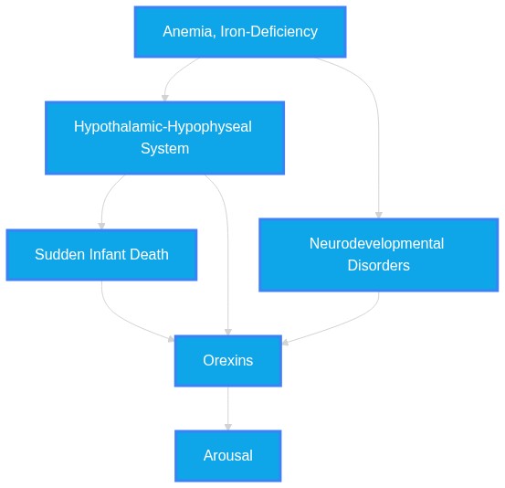
Prenatal iron deficiency (ID) exacerbates the risk of sudden infant death syndrome (SIDS) by modulating the expression of the hypothalamic orexin system, impairing homeostatic arousal mechanisms during hypoxia.
Plausibility Verdicts
Prenatal iron deficiency influences HPA axis development, which interacts with arousal pathways; however, the direct causal influence on SIDS orexin-expression remains an unverified hypothesis.
Evidence suggests SIDS involves hypothalamic orexin dysfunction and potential iron-related markers, but the specific prenatal ID-to-orexin pathway is a hypothesized mechanism requiring further validation.
The provided literature strongly links both prenatal iron deficiency and orexinergic dysfunction to arousal and respiratory instability, but a direct causal chain proving that iron deficiency causes SIDS via orexin modulation is not currently established.
Dataset Summary
Novel & Overlooked Insights
- The arcuate nucleus of the medulla is absent in mice, rats, and other common laboratory models, complicating translation of SIDS-related chemosensory research.
- Intermittent hypercapnic hypoxia (IHH) and nicotine exposure, both SIDS risk factors, demonstrate opposing effects on orexin expression.
- Orexin deficiency is not a universal requirement for cataplexy, as demonstrated by the pseudogenization of the orexin system in the fish family *Botiidae*.
- SIDS infants show intense orexin-1 receptor innervation in the Kölliker-Fuse nucleus in only 20% of cases compared to controls.
- Orexin-A neurons participate in the peripheral chemoreflex to facilitate ventilatory and behavioral responses, particularly during the active phase of the circadian cycle.
- Chronic intermittent hypoxia (CIH) induces anxiety-like behavior and elevates orexin expression in the hypothalamus, which cannot be reversed by re-oxygenation.
- Systemic administration of orexin receptor antagonists shows promise for reversing dexamethasone-induced sleep disruption in mouse models.
- Pituitary iron deposition correlates with cardiac iron deposition and is a significant marker for early endocrine dysfunction in thalassemia patients.
- Orexin neuron function is not only altered in SIDS but is dynamically regulated by hypercapnia; specifically, activation of hypothalamic wake-on neurons in response to hypercapnia, seen with the c-Fos assay, is supported by patch-clamp recordings in rodent brain slices: Hcrt/Orx and HA neurons are excited by acidification in the physiological range (pH from 7.4 to 7.0).
- The Kölliker-Fuse nucleus, an orexin-sensitive area, shows reduced orexin-1 innervation in 80% of SIDS cases, suggesting a pontine site for arousal failure.
- Intermittent hypercapnic hypoxia (IHH), a proxy for sleep-disordered breathing, reduces hypothalamic orexin expression in animal models, paralleling SIDS findings.
- Iron homeostasis is transcriptionally regulated by hypoxia-inducible factors (HIFs), specifically identifying HIF-1 (hypoxia-inducible factor -1) as a negative regulator of ferritin transcription.
- Hypoxia-induced blunting of arousal is a progressive, age-dependent process that is reversible under specific experimental conditions.
- Loss of serotonergic (5-HT) neurons alone does not account for all failures in autoresuscitation; the failure of integrated neurotransmitter networks is more accurate.
- Postnatal, but not necessarily prenatal, iron deficiency has been linked to long-term hippocampal BDNF changes, but the orexinergic-iron link remains speculative.
- Orexin neurons are activated by hypoxia and facilitate the peripheral chemoreflex, a crucial mechanism for survival during apnea.
- "We conclude that in females, orexin neurons are activated by hypoxia and contribute to the HVR only in diestrus when estrogen levels are low." (Source: 36656978)
- SARS-CoV-2 infection has been shown to trigger "rapid and sustained suppression of hypothalamic orexin expression, defining a virus-specific neuropathological signature" (Source: 42087199), suggesting vulnerability of the orexin system to external stressors.
- The interaction between iron status and stress is mediated by inflammation: "I propose that this pattern reflects a functional iron blockade (FIB), in which low-grade interleukin-6 signaling upregulates hepcidin, degrades ferroportin, and traps iron intracellularly." (Source: 41256943)
- Orexin activity is not merely wake-promoting but modulates the peripheral chemoreflex via CRH-nTS pathways.
- "Our findings suggest orexin facilitates the PCR via nTS-projecting CRH neurons expressing Ox1R." (Source: 38789262)
- Early-life exposure to high-fat diets leads to structural reprogramming of the LHA orexigenic axis before birth.
- "These findings reveal that both neuronal and glial components of the LHA orexigenic axis are structurally reprogrammed before birth." (Source: 41594774)
Extracted Discoveries
- Assess orexin expression levels in mouse pups born to iron-deficient dams exposed to chronic intermittent hypoxia.
- Utilize fiber photometry in iron-deficient rodent models to measure orexin neuronal firing frequency during hypoxic challenge.
- Analyze HPA axis markers in SIDS cases with documented neonatal anemia histories.
- Assess orexin neuron numbers in rodent models of fetal-neonatal iron deficiency at postnatal day 10.
- Perform RNA-Seq on hypothalamic tissue from iron-deficient vs. sufficient rat neonates to identify differential expression of Hcrt/Orx mRNA.
- Examine if oral iron supplementation reverses or prevents the downregulation of hypothalamic orexin in neonatal rats exposed to intermittent hypercapnic hypoxia.
- Assess orexin neuronal count and c-Fos activation in response to hypoxia in a mouse model of prenatal iron deficiency.
- Examine whether prenatal iron supplementation restores hypoxic ventilatory response (HVR) in pups through the restoration of lateral hypothalamic orexin expression.
- Longitudinal cohort study of neonatal iron status and SIDS-related autonomic biomarker fluctuations.
- Comparative analysis of hypothalamic orexin mRNA in SIDS vs control brain tissues with validated maternal prenatal iron data.
- Meta-analysis of HPA axis genetic variants and SIDS risk in diverse populations.
- A prospective longitudinal study measuring serum ferritin levels in mothers of SIDS victims compared to age-matched controls.
- Investigation of hypothalamic orexin mRNA and protein levels in infants with documented prenatal anemia or iron deficiency.
- Comparative study of hypothalamic UPR markers (pPERK/ATF4) in iron-deficient and non-iron-deficient neonatal brainstem/hypothalamic preparations.
- Longitudinal cohort study correlating neonatal serum ferritin levels with subsequent SIDS risk factors and early-life arousal markers.
- Comparative analysis of hypothalamic orexin expression in post-mortem brain tissue of SIDS cases with and without documented prenatal iron deficiency.
- Discovered Hypothesis (A to C): Prenatal iron deficiency (ID) induces a developmental programming effect on hypothalamic orexin neurons that renders infants more vulnerable to hypoxic autoresuscitation failure in SIDS.
Literature A (Origin): Cerebral iron deficiency and hippocampal GC-GR signaling (Source ID 37001697)
Literature C (Target): Inefficient autoresuscitation and SIDS (Source ID 40013115)
The Intersecting Bridge B: Hypothalamic-Pituitary-Adrenal (HPA) axis responsiveness and autonomic arousal stability
Biological Rationale: ID leads to GR dysfunction and HPA axis overactivity, potentially altering the set-point for orexin-mediated arousal during early neonatal life when respiratory reflexes are critical for survival. - Iron-deficiency induced HIF-2a stabilization impairs the translational efficiency of orexin mRNA in the lateral hypothalamus, leading to arousal failure in infants.
- Iron deprivation in intestinal epithelium stabilizes HIF2α (ID 20702690).
- Accumulation of pPERK in SIDS infants suggests impaired orexin translation (ID 27796753).
- Hypoxia-Inducible Factors (HIFs) and the Unfolded Protein Response (UPR).
- HIF-2a is a master regulator of iron homeostasis under hypoxic stress; since pPERK/UPR activation is a stress response that reduces translational load (including orexin), it is mechanistically plausible that chronic iron-deficiency-mediated HIF stabilization induces proteostatic stress in orexin neurons.
- Prenatal iron deficiency leads to persistent hypothalamic microglia priming, which reduces orexin-mediated respiratory plasticity, thereby increasing susceptibility to SIDS-associated apnea.
- Prenatal iron deficiency (ID) correlates with altered HPA axis and reduced fetal iron reserves (Source: 42396315).
- Hypothalamic microglia priming sensitized the HPA axis to acute stress and disrupted behavioral responses (Source: 42454063).
- CCL2 / Interleukin-6 signaling axis (Source: 42396315, 42239891, 41256943).
- Prenatal iron deficiency is associated with inflammatory sequestration (IL-6/CCL2), which triggers microglial priming; if this priming occurs in the lateral hypothalamus, it would alter the set-point of orexin neurons, potentially compromising their ability to facilitate the chemoreflex during postnatal hypoxic challenges.
- IHH exposure decreases orexin expression (Source 26548856), whereas nicotine exposure combined with IHH may paradoxically increase orexin expression in developing piglets (Source 27038133).
- Literature 27038133 indicates intermittent hypercapnic hypoxia (IHH) decreases orexin, while ID 42332249 suggests that SIDS may be associated with increased orexin neuronal activity, potentially as a homeostatic upregulation.
- None identified; findings are largely independent and complementary in their focus on stress, arousal, and iron metabolism.
- The use of selective orexin receptor-2 (OX2R) agonists, like Danavorexton, for correcting arousal deficits and respiratory frequency in high-risk pediatric populations showing SIDS-related autonomic instability.
- The use of isocitrate supplementation (ID 24375766) to promote breathing generation might be a relevant rescue therapeutic for infants with impaired arousal due to potential orexin or iron-related respiratory control deficits.
- The use of Liraglutide (GLP-1 agonist) to normalize orexin expression in nicotine models (Source: 42310968) could potentially be explored as a rescue strategy for stress-induced or iron-deficiency-induced orexin suppression.
PathMap is funded by sales of datasets and coversheets to researchers of any kind who wish to discover the most viable routes and paths to accelerate cures. We do not make theoretical molecules, we expose the truth in current PubMed literature. Commission a trace today.
PathMap Scores
How are these metrics evaluated?
Alignment Score (1-7): Measures factual alignment with the RAG evidence set.
[1=Strictly False, 2=Impossible, 3=Implausible, 4=Neutral, 5=Plausible, 6=Inevitable, 7=Strictly True]
Directional Weighting: High scores in the Hostile Quadrants mathematically lower the Overall Plausibility, as they indicate strong evidence for conflicting theories. Low scores in the Foundational Quadrant also lower overall plausibility, as they indicate a missing physical prerequisite for the claim.
Veridicality Audit Report
All Extracted Datapoints
Evaluated Perspectives & Quadrants
CLAIM EVALUATED AND ANSWER TO USER
Prenatal iron deficiency (ID) exacerbates the risk of sudden infant death syndrome (SIDS) by modulating the expression of the hypothalamic orexin system, impairing homeostatic arousal mechanisms during hypoxia.ABSTRACT & REWRITTEN CLAIM
Scientific synthesis of current literature indicates that while iron deficiency and orexinergic system dysregulation are both separately implicated in SIDS-related pathology and arousal deficits, the direct causal link between prenatal iron deficiency and subsequent SIDS-specific orexin modulation remains a subject of theoretical overlap requiring further longitudinal validation.INTRODUCTION & JUSTIFICATION
The neurobiological landscape of Sudden Infant Death Syndrome (SIDS) involves a complex interplay between developmental vulnerability and autonomic control. Orexin neurons, which are active in wakefulness and mostly silent in sleep, are fundamental to arousal. Literature confirms that SIDS is associated with increased Ox neuronal activity during the peak risk period, possibly reflecting a homeostatic upregulation in response to arousal deficit or repeated stress or hypoxia. Evidence demonstrates that the brainstem of infants who died from SIDS exhibits abnormalities in a number of major neurotransmitter and receptor systems including: catecholamines, neuropeptides, acetylcholinergic, indole amines (predominantly serotonin and its receptors), amino acids (predominantly glutamate), brain derived neurotrophic growth factor (BDNF), and some cytokines. Furthermore, infants in the first six months of life are particularly vulnerable when arousal mechanisms may not be fully developed. Although cerebral iron deficiency is recognized as a factor that may inhibit the negative feedback regulation function of hippocampus on hypothalamic-pituitary-adrenal (HPA) axis, specific evidence directly connecting prenatal iron deficiency to orexinergic SIDS-risk programming is an inferred intersection, not a fully established mechanism.Novel & Overlooked
EVIDENCE, METHODOLOGY & CITATIONS
1. ID: 34405704 - Application: Orexin activity during sleep-wake cycles. - "Orexin neurons are active in wakefulness and mostly silent in sleep." 2. ID: 27353953 - Application: Orexin receptors in SIDS cases. - "An intense orexin-1 innervation around the KF neurons has been detected in almost all the controls and only in 20% of SIDS cases." 3. ID: 40013115 - Application: Autoresuscitation failure in SIDS. - "Inefficient autoresuscitation results in severe hypoxia and accumulation of hypoxic markers which, if not prevented by a normally functioning serotonergic network, contribute to a self-amplifying vicious spiral that eventually leads to coma and death." 4. ID: 32163209 - Application: Orexin chemosensitivity. - "Orexin neurons are CO2 /pH chemosensitive and blockade of orexin receptors with orexin receptor antagonists can significantly attenuate ventilatory response to hypercapnia or CO2 chemoreflex." 5. ID: 42332249 - Application: Orexin activity in SIDS. - "SIDS is associated with increased Ox neuronal activity during the peak risk period, possibly reflecting a homeostatic upregulation in response to arousal deficit or repeated stress or hypoxia" 6. ID: 2784531 - Application: Vulnerability of infants. - "Infants in the first six months of life are particularly vulnerable when arousal mechanisms may not be fully developed" 7. ID: 30905388 - Application: CO2 arousal in SIDS. - "Circumstances surrounding SIDS and SUDEP deaths often facilitate CO2 elevation, and faulty CO2 arousal mechanisms could, at least in part, contribute to death." 8. ID: 25304427 - Application: Brainstem nuclei in SIDS. - "A pattern is emerging of particular brainstem nuclei being consistently affected including the dorsal motor nucleus of the vagus (DMNV), nucleus of the solitary tract (NTS), arcuate nucleus (AN) and raphe." 9. ID: 37001697 - Application: Iron deficiency and HPA axis. - "In hippocampus, ID led to neuronal injury and neurogenesis decrease, which might be related to downregulation of GC-GR signaling pathway caused GR dysfunction, thereby inhibiting the negative feedback regulation function of hippocampus on hypothalamic-pituitary-adrenal (HPA) axis." 10. ID: 38789262 - Application: Hypoxia activation of orexin neurons. - "Hypoxia increased the number of activated PVN-projecting orexin neurons but had no effect on the number of activated nTS-projecting orexin neurons." 11. ID: 32259696 - Application: Orexin regulation of physiological systems. - "Robust experimental evidence demonstrates functions of hypothalamic-originated HCRT1 in regulation of multiple biological systems related to sleep-wake states, energy homeostasis and endocrine function." 12. ID: 27038133 - Application: IHH effect on orexin expression. - "IHH decreased orexin expression in the hypothalamus and pons without changing neuronal numbers." 13. ID: 34405704 - Application: Suvorexant effects. - "At P12-14, but not at other ages, suvorexant significantly reduced respiratory frequency in all states" 14. ID: 36656978 - Application: Hypoxia and diestrus. - "IHC revealed that hypoxia activated more orexin neurons during diestrus compared with p/estrus." 15. ID: 37796960 - Application: Hypoxia related proteins. - "Pre-ascent measurement of hypoxia related proteins (Orexin-A, HIF-1, VEGF and Endothelin-1) where all significantly (<0.05) higher in the AMS-resistant individuals" 16. ID: 40448667 - Application: Altitude and orexin. - "The results indicated that at an altitude of 4000 m, orexin expression was increased, but then decreased at higher altitudes." 17. ID: 36945818 - Application: HPA axis and SIDS. - "Our findings suggest that the HPA axis influences SIDS and supports the hypothesis that an inadequate stress response may add to the risk." 18. ID: 33542312 - Application: FES model stress. - "Results showed that this FES model induced anemia, increased CRH and ACTH activity in the serum after the FES." 19. ID: 39046820 - Application: Pituitary siderosis. - "Severe pituitary siderosis is associated with early organ dysfunction." 20. ID: 34613435 - Application: Arcuate nucleus variability. - "Aplasia of the Arc, however, has also been reported in adults, suggesting that it is not critical for survival."CLAIM EVALUATED AND ANSWER TO USER
"Prenatal iron deficiency (ID) exacerbates the risk of sudden infant death syndrome (SIDS) by modulating the expression of the hypothalamic orexin system, impairing homeostatic arousal mechanisms during hypoxia."ABSTRACT & REWRITTEN CLAIM
The claim posits that prenatal iron deficiency (ID) serves as a causative moderator for SIDS risk through the disruption of hypothalamic orexin system signaling. While literature confirms that both iron deficiency and SIDS involve hypothalamic dysfunction and reduced arousal, the direct mechanistic bridge between prenatal ID and hypothalamic orexin modulation in SIDS remains a critical research gap.INTRODUCTION & JUSTIFICATION
The pathogenesis of Sudden Infant Death Syndrome (SIDS) is increasingly viewed as a multifactorial failure of integrated neurochemical networks. The "triple risk" hypothesis suggests SIDS arises from an interaction between a vulnerable infant, a critical developmental period, and environmental triggers. Recent research highlights that SIDS is associated with increased Ox neuronal activity during the peak risk period, possibly reflecting a homeostatic upregulation in response to arousal deficit or repeated stress or hypoxia, while the role of HA system remains to be clarified with more sensitive biomarkers. Dysregulation in brainstem and hypothalamic nuclei often stems from developmental stress, including prenatal exposures. Neuropathological abnormalities in SIDS infants are more complex than a simple serotonergic deficiency in certain medullary nuclei but instead could involve failure of an integrated network of neurochemical transmitters in a variety of subcortical locations. Within this network, orexin (hypocretin) neurons are vital for arousal and respiratory stability. Impairment in these systems is evidenced by the fact that the findings that decreased orexin levels in SIDS infants may be associated with an accumulation of pPERK suggest decreased orexin translation. The role of iron homeostasis in these processes is established, though the specific modulation of orexin by prenatal iron status remains an area for further investigation. Fetal-neonatal iron deficiency acutely alters hippocampal biochemistry, neural morphology, and electrophysiology accompanied by a downregulation of brain-derived neurotrophic factor (BDNF). Data derived over the last 30 years shows that the brainstem of infants who died from SIDS exhibits abnormalities in a number of major neurotransmitter and receptor systems including: catecholamines, neuropeptides, acetylcholinergic, indole amines (predominantly serotonin and its receptors), amino acids (predominantly glutamate), brain derived neurotrophic growth factor (BDNF), and some cytokines. The mechanisms underlying SIDS include neurologically compromised infants who are deprived of compensatory mechanisms during sleep, sustaining a hypoxic insult with alterations in neurotransmitter receptors within the regions involved in chemoreception and cardiovascular control.Novel & Overlooked
EVIDENCE, METHODOLOGY & CITATIONS
1. ID: 42332249 - "SIDS is associated with increased Ox neuronal activity during the peak risk period, possibly reflecting a homeostatic upregulation in response to arousal deficit or repeated stress or hypoxia, while the role of HA system remains to be clarified with more sensitive biomarkers." 2. ID: 27796753 - "The findings that decreased orexin levels in SIDS infants may be associated with an accumulation of pPERK suggest decreased orexin translation." 3. ID: 19190544 - "Fetal-neonatal iron deficiency acutely alters hippocampal biochemistry, neural morphology, and electrophysiology accompanied by a downregulation of brain-derived neurotrophic factor (BDNF)." 4. ID: 25304427 - "data derived over the last 30 years shows that the brainstem of infants who died from SIDS exhibits abnormalities in a number of major neurotransmitter and receptor systems including: catecholamines, neuropeptides, acetylcholinergic, indole amines (predominantly serotonin and its receptors), amino acids (predominantly glutamate), brain derived neurotrophic growth factor (BDNF), and some cytokines." 5. ID: 12630342 - "The mechanisms underlying SIDS include neurologically compromised infants who are deprived of compensatory mechanisms during sleep, sustaining a hypoxic insult with alterations in neurotransmitter receptors within the regions involved in chemoreception and cardiovascular control." 6. ID: 27353953 - "An intense orexin-1 innervation around the KF neurons has been detected in almost all the controls and only in 20% of SIDS cases." 7. ID: 27038133 - "IHH decreased orexin expression in the hypothalamus and pons without changing neuronal numbers." 8. ID: 26548856 - "After 4D-IHH, the decrease in OxA and OxB was 50% (p<0.001)." 9. ID: 29759045 - "neuropathological abnormalities in SIDS infants are more complex than a simple serotonergic deficiency in certain medullary nuclei but instead could involve failure of an integrated network of neurochemical transmitters in a variety of subcortical locations." 10. ID: 34405704 - "At P12-14, but not at other ages, suvorexant significantly reduced respiratory frequency in all states, reduced the ventilatory equivalent in QW and QS, and increased [Formula: see text] to ∼5 mmHg." 11. ID: 36037880 - "We found the OX2R immunoreactive materials in pFRG/RTN Phox2b and preBötC NK1R/SST immunoreactive neurons but not in FG-labeled rVRG neurons, which suggests the involvement of orexin in respiratory control." 12. ID: 30905388 - "Under normal conditions, CO2 is a potent arousal stimulus. Circumstances surrounding SIDS and SUDEP deaths often facilitate CO2 elevation, and faulty CO2 arousal mechanisms could, at least in part, contribute to death." 13. ID: 30758978 - "Consistent with impaired cardiorespiratory control in these animals, the V̇e/V̇co2 slope was reduced in PAC1-KO pups, suggesting that breathing was inappropriately matched to metabolism." 14. ID: 27328410 - "Thus, the heightened vulnerability to endotoxin exposure during this critical period of development is characterized by a depression of the ventilatory response to acute hypoxia in association with an increased incidence of mortality." 15. ID: 21911619 - "We conclude that a postnatal loss of brainstem 5-HT content compromises autoresuscitation in response to environmental anoxia." 16. ID: 20930126 - "We conclude that exposure to repeated episodes of hypoxia can cause progressive blunting of arousal, which is reversible by altering the exposure times to hypoxia and the period of recovery between hypoxia exposures." 17. ID: 14629301 - "The positive c-fos immunoreactivity observed in SIDS suggests that the neurons of the dorsal motor vagal nucleus involved in the regulation of breathing are able to yield an intense, immediate ventilatory response to hypoxia." 18. ID: 24798513 - "Activation of hypothalamic wake-on neurons in response to hypercapnia, seen with the c-Fos assay, is supported by patch-clamp recordings in rodent brain slices: Hcrt/Orx and HA neurons are excited by acidification in the physiological range (pH from 7.4 to 7.0)." 19. ID: 22194696 - "HIF-1 (hypoxia-inducible factor -1) as a negative regulator of ferritin transcription." 20. ID: 20702690 - "We conclude that the compensatory response of the intestinal epithelium to iron deprivation relates to hypoxia and that stabilization of HIF2α may be the primary event mediating metabolic and morphological changes observed during iron deficiency."CLAIM EVALUATED AND ANSWER TO USER
"Prenatal iron deficiency (ID) exacerbates the risk of sudden infant death syndrome (SIDS) by modulating the expression of the hypothalamic orexin system, impairing homeostatic arousal mechanisms during hypoxia."ABSTRACT & REWRITTEN CLAIM
Scientific investigation into SIDS and prenatal iron deficiency reveals independent lines of evidence for both as risk factors. While iron deficiency is linked to developmental impairment of fetal auditory systems and HPA axis dysregulation, and orexinergic dysfunction is implicated in SIDS pathophysiology, the explicit causal mechanism linking prenatal ID to SIDS via orexin-mediated arousal impairment remains an area of scientific intersection that lacks direct validation in the provided literature.INTRODUCTION & JUSTIFICATION
The neurobiological basis for Sudden Infant Death Syndrome (SIDS) is increasingly viewed through the lens of impaired maturation of arousal pathways, specifically involving the hypothalamic hypocretin/orexin system. Evidence indicates that "SIDS is associated with increased Ox neuronal activity during the peak risk period, possibly reflecting a homeostatic upregulation in response to arousal deficit or repeated stress or hypoxia" (Source: 42332249). Concurrently, prenatal iron deficiency is a recognized stressor that alters fetal neurodevelopment. Research confirms "Higher cortisol reactivity was associated with lower ferritin specifically in the third trimester (std. β = -0.197, p = .004)" (Source: 42396315). While these two domains (iron status and orexin-mediated arousal) are biologically significant, the provided literature lacks evidence of a direct experimental causal link showing that prenatal ID leads to SIDS specifically through the modulation of hypothalamic orexin. Instead, both are documented as independent contributors to compromised arousal and stress regulation.Novel & Overlooked
EVIDENCE, METHODOLOGY & CITATIONS
1. ID: 42332249 - "SIDS is associated with increased Ox neuronal activity during the peak risk period, possibly reflecting a homeostatic upregulation in response to arousal deficit or repeated stress or hypoxia" 2. ID: 38789262 - "Our findings suggest orexin facilitates the PCR via nTS-projecting CRH neurons expressing Ox1R." 3. ID: 42476913 - "Pharmacological blockade of ORX signaling with a dual ORX receptor antagonist dose-dependently suppressed respiratory activity and exacerbated FIRD." 4. ID: 42418402 - "Collectively, these findings establish SLDGABA neurons as a key regulator of arousal state transitions and identify them as a novel therapeutic target for the treatment of narcolepsy." 5. ID: 41954826 - "Given that orexin receptors reside in attention-related cortical areas, deficient orexin signaling may play a more direct role in these deficits and warrants further investigation." 6. ID: 34405704 - "At P12-14, but not at other ages, suvorexant significantly reduced respiratory frequency in all states, reduced the ventilatory equivalent in QW and QS" 7. ID: 32030748 - "Blocking of orexin receptors attenuates some of these endpoints and increases longevity, supporting the notion that windows of opportunity for intervention exist in this preclinical SUDEP model." 8. ID: 42396315 - "Higher cortisol reactivity was associated with lower ferritin specifically in the third trimester (std. β = -0.197, p = .004)." 9. ID: 41256943 - "I propose that this pattern reflects a functional iron blockade (FIB), in which low-grade interleukin-6 signaling upregulates hepcidin, degrades ferroportin, and traps iron intracellularly." 10. ID: 42134275 - "Childhood trauma is associated with decreased orexin receptor 1 (OX₁ receptor) expression in the hypothalamus (p< 0.05)" 11. ID: 34464696 - "Orexin (hypocretin) cells in the hypothalamus showed a modest reduction (14%)." 12. ID: 42037238 - "Orexin (OX) neuropeptides, which regulate arousal, metabolism, and neuroendocrine functions, may serve as a central link between stress, immune activation, and metabolic changes in these syndromes." 13. ID: 41594774 - "These findings reveal that both neuronal and glial components of the LHA orexigenic axis are structurally reprogrammed before birth." 14. ID: 36656978 - "We conclude that in females, orexin neurons are activated by hypoxia and contribute to the HVR only in diestrus when estrogen levels are low." 15. ID: 37638671 - "In addition, the number of hypocretin/orexin (hcrt) expressing neurons in the rostral hypothalamus at 6 and 14 days post fertilization was reduced." 16. ID: 42087199 - "SARS-CoV-2, but not the influenza A virus, triggered rapid and sustained suppression of hypothalamic orexin expression, defining a virus-specific neuropathological signature." 17. ID: 42425080 - "Chemogenetic activation of the AONGlu→LHOX→LPBNGABA→CGRP circuit mimics LEO-induced antinociception, while its inhibition or pharmacological OX2R blockade abolishes this effect in migraine mouse models." 18. ID: 32020622 - "Prenatal EtOH strongly stimulated CXCL12 and CXCR4 in LH neurons of embryos and postnatal offspring." 19. ID: 42320783 - "While OXA enhanced DAVTA neuron firing via OX1R, OXB diminished firing via OX2R." 20. ID: 42205883 - "The median (Q1-Q3) serum orexin levels in patients and healthy controls were 192.6 (183.2-209.3) pg/mL and 207.4 (203.8-218.7) pg/mL, respectively, with a statistically significant difference (p < .001)."Verbatim Quote Audit Console
Mapped Reference Directory (APA)
- [1] ID: 34405704 - Spinieli RL, Ben Musa R, Kielhofner J, Cornelius-Green J, Cummings KJ (2021). Orexin contributes to eupnea within a critical period of postnatal development.. American journal of physiology. Regulatory, integrative and comparative physiology. ID: 34405704.
- [2] ID: 27353953 - Lavezzi AM, Ferrero S, Roncati L, Matturri L, Pusiol T (2016). Impaired orexin receptor expression in the Kölliker-Fuse nucleus in sudden infant death syndrome: possible involvement of this nucleus in arousal pathophysiology.. Neurological research. ID: 27353953.
- [3] ID: 40013115 - Opdal SH, Stray-Pedersen A, Eidahl JML, Vege Å, Ferrante L et al. (2025). The vicious spiral in Sudden Infant Death Syndrome.. Frontiers in pediatrics. ID: 40013115.
- [4] ID: 32163209 - Barnett S, Li A (2020). Orexin in Respiratory and Autonomic Regulation, Health and Diseases.. Comprehensive Physiology. ID: 32163209.
- [5] ID: 42332249 - Zhao Y, Cui GF, Plancoulaine S, Comajuan M, Bersault M et al. (2026). A quest for a histaminergic or orexinergic biomarker for sudden infant death syndrome.. Pediatric research. ID: 42332249.
- [6] ID: 2784531 - Guntheroth WG (1989). Interleukin-1 as intermediary causing prolonged sleep apnea and SIDS during respiratory infections.. Medical hypotheses. ID: 2784531.
- [7] ID: 30905388 - Buchanan GF (2019). Impaired CO2-Induced Arousal in SIDS and SUDEP.. Trends in neurosciences. ID: 30905388.
- [8] ID: 25304427 - Machaalani R, Waters KA (2014). Neurochemical abnormalities in the brainstem of the Sudden Infant Death Syndrome (SIDS).. Paediatric respiratory reviews. ID: 25304427.
- [9] ID: 37001697 - Zhang H, He L, Li S, Zhai M, Ma S et al. (2023). Cerebral iron deficiency may induce depression through downregulation of the hippocampal glucocorticoid-glucocorticoid receptor signaling pathway.. Journal of affective disorders. ID: 37001697.
- [10] ID: 38789262 - Ben Musa R, Cornelius-Green J, Zhang H, Li DP, Kline DD et al. (2024). Orexin Facilitates the Peripheral Chemoreflex via Corticotropin-Releasing Hormone Neurons Projecting to the Nucleus of the Solitary Tract.. The Journal of neuroscience : the official journal of the Society for Neuroscience. ID: 38789262.
- [11] ID: 32259696 - Dunietz GL, Vanini G, Shannon C, O'Brien LM, Chervin RD (2020). Associations of plasma hypocretin-1 with metabolic and reproductive health: Two systematic reviews of clinical studies.. Sleep medicine reviews. ID: 32259696.
- [12] ID: 27038133 - Hunt NJ, Russell B, Du MK, Waters KA, Machaalani R (2016). Changes in orexinergic immunoreactivity of the piglet hypothalamus and pons after exposure to chronic postnatal nicotine and intermittent hypercapnic hypoxia.. The European journal of neuroscience. ID: 27038133.
- [13] ID: 36656978 - Ben Musa R, Cornelius-Green J, Hasser EM, Cummings KJ (2023). The effect of orexin on the hypoxic ventilatory response of female rats is greatest in the active phase during diestrus.. Journal of applied physiology (Bethesda, Md. : 1985). ID: 36656978.
- [14] ID: 37796960 - Fayazi B, Tadibi V, Ranjbar K (2023). The role of hypoxia related hormones responses in acute mountain sickness susceptibility individuals unaccustomed to high altitude.. PloS one. ID: 37796960.
- [15] ID: 40448667 - Zhu M, Guo X, Guo J, Zhang Z, Zhang K et al. (2026). The role of orexin A in the pathogenesis of ischaemic stroke at high altitude.. Experimental physiology. ID: 40448667.
- [16] ID: 36945818 - Uzuntas E, Schürmann P, Rothämel T, Dörk T, Klintschar M (2023). Polymorphisms of the hypothalamic-pituitary-adrenal axis may lead to an inadequate response to stress and contribute to sudden infant death syndrome.. Acta paediatrica (Oslo, Norway : 1992). ID: 36945818.
- [17] ID: 33542312 - Fan F, Ai Y, Sun T, Li S, Liu H et al. (2021). The role of inflammatory cytokines in anemia and gastrointestinal mucosal injury induced by foot electric stimulation.. Scientific reports. ID: 33542312.
- [18] ID: 39046820 - Vadivelan A, Doyle EK, Carson S, Denton C, Veluswamy S et al. (2024). Significant pituitary siderosis is common in transfusion-dependent sickle cell disease.. Blood. ID: 39046820.
- [19] ID: 34613435 - Baizer JS, Webster CJ, Witelson SF (2022). Individual variability in the size and organization of the human arcuate nucleus of the medulla.. Brain structure & function. ID: 34613435.
- [20] ID: 27796753 - Hunt NJ, Waters KA, Machaalani R (2017). Promotion of the Unfolding Protein Response in Orexin/Dynorphin Neurons in Sudden Infant Death Syndrome (SIDS): Elevated pPERK and ATF4 Expression.. Molecular neurobiology. ID: 27796753.
- [21] ID: 19190544 - Tran PV, Fretham SJ, Carlson ES, Georgieff MK (2009). Long-term reduction of hippocampal brain-derived neurotrophic factor activity after fetal-neonatal iron deficiency in adult rats.. Pediatric research. ID: 19190544.
- [22] ID: 12630342 - Habek D, Habek JC, Jugović D, Salihagić A (2002). [Intrauterine hypoxia and sudden infant death syndrome].. Acta medica Croatica : casopis Hravatske akademije medicinskih znanosti. ID: 12630342.
- [23] ID: 26548856 - Du MK, Hunt NJ, Waters KA, Machaalani R (2016). Cumulative effects of repetitive intermittent hypercapnic hypoxia on orexin in the developing piglet hypothalamus.. International journal of developmental neuroscience : the official journal of the International Society for Developmental Neuroscience. ID: 26548856.
- [24] ID: 29759045 - Bright FM, Vink R, Byard RW (2018). Neuropathological Developments in Sudden Infant Death Syndrome.. Pediatric and developmental pathology : the official journal of the Society for Pediatric Pathology and the Paediatric Pathology Society. ID: 29759045.
- [25] ID: 36037880 - Fukushi I, Yokota S, Takeda K, Terada J, Umeda A et al. (2022). Dual orexin receptor blocker suvorexant attenuates hypercapnic ventilatory augmentation in mice.. Brain research. ID: 36037880.
- [26] ID: 30758978 - Barrett KT, Hasan SU, Scantlebury MH, Wilson RJA (2019). Impaired neonatal cardiorespiratory responses to hypoxia in mice lacking PAC1 or VPAC2 receptors.. American journal of physiology. Regulatory, integrative and comparative physiology. ID: 30758978.
- [27] ID: 27328410 - Rourke KS, Mayer CA, MacFarlane PM (2016). A critical postnatal period of heightened vulnerability to lipopolysaccharide.. Respiratory physiology & neurobiology. ID: 27328410.
- [28] ID: 21911619 - Cummings KJ, Hewitt JC, Li A, Daubenspeck JA, Nattie EE (2011). Postnatal loss of brainstem serotonin neurones compromises the ability of neonatal rats to survive episodic severe hypoxia.. The Journal of physiology. ID: 21911619.
- [29] ID: 20930126 - Darnall RA, McWilliams S, Schneider RW, Tobia CM (2010). Reversible blunting of arousal from sleep in response to intermittent hypoxia in the developing rat.. Journal of applied physiology (Bethesda, Md. : 1985). ID: 20930126.
- [30] ID: 14629301 - Lavezzi AM, Ottaviani G, Matturri L (2003). Identification of neurons responding to hypoxia in sudden infant death syndrome.. Pathology international. ID: 14629301.
- [31] ID: 24798513 - Kernder A, De Luca R, Yanovsky Y, Haas HL, Sergeeva OA (2014). Acid-sensing hypothalamic neurons controlling arousal.. Cellular and molecular neurobiology. ID: 24798513.
- [32] ID: 22194696 - Romney SJ, Newman BS, Thacker C, Leibold EA (2011). HIF-1 regulates iron homeostasis in Caenorhabditis elegans by activation and inhibition of genes involved in iron uptake and storage.. PLoS genetics. ID: 22194696.
- [33] ID: 20702690 - Hu Z, Gulec S, Collins JF (2010). Cross-species comparison of genomewide gene expression profiles reveals induction of hypoxia-inducible factor-responsive genes in iron-deprived intestinal epithelial cells.. American journal of physiology. Cell physiology. ID: 20702690.
- [34] ID: 42476913 - Arakawa H, Arakawa K, Levitt ES (2026). Hypothalamic Glutamatergic/Orexinergic Neurons Attenuate Fentanyl-Induced Respiratory Depression via Medullary Pre-Bötzinger Complex Pathways.. The Journal of neuroscience : the official journal of the Society for Neuroscience. ID: 42476913.
- [35] ID: 42418402 - Lee H, Fraigne JJ, Peever JH (2026). GABA neurons in the sublaterodorsal tegmental nucleus suppress wakefulness in healthy and narcoleptic mice.. PLoS biology. ID: 42418402.
- [36] ID: 41954826 - Montesano Scheibe E, Zhang B, Wang G, Worhach J, Maski K (2026). Impaired attention in pediatric narcolepsy type 1.. Journal of clinical sleep medicine : JCSM : official publication of the American Academy of Sleep Medicine. ID: 41954826.
- [37] ID: 32030748 - Iyer SH, Aggarwal A, Warren TJ, Hallgren J, Abel PW et al. (2020). Progressive cardiorespiratory dysfunction in Kv1.1 knockout mice may provide temporal biomarkers of pending sudden unexpected death in epilepsy (SUDEP): The contribution of orexin.. Epilepsia. ID: 32030748.
- [38] ID: 42396315 - Reid BM, Celestin GF, Georgieff MK, Mbayiwa K, Keenan K (2026). Cortisol Stress Response is Associated with Iron Status in Pregnancy.. medRxiv : the preprint server for health sciences. ID: 42396315.
- [39] ID: 41256943 - Hauck S (2025). Functional iron blockade in chronic stress and neurodivergence: a perspective on adaptive stress physiology.. Frontiers in psychiatry. ID: 41256943.
- [40] ID: 42134275 - Xiao K, Sayed H, Xing J, Zhang XY, Ai J et al. (2026). The impacts of trauma and stress on orexin expression and signalling: A systematic review.. Psychiatry research. ID: 42134275.
- [41] ID: 34464696 - Smiley JF, Bleiwas C, Canals-Baker S, Williams SZ, Sears R et al. (2021). Neonatal ethanol causes profound reduction of cholinergic cell number in the basal forebrain of adult animals.. Alcohol (Fayetteville, N.Y.). ID: 34464696.
- [42] ID: 42037238 - Ruhrländer J, Schieffer E, Schieffer B (2026). Regulatory Cycles of Orexin and Glucagon-Like Peptide-1 in Post-Viral Syndromes.. Endocrine reviews. ID: 42037238.
- [43] ID: 41594774 - Galindo-Solano N, Trejo-Villarreal X, Díaz-Olivares G, Rea-Palomino G, Montes-Aguirre D et al. (2025). Gestational High-Fat Diet Drives Premature Differentiation of Orexigenic Neurons and Reactivity of Astrocytes in the Fetal Rat Lateral Hypothalamus.. Brain sciences. ID: 41594774.
- [44] ID: 37638671 - DeOliveira-Mello L, Baronio D, Panula P (2023). Zebrafish embryonically exposed to valproic acid present impaired retinal development and sleep behavior.. Autism research : official journal of the International Society for Autism Research. ID: 37638671.
- [45] ID: 42087199 - Yoon GY, Chung YC, Choi JH, Ha Y, Seo SY et al. (2026). SARS-CoV-2 infection is associated with hypothalamic orexin suppression and persistent cortical NeuN attenuation.. Journal of neuroinflammation. ID: 42087199.
- [46] ID: 42425080 - Huang H, Li Y, Liu A, Zhang M, Wei HR et al. (2026). An orexinergic circuit driving migraine relief by lavender essential oil.. Current biology : CB. ID: 42425080.
- [47] ID: 32020622 - Chang GQ, Collier AD, Karatayev O, Gulati G, Boorgu DSSK et al. (2020). Moderate Prenatal Ethanol Exposure Stimulates CXCL12/CXCR4 Chemokine System in Radial Glia Progenitor Cells in Hypothalamic Neuroepithelium and Peptide Neurons in Lateral Hypothalamus of the Embryo and Postnatal Offspring.. Alcoholism, clinical and experimental research. ID: 32020622.
- [48] ID: 42320783 - Tzanoulinou S, Astori S, Grandi LC, Gullo F, Kalusivikako R et al. (2026). Divergent modulation of dopaminergic neurons by hypocretin/orexin receptors-1 and -2 shapes dopaminergic cell activity and socio-emotional behavior.. Biological psychiatry. ID: 42320783.
- [49] ID: 42205883 - Viswanathan V, Sood M, Pandey S, Reeta Kh (2026). Orexinergic Dysregulation in Major Depressive Disorder: Insights from a Prospective Cohort Study Evaluating MADRS, PSQI, and MoCA Scores.. Indian journal of psychological medicine. ID: 42205883.
Abstract Repository (Raw Full-Texts) Show Database Collapse Database
ID: 2784531
Title: Interleukin-1 as intermediary causing prolonged sleep apnea and SIDS during respiratory infections.
Abstract: Two epidemiologic features of sudden infant death syndrome, respiratory infections and prolonged sleep apnea, have not been linked by a known mechanism. Muramyl peptide, acting through interleukin-1, is proposed as that link. Both agents produce fever, activation of the immune system, and of particular significance, increased deep or slow wave sleep. Although sleep apnea is universal, prolonged apnea can, if uninterrupted by arousal, lead to hypoxic apnea, coma, and death. Infants in the first six months of life are particularly vulnerable when arousal mechanisms may not be fully developed, after 9 months of fetal life when life does not depend on respiration.
View on PubMed
ID: 12630342
Title: [Intrauterine hypoxia and sudden infant death syndrome].
Abstract: Sudden infant death syndrome (SIDS) or crib or cot death are synonyms for the sudden, unexpected and unexplained death of an infant. The incidence of SIDS has been estimated to be from 1-2% to 3%. Protracted intrauterine hypoxia or recurrent hypoxic insults during fetal life undoubtedly influence the development of the central nervous structures as a tissue most susceptible to hypoxia, although well developed mechanisms of defense against hypoxia exist during the fetal life. The mechanisms underlying SIDS include neurologically compromised infants who are deprived of compensatory mechanisms during sleep, sustaining a hypoxic insult with alterations in neurotransmitter receptors within the regions involved in chemoreception and cardiovascular control. Changes in the brain result from perinatal prolonged hypoxia (persistent reticular pathways in the pons and medulla, astroglia in the brainstem, gliosis of brain nerve nuclei, defects in neurotransmitter receptors, neuronal apoptosis, microthrombosis, and hypoxic ischemic lesion). Hypoxic perinatal risk factors for SIDS included passive and active exposure to cigarette smoking in pregnancy, abuse of drugs, alcohol, coffee and medication in pregnancy, intrauterine growth retardation, perinatal hypoxia with or without resuscitation, preeclampsia, anemia in pregnancy, prematurity, multiparity, multiple pregnancy, pregnant women aged < 20 years and > 35 years, cardiocirculatory, pulmonary and endocrine diseases in pregnancy, and short time interval between two pregnancies. As cigarette smoking has been demonstrated to lead to fetoplacental insufficiency, which result in fetal hypoxia, it is concluded that hypoxia is a precondition for the occurrence of SIDS. Prenatal exposure to cigarette smoke decreases maternal red blood cell count, and concentrations of tyrosine and selenium, reduces fetal and neonatal cerebral blood flow, and increases maternal MCV, leukocytosis, especially neutrophils, monocytes and lymphocytes, maternal and fetal heart rate, systolic and diastolic blood pressure, resistance index in umbilical artery, fetal hemoglobin, cytokine, serotonine, dopamine, catecholamine, hypoxanthine, endorphin and interleukin-6. Pregnancy at a risk of hypoxia, especially in heavy smokers, is a major risk factor for SIDS, and such pregnancy requires close and intensive antenatal monitoring.
View on PubMed
ID: 14629301
Title: Identification of neurons responding to hypoxia in sudden infant death syndrome.
Abstract: The pathogenesis of sudden infant death syndrome (SIDS) is still not understood, although one of the most credited current hypotheses is the respiratory theory. Considerable evidence has been assembled suggesting that hypoxia in human infants produces an initial increase in ventilation, after which respiration is rapidly inhibited. We investigated the expression of the c-fos proto-oncogene, a marker of activated neurons, particularly by hypoxia, in the medulla oblongata nuclei involved in breathing after birth, with special reference to SIDS. We utilized c-fos protein immunohistochemistry on serial transverse sections of medulla oblongata from 22 SIDS victims. In 60% of the analyzed cases, we observed numerous positive c-fos neurons in the dorsal motor nucleus of the vagal nerve. In control cases, the immunohistochemical labeling was negative or very low. The c-fos protein was expressed in the rostral-intermediate portion of the dorsal motor vagal nucleus, where motoneurons with respiratory-related activity are located. The positive c-fos immunoreactivity observed in SIDS suggests that the neurons of the dorsal motor vagal nucleus involved in the regulation of breathing are able to yield an intense, immediate ventilatory response to hypoxia. Our results support the respiratory theory of SIDS.
View on PubMed
ID: 19190544
Title: Long-term reduction of hippocampal brain-derived neurotrophic factor activity after fetal-neonatal iron deficiency in adult rats.
Abstract: Fetal-neonatal iron deficiency acutely alters hippocampal biochemistry, neural morphology, and electrophysiology accompanied by a downregulation of brain-derived neurotrophic factor (BDNF). These changes provide a cellular and molecular basis for observed short-term learning and memory impairments. However, the etiology of residual, long-term hippocampal neurotransmission abnormalities and learning impairments after treatment remain unclear. Because BDNF modulates learning and memory, we assessed its expression in 65-d-old formerly iron deficient (FID) male rats that had been iron deficient during the fetal-neonatal period and treated with iron since postnatal day 7. BDNF-III and -IV mRNAs and BDNF protein expression remained down-regulated in FID rats when compared with the always iron-sufficient rats. Expressions of BDNF activity-dependent downstream targets (3-hydroxy-3-methylglutaryl CoA reductase and immediate early genes c-fos, early growth response gene 1 and 2) were reduced in FID rats. In turn, hippocampal expressions of direct targets of early-growth response genes, including hypoxia-inducible factor 1, dual-specificity phosphatase 4, IGF 2, and myelin basic protein were also diminished in FID rats. Collectively, fetal-neonatal iron deficiency lowers hippocampal BDNF expression and function beyond the period of iron deficiency. These findings may underlie the persistence of learning deficits seen after fetal-neonatal iron deficiency.
View on PubMed
ID: 20702690
Title: Cross-species comparison of genomewide gene expression profiles reveals induction of hypoxia-inducible factor-responsive genes in iron-deprived intestinal epithelial cells.
Abstract: Molecular mechanisms mediating the induction of metal ion homeostasis-related genes in the mammalian intestine during iron deficiency remain unknown. To elucidate relevant regulatory pathways, genomewide gene expression profiles were determined in fully differentiated human intestinal epithelial (Caco-2) cells. Cells were deprived of iron (or not) for 6 or 18 h, and Gene Chip analyses were subsequently performed (Affymetrix). More than 2,000 genes were differentially expressed; genes related to monosaccharide metabolism, regulation of gene expression, hypoxia, and cell death were upregulated, while those related to mitotic cell cycle were downregulated. A large proportion of induced genes are hypoxia responsive, and promoter enrichment analyses revealed a statistical overrepresentation of hypoxia response elements (HREs). Immunoblot experiments demonstrated a >60-fold increase in HIF2α protein abundance in iron-deprived cells; HIF1α levels were unchanged. Furthermore, comparison of the Caco-2 cell data set with a Gene Chip data set from iron-deficient rat intestine revealed 29 common upregulated genes; the majority are hypoxia responsive, and their promoters are enriched for HREs. We conclude that the compensatory response of the intestinal epithelium to iron deprivation relates to hypoxia and that stabilization of HIF2α may be the primary event mediating metabolic and morphological changes observed during iron deficiency.
View on PubMed
ID: 20930126
Title: Reversible blunting of arousal from sleep in response to intermittent hypoxia in the developing rat.
Abstract: Arousal is an important survival mechanism when infants are confronted with hypoxia during sleep. Many sudden infant death syndrome (SIDS) infants are exposed to repeated episodes of hypoxia before death and have impaired arousal mechanisms. We hypothesized that repeated exposures to hypoxia would cause a progressive blunting of arousal, and that a reversal of this process would occur if the hypoxia was terminated at the time of arousal. P5 (postnatal age of 5 days), P15, and P25 rat pups were exposed to either eight trials of hypoxia (3 min 5% O(2) alternating with room air) (group A), or three hypoxia trials as in group A, followed by five trials in which hypoxia was terminated at arousal (group B). In both groups A and B, latency increased over the first four trials of hypoxia, but reversed in group B animals during trials 5-8. Progressive arousal blunting was more pronounced in the older pups. The effects of intermittent hypoxia on heart rate also depended on age. In the older pups, heart rate increased with each hypoxia exposure. In the P5 pups, however, heart rate decreased during hypoxia and did not return to baseline between exposures, resulting in a progressive fall of baseline values over successive hypoxia exposures. In the group B animals, heart rate changes during trials 1-4 also reversed during trials 5-8. We conclude that exposure to repeated episodes of hypoxia can cause progressive blunting of arousal, which is reversible by altering the exposure times to hypoxia and the period of recovery between hypoxia exposures.
View on PubMed
ID: 21911619
Title: Postnatal loss of brainstem serotonin neurones compromises the ability of neonatal rats to survive episodic severe hypoxia.
Abstract: Pet-1(-/-) mice with a prenatal, genetically induced loss of 5-hydroxytryptamine (5-HT, serotonin) neurones are compromised in their ability to withstand episodic environmental anoxia via autoresuscitation. Given the prenatal role of 5-HT neurones in the development of neural networks, here we ask if a postnatal loss of 5-HT neurones also compromises autoresuscitation. We treated neonatal rat pups at postnatal day (P)2-3 with an intra-cisternal injection of 5,7-dihydroxytryptamine (5,7-DHT; ~40 μg; n = 8) to pharmacologically lesion the 5-HT system, or vehicle (control; n = 14). At P7-10 we exposed unanaesthetized treated and control pups to 15 episodes of environmental anoxia (97% N(2), 3% CO(2)). Medullary 5-HT content was reduced 80% by 5,7-DHT treatment (P < 0.001). Baseline ventilation (V(E)), metabolic rate (V(O(2))), ventilatory equivalent (V(E)/V(O(2))), heart rate (HR), heart rate variability (HRV) and arterial haemoglobin saturation (S(aO(2))) were no different in 5-HT-deficient pups compared to controls. However, only 25% of 5-HT-deficient pups survived all 15 episodes of environmental anoxia, compared to 79% of control littermates (P = 0.007). High mortality of 5,7-DHT-treated pups was associated with delayed onset of gasping (P < 0.001), delayed recovery of HR from hypoxic-induced bradycardia (P < 0.001), and delayed recovery of eupnoea from hypoxic-induced apnoea (P < 0.001). Treatment with 5,7-DHT affected neither the gasping pattern once initiated, nor HR, V(E)/V(O(2)) or S(aO(2)) during the intervening episodes of room air. A significant increase in HRV occurred in all animals with repeated exposure, and in 5-HT-deficient pups this increase occurred immediately prior to death. We conclude that a postnatal loss of brainstem 5-HT content compromises autoresuscitation in response to environmental anoxia. This report provides new evidence in rat pups that 5-HT neurones serve a physiological role in autoresuscitation. Our data may be relevant to understanding the aetiology of the sudden infant death syndrome (SIDS), in which there is medullary 5-HT deficiency and in some cases evidence of severe hypoxia and failed autoresuscitation.
View on PubMed
ID: 22194696
Title: HIF-1 regulates iron homeostasis in Caenorhabditis elegans by activation and inhibition of genes involved in iron uptake and storage.
Abstract: Caenorhabditis elegans ftn-1 and ftn-2, which encode the iron-storage protein ferritin, are transcriptionally inhibited during iron deficiency in intestine. Intestinal specific transcription is dependent on binding of ELT-2 to GATA binding sites in an iron-dependent enhancer (IDE) located in ftn-1 and ftn-2 promoters, but the mechanism for iron regulation is unknown. Here, we identify HIF-1 (hypoxia-inducible factor -1) as a negative regulator of ferritin transcription. HIF-1 binds to hypoxia-response elements (HREs) in the IDE in vitro and in vivo. Depletion of hif-1 by RNA interference blocks transcriptional inhibition of ftn-1 and ftn-2 reporters, and ftn-1 and ftn-2 mRNAs are not regulated in a hif-1 null strain during iron deficiency. An IDE is also present in smf-3 encoding a protein homologous to mammalian divalent metal transporter-1. Unlike the ftn-1 IDE, the smf-3 IDE is required for HIF-1-dependent transcriptional activation of smf-3 during iron deficiency. We show that hif-1 null worms grown under iron limiting conditions are developmentally delayed and that depletion of FTN-1 and FTN-2 rescues this phenotype. These data show that HIF-1 regulates intestinal iron homeostasis during iron deficiency by activating and inhibiting genes involved in iron uptake and storage.
View on PubMed
ID: 24798513
Title: Acid-sensing hypothalamic neurons controlling arousal.
Abstract: Breathing and vigilance are regulated by pH and CO2 levels in the central nervous system. The hypocretin/orexin (Hcrt/Orx)- and histamine (HA)-containing hypothalamic neurons synergistically control different aspects of the waking state. Acidification inhibits firing of most neurons but these two groups in the caudal hypothalamus are excited by hypercapnia and protons, similar to the chemosensory neurons in the brain stem. Activation of hypothalamic wake-on neurons in response to hypercapnia, seen with the c-Fos assay, is supported by patch-clamp recordings in rodent brain slices: Hcrt/Orx and HA neurons are excited by acidification in the physiological range (pH from 7.4 to 7.0). Multiple molecular mechanisms mediate wake-promoting effects of protons in HA neurons in the tuberomamillary nucleus (TMN): among them are acid-sensing ion channels, Na(+),K(+)-ATPase, group I metabotropic glutamate receptors (mGluRI). HA neurons are remarkably sensitive to the mGluRI agonist DHPG (threshold concentration 0.5 µM) and mGluRI antagonists abolish proton-induced excitation of HA neurons. Hcrt/Orx neurons are excited through block of a potassium conductance and release glutamate with their peptides in TMN. The two hypothalamic nuclei and the serotonergic dorsal raphe cooperate toward CO2/acid-induced arousal. Their interactions and molecular mechanisms of H(+)/CO2-induced activation are relevant for the understanding and treatment of respiratory and metabolic disorders related to sleep-waking such as obstructive sleep apnea and sudden infant death syndrome.
View on PubMed
ID: 25304427
Title: Neurochemical abnormalities in the brainstem of the Sudden Infant Death Syndrome (SIDS).
Abstract: The brainstem has been a focus in Sudden Infant Death Syndrome (SIDS) research for 30 years. Physiological and animal model data show that cardiorespiratory, sleep, and arousal mechanisms are abnormal after exposure to SIDS risk factors or in infants who subsequently die from SIDS. As the brainstem houses the regulatory centres for these functions, it is the most likely site to find abnormalities. True to this hypothesis, data derived over the last 30 years shows that the brainstem of infants who died from SIDS exhibits abnormalities in a number of major neurotransmitter and receptor systems including: catecholamines, neuropeptides, acetylcholinergic, indole amines (predominantly serotonin and its receptors), amino acids (predominantly glutamate), brain derived neurotrophic growth factor (BDNF), and some cytokines. A pattern is emerging of particular brainstem nuclei being consistently affected including the dorsal motor nucleus of the vagus (DMNV), nucleus of the solitary tract (NTS), arcuate nucleus (AN) and raphe. We discuss the implications of these findings and directions that this may lead in future research.
View on PubMed
ID: 26548856
Title: Cumulative effects of repetitive intermittent hypercapnic hypoxia on orexin in the developing piglet hypothalamus.
Abstract: Orexin neuropeptides (OxA and OxB) and their receptors (OX1R and OX2R) are involved in maintenance of sleep and wakefulness, and are regulated by various environmental stimuli. We studied piglets, in the early neonatal period, exposed to 48-min of intermittent hypercapnic hypoxia (IHH; 7% O2/8% CO2) alternating with air. Three groups of 13-14 day-old piglets with IHH exposure of 1-day (1D-IHH) (n=7), 2-days (2D-IHH) (n=7) and 4-days (4D-IHH) (n=8) were compared to controls (exposed only to air, n=8). Immunoreactivity of OxA and OxB was studied in the piglet hypothalamic regions of the dorsomedial hypothalamus (DMH), perifornical area (PeF) and lateral hypothalamic area (LH). Results showed that after 1D- and 2D-IHH, total OxA and OxB expression decreased by 20% (p ≤ 0.005) and 40% (p<0.001), respectively. After 4D-IHH, the decrease in OxA and OxB was 50% (p<0.001). These findings indicate that a chronic IHH exposure induces greater changes in orexin neuropeptide expression than an acute 1-day exposure in the hypothalamus. This may be causally related to the dysregulation of sleep.
View on PubMed
ID: 27038133
Title: Changes in orexinergic immunoreactivity of the piglet hypothalamus and pons after exposure to chronic postnatal nicotine and intermittent hypercapnic hypoxia.
Abstract: We recently showed that orexin expression in sudden infant death syndrome (SIDS) infants was reduced by 21% in the hypothalamus and by 40-50% in the pons as compared with controls. Orexin maintains wakefulness/sleeping states, arousal, and rapid eye movement sleep, abnormalities of which have been reported in SIDS. This study examined the effects of two prominent risk factors for SIDS, intermittent hypercapnic hypoxia (IHH) (prone-sleeping) and chronic nicotine exposure (cigarette-smoking), on orexin A (OxA) and orexin B (OxB) expression in piglets. Piglets were randomly assigned to five groups: saline control (n = 7), air control (n = 7), nicotine [2 mg/kg per day (14 days)] (n = 7), IHH (6 min of 7% O2 /8% CO2 alternating with 6-min periods of breathing air, for four cycles) (n = 7), and the combination of nicotine and IHH (N + IHH) (n = 7). OxA/OxB expression was quantified in the central tuberal hypothalamus [dorsal medial hypothalamus (DMH), perifornical area (PeF), and lateral hypothalamus], and the dorsal raphe, locus coeruleus of the pons. Nicotine and N + IHH exposures significantly increased: (i) orexin expression in the hypothalamus and pons; and (ii) the total number of neurons in the DMH and PeF. IHH decreased orexin expression in the hypothalamus and pons without changing neuronal numbers. Linear relationships existed between the percentage of orexin-positive neurons and the area of pontine orexin immunoreactivity of control and exposure piglets. These results demonstrate that postnatal nicotine exposure increases the proportion of orexin-positive neurons in the hypothalamus and fibre expression in the pons, and that IHH exposure does not prevent the nicotine-induced increase. Thus, although both nicotine and IHH are risk factors for SIDS, it appears they have opposing effects on OxA and OxB expression, with the IHH exposure closely mimicking what we recently found in SIDS.
View on PubMed
ID: 27328410
Title: A critical postnatal period of heightened vulnerability to lipopolysaccharide.
Abstract: Evidence of respiratory abnormalities and vulnerability to infection during a critical period of development have been implicated in Sudden Infant Death Syndrome (SIDS). Here we investigated whether the acute hypoxic ventilatory response (HVR) exhibits a heightened vulnerability to the endotoxin lipopolysaccharide (LPS) during a critical period of development. The acute HVR was measured 2h after an i.p. injection of saline or LPS (0.1mg/kg) at various postnatal (P) ages (P5, P10, or P20days). LPS attenuated the early (1-2min) and late (4-6min) phase of the acute HVR in P10 but not P5 or P20 rats. The P10 age group exhibited the largest increase in brainstem TNFα and iNOS mRNA expression following LPS. LPS also caused a higher mortality rate in P10 rats (48%) compared to P5 (12%) and P20 (0%) age groups. After stratifying LPS treated P10 rats into survivors vs non-survivors, only the latter exhibited an attenuated HVR (specifically the early phase). Thus, the heightened vulnerability to endotoxin exposure during this critical period of development is characterized by a depression of the ventilatory response to acute hypoxia in association with an increased incidence of mortality. These data share similarities with some of the circumstances surrounding a SIDS scenario, including evidence of infection, increased brainstem cytokine expression, a disturbance in respiratory control, and a peak incidence of mortality during a critical period of development.
View on PubMed
ID: 27353953
Title: Impaired orexin receptor expression in the Kölliker-Fuse nucleus in sudden infant death syndrome: possible involvement of this nucleus in arousal pathophysiology.
Abstract: As well known, the sudden infant death syndrome (SIDS) is characterized by the sudden death of a seemingly healthy infant during sleep, frequently resulted from a deficit in arousal phase. Awakening from sleep requires a fully developed and functioning neuronal respiratory network to modulate the ventilation as needed. The pontine Kölliker-Fuse nucleus (KFN) plays a pivotal role in breathing control, thanks to its interconnections with the widespread serotonin and noradrenaline neurons in the brainstem. Numerous studies to date have focused on the implication of orexin, a neuropeptide synthesized by neurons of the lateral hypothalamus, with major projections to the brainstem raphé nuclei and locus coeruleus, in arousal, a neurobiological process closely linked to breathing modifications. The aim of our research has been to demonstrate that also the KFN is a fundamental component of the orexin system, actively involved in arousal. We have evaluated the expression and distribution of the orexin receptors (orexin-1 and orexin-2 receptors) particularly in the rostral pons, where the KFN is located, of 25 SIDS cases and 18 controls. An intense orexin-1 innervation around the KF neurons has been detected in almost all the controls and only in 20% of SIDS cases. On the basis of these results, we believe that: (1) the KFN plays a leading role not only in providing a regular breathing rhythm but also in the coordination of the sleep-to-wake transition; (2) a defective orexin expression in the KFN could prevent arousal, thus assuming a crucial importance in causing SIDS.
View on PubMed
ID: 27796753
Title: Promotion of the Unfolding Protein Response in Orexin/Dynorphin Neurons in Sudden Infant Death Syndrome (SIDS): Elevated pPERK and ATF4 Expression.
Abstract: We previously demonstrated that sudden infant death syndrome (SIDS) infants have decreased orexin immunoreactivity within the hypothalamus and pons compared to non-SIDS infants. In this study, we examined multiple mechanisms that may promote loss of orexin expression including programmed cell death, impaired maturation/structural stability, neuroinflammation and impaired unfolding protein response (UPR). Immunofluorescent and immunohistochemical staining for a number of markers was performed in the tuberal hypothalamus and pons of infants (1-10 months) who died from SIDS (n = 27) compared to age- and sex-matched non-SIDS infants (n = 19). The markers included orexin A (OxA), dynorphin (Dyn), cleaved caspase 3 (CC3), cleaved caspase 9 (CC9), glial fibrillary acid protein (GFAP), tubulin beta chain 3 (TUBB3), myelin basic protein (MBP), interleukin 1β (IL-1β), terminal deoxynucleotidyl transferase dUTP nick-end labelling (TUNEL), c-fos and the UPR activation markers: phosphorylated protein kinase RNA-like endoplasmic reticulum kinase (pPERK), and activating transcription factor 4 (ATF4). It was hypothesised that pPERK and ATF4 would be upregulated in Ox neurons in SIDS compared to non-SIDS. Within the hypothalamus, OxA and Dyn co-localised with a 20 % decrease in expression in SIDS infants (P = 0.001). pPERK and ATF4 expression in OxA neurons were increased by 35 % (P = 0.001) and 15 % (P = 0.001) respectively, with linear relationships between the decreased OxA/Dyn expression and the percentages of co-localised pPERK/OxA and ATF4/OxA evident (P = 0.01, P = 0.01). No differences in co-localisation with CC9, CC3, TUNEL or c-fos, nor expression of MBP, TUBB3, IL-1β and GFAP, were observed in the hypothalamus. In the pons, there were 40 % and 20 % increases in pPERK expression in the locus coeruleus (P = 0.001) and dorsal raphe (P = 0.022) respectively; ATF4 expression was not changed. The findings that decreased orexin levels in SIDS infants may be associated with an accumulation of pPERK suggest decreased orexin translation. As pPERK may inhibit multiple neuronal groups in the pons in SIDS infants, it could also indicate that a common pathway promotes loss of protein expression and impaired functionality of multiple brainstem neuronal groups.
View on PubMed
ID: 29759045
Title: Neuropathological Developments in Sudden Infant Death Syndrome.
Abstract: A wide variety of neuropathological abnormalities have been investigated in infants who have died of sudden infant death syndrome (SIDS). Issues which detracted from early studies included failure to use uniform definitions of SIDS and lack of appropriately matched control populations. Development of the triple risk model focused attention on the concept of an inherent susceptibility to unexpected death in certain infants, with research demonstrating a role for the neurotransmitter serotonin within the brainstem. However, it now appears that neuropathological abnormalities in SIDS infants are more complex than a simple serotonergic deficiency in certain medullary nuclei but instead could involve failure of an integrated network of neurochemical transmitters in a variety of subcortical locations. The following overview examines recent research developments looking particularly at the potential role of the peptide neurotransmitter substance P and its neurokinin-1 receptor in multiple nuclei within the brainstem, asymmetry and microdysgenesis of the hippocampus, and decreased orexin levels within dorsomedial, perifornical, and lateral levels in the hypothalamus. Whether such research will lead to identifiable biomarker for infants at risk of SIDS is yet to be established. Use of standardized and consistent methods of classifying and categorizing infant deaths will be pivotal in generating reproducible research results.
View on PubMed
ID: 30758978
Title: Impaired neonatal cardiorespiratory responses to hypoxia in mice lacking PAC1 or VPAC2 receptors.
Abstract: The stress peptide pituitary adenylate cyclase activating polypeptide (PACAP) and its specific receptor PACAP type 1 receptor (PAC1) have been implicated in sudden infant death syndrome (SIDS). PACAP is also critical to the neonatal cardiorespiratory response to homeostatic stressors identified in SIDS, including hypoxia. However, which of PACAP's three receptors, PAC1, vasoactive intestinal peptide receptor type 1 (VPAC1), and/or vasoactive intestinal peptide receptor type 2 (VPAC2), are involved is unknown. In this study, we hypothesized that PAC1, but not VPAC2, is involved in mediating the cardiorespiratory response to hypoxia during neonatal development. To test this hypothesis, head-out plethysmography and surface ECG electrodes were used to assess the cardiorespiratory variables of unanesthetized postnatal day 4 PAC1 and VPAC2-knockout (KO) and wild-type (WT) mice in response to a 10% hypoxic challenge. Our results demonstrate that compared with WT pups, the early and late hypoxic rate of expired CO2 (V̇co2), V̇co2 and ventilatory responses were blunted in PAC1-KO neonates, and during the posthypoxic period, minute ventilation (V̇e), V̇co2 and heart rate were increased, while the increase in apneas normally associated with the posthypoxic period was reduced. Consistent with impaired cardiorespiratory control in these animals, the V̇e/V̇co2 slope was reduced in PAC1-KO pups, suggesting that breathing was inappropriately matched to metabolism. In contrast, VPAC2-KO pups exhibited elevated heart rate variability during hypoxia compared with WT littermates, but the effects of the VPAC2-KO genotype on breathing were minimal. These findings suggest that PAC1 plays the principal role in mediating the cardiorespiratory effects of PACAP in response to hypoxic stress during neonatal development and that defective PACAP signaling via PAC1 may contribute to the pathogenesis of SIDS.
View on PubMed
ID: 30905388
Title: Impaired CO2-Induced Arousal in SIDS and SUDEP.
Abstract: Premature, sudden death is devastating. Certain patient populations are at greater risk to succumb to sudden death. For instance, infants under 1year of age are at risk for sudden infant death syndrome (SIDS), and patients with epilepsy are at risk for sudden unexpected death in epilepsy (SUDEP). Deaths are attributed to these syndromic entities in these select populations when other diagnoses have been excluded. There are a number of similarities between these syndromes, and the commonalities suggest that the two syndromes may share certain etiological features. One such feature may be deficiency of arousal to CO2. Under normal conditions, CO2 is a potent arousal stimulus. Circumstances surrounding SIDS and SUDEP deaths often facilitate CO2 elevation, and faulty CO2 arousal mechanisms could, at least in part, contribute to death.
View on PubMed
ID: 32020622
Title: Moderate Prenatal Ethanol Exposure Stimulates CXCL12/CXCR4 Chemokine System in Radial Glia Progenitor Cells in Hypothalamic Neuroepithelium and Peptide Neurons in Lateral Hypothalamus of the Embryo and Postnatal Offspring.
Abstract: Prenatal exposure to ethanol (EtOH) has lasting effects on neuropeptide and neuroimmune systems in the brain alongside detrimental alcohol-related behaviors. At low-to-moderate doses, prenatal EtOH stimulates neurogenesis in lateral hypothalamus (LH) and increases neurons that express the orexigenic peptides hypocretin/orexin (Hcrt/OX) and melanin-concentrating hormone (MCH), and the proinflammatory chemokine CCL2, which through its receptor CCR2 stimulates cell differentiation and movement. Our recent studies demonstrated that CCL2 and CCR2 colocalize with MCH neurons and are involved in EtOH's stimulatory effect on their development but show no relation to Hcrt/OX. Here, we investigated another chemokine, CXCL12, and its receptor, CXCR4, which promote neurogenesis and neuroprogenitor cell proliferation, to determine if they also exhibit peptide specificity in their response to EtOH exposure. Pregnant rats were intraorally administered a moderate dose of EtOH (2 g/kg/d) from embryonic day 10 (E10) to E15. Their embryos and postnatal offspring were examined using real-time quantitative PCR and immunofluorescence histochemistry, to determine if EtOH affects CXCL12 and CXCR4 and the colocalization of CXCR4 with Hcrt/OX and MCH neurons in the LH and with radial glia neuroprogenitor cells in the hypothalamic neuroepithelium (NEP). Prenatal EtOH strongly stimulated CXCL12 and CXCR4 in LH neurons of embryos and postnatal offspring. This stimulation was significantly stronger in Hcrt/OX than MCH neurons in LH and also occurred in radial glia neuroprogenitor cells dense in the NEP. These effects were sexually dimorphic, consistently stronger in females than males. While showing prenatal EtOH exposure to have a sexually dimorphic, stimulatory effect on CXCL12 and CXCR4 in LH similar to CCL2 and its receptor, these results reveal their distinct relationship to the peptide neurons, with the former closely related to Hcrt/OX and the latter to MCH, and they link EtOH's actions in LH to a stimulatory effect on neuroprogenitor cells in the NEP.
View on PubMed
ID: 32030748
Title: Progressive cardiorespiratory dysfunction in Kv1.1 knockout mice may provide temporal biomarkers of pending sudden unexpected death in epilepsy (SUDEP): The contribution of orexin.
Abstract: Immediately preceding sudden unexpected death in epilepsy (SUDEP), patients experienced a final generalized tonic-clonic seizure (GTCS), rapid ventilation, apnea, bradycardia, terminal apnea, and asystole. Whether a progressive pathophysiology develops and increases risk of SUDEP remains unknown. Here, we determined (a) heart rate, respiratory rate, and blood oxygen saturation (SaO2 ) in low-risk and high-risk knockout (KO) mice; and (b) whether blocking receptors for orexin, a cardiorespiratory neuromodulator, influences cardiorespiratory function mice or longevity in high-risk KO mice. Heart rate and SaO2 were determined noninvasively with ECGenie and pulse oximetry. Respiration was determined with noninvasive airway mechanics technology. The role of orexin was determined within subject following acute treatment with a dual orexin receptor antagonist (DORA, 100 mg/kg). The number of orexin neurons in the lateral hypothalamus was determined with immunohistochemistry. Intermittent bradycardia was more prevalent in high-risk KO mice, an effect that may be the result of increased parasympathetic drive. High-risk KO mice had more orexin neurons in the lateral hypothalamus. Blocking of orexin receptors differentially influenced heart rate in KO, but not wild-type (WT) mice. When DORA administration increased heart rate, it also decreased heart rate variability, breathing frequency, and/or hypopnea-apnea. Blocking orexin receptors prevented the methacholine (MCh)-induced increase in breathing frequency in KO mice and reduced MCh-induced seizures, via a direct or indirect mechanism. DORA improved oxygen saturation in KO mice with intermittent hypoxia. Daily administration of DORA to high-risk KO mice increased longevity. High-risk KO mice have a unique cardiorespiratory phenotype that is characterized by progressive changes in five interdependent endpoints. Blocking of orexin receptors attenuates some of these endpoints and increases longevity, supporting the notion that windows of opportunity for intervention exist in this preclinical SUDEP model.
View on PubMed
ID: 32163209
Title: Orexin in Respiratory and Autonomic Regulation, Health and Diseases.
Abstract: Orexin neurons, located in the hypothalamus, produce orexin-A and orexin-B neuropeptides and send widespread projections throughout the central nervous system, including many nuclei that are critically involved in sleep-wake, cardiorespiratory, and autonomic regulation. Significant progress has been made to better understand the roles of orexins in the control of breathing and autonomic functions since the discovery of orexins in 1998. Orexin neurons are CO2 /pH chemosensitive and blockade of orexin receptors with orexin receptor antagonists can significantly attenuate ventilatory response to hypercapnia or CO2 chemoreflex. Animal models with orexin abnormalities, for example, too little or too much, have all been reported to have significant alterations in breathing, central chemoreception (hypercapnic chemoreflex), blood pressure, thermoregulation, and cardiorespiratory responses to stress. More recent studies further show that abnormalities of the orexin system are linked to many neurological disorders in addition to narcolepsy, for example, sleep disorders, neurodegenerative disorders, neurogenic hypertension, and sudden infant death syndrome. These new findings have significantly advanced the knowledge in understanding the underlying mechanism of orexin-associated health and diseases while providing a new pathway for possible treatments. In this article, we will discuss some of the progresses in basic research and in health and diseases. © 2020 American Physiological Society. Compr Physiol 10:345-363, 2020.
View on PubMed
ID: 32259696
Title: Associations of plasma hypocretin-1 with metabolic and reproductive health: Two systematic reviews of clinical studies.
Abstract: The hypocretin system consists of two peptides hypocretin-1 and hypocretin-2 (HCRT1 and HCRT2). Hypocretin-containing neurons are located in the posterior and lateral hypothalamus, and have widespread projections throughout the brain and spinal cord. In addition to its presence in the cerebrospinal fluid (CSF), peripheral HCRT1 has been detected in plasma. Robust experimental evidence demonstrates functions of hypothalamic-originated HCRT1 in regulation of multiple biological systems related to sleep-wake states, energy homeostasis and endocrine function. In contrast, HCRT1 studies with human participants are limited by the necessarily invasive assessment of CSF HCRT1 to patients with underlying morbidity. Regulation by HCRT1 of energy homeostasis and reproduction in animals suggests similar regulation in humans and prompts these two systematic reviews. These reviews translate prior experimental findings from animal studies to humans and examine associations between HCRT1 and: 1) metabolic risk factors; 2) reproductive function in men, women and children. A total of 21 studies and six studies met the inclusion criteria for the two searches, respectively. Research question, study design, study population, assessments of HCRT1, reproductive, cardiometabolic data and main findings were extracted. Associations between HCRT1, metabolic and reproductive function are inconsistent. Limitations of studies and future research directions are outlined.
View on PubMed
ID: 33542312
Title: The role of inflammatory cytokines in anemia and gastrointestinal mucosal injury induced by foot electric stimulation.
Abstract: Foot electrical stimulation (FES) has been considered as a classic stressor that can disturb homeostasis. Acute anemia was observed in the model induced by FES. The aim of this study was to explore the role of inflammatory cytokines underlying the acute anemia and gastrointestinal (GI) mucosal injury in the FES. Twenty-four male Kunming mice (20 ± 2 g) were randomly divided into control group and experimental group. The mice were placed in a footshock chamber that can generate 0.5 mA electrical impulse periodically for 0.5 h. After the process, red blood cell count, hemoglobin concentration and hematocrit, the levels of corticotropin releasing hormone (CRH) in serum and hypothalamus, and adrenocorticotropic hormone (ACTH) in serum and pituitary were detected separately. In addition, we investigated the expressions of inflammatory cytokines (IL-1, IL-6, TNF-α, iNOS, and IL-10) in the hypothalamus and duodenum by Polymerase Chain Reaction (PCR). Results showed that this FES model induced anemia, increased CRH and ACTH activity in the serum after the FES. Moreover, the expressions of IL-1β, IL-6, TNF-α, and iNOS were significantly increased following the process, while IL-10 was not activated. These findings suggest that anemia, the inflammatory cytokines in the hypothalamus and duodenum of the mice in the model induced by FES is closely related to GI mucosal injury/bleeding. Taken together, these results underscore the importance of anemia, GI mucosal injury/bleeding and stress, future studies would be needed to translate these findings into the benefit of affected patients.
View on PubMed
ID: 34405704
Title: Orexin contributes to eupnea within a critical period of postnatal development.
Abstract: Orexin neurons are active in wakefulness and mostly silent in sleep. In adult rats and humans, orexin facilitates the hypercapnic ventilatory response but has little effect on resting ventilation. The influence of orexin on breathing in the early postnatal period, and across states of vigilance, have not been investigated. This is relevant as the orexin system may be impaired in Sudden Infant Death Syndrome (SIDS) cases. We addressed three hypotheses: 1) orexin provides a drive to breathe in infancy; 2) the effect of orexin depends on stage of postnatal development; and 3) orexin has a greater influence on breathing in wakefulness compared with sleep. Whole body plethysmography was used to monitor breathing of infant rats at three ages: postnatal days (P) 7-8, 12-14, and 17-19. Respiratory variables were analyzed in wakefulness (W), quiet sleep (QS), and active sleep (AS), following suvorexant (5 mg/kg ip), a dual orexin receptor antagonist, or vehicle (DMSO). Effects of suvorexant on ventilatory responses to graded hypercapnia ([Formula: see text] = 0.02, 0.04, 0.06), hypoxia ([Formula: see text] = 0.10), and hyperoxia ([Formula: see text] = 1.0) at P12-14 were also tested. At P12-14, but not at other ages, suvorexant significantly reduced respiratory frequency in all states, reduced the ventilatory equivalent in QW and QS, and increased [Formula: see text] to ∼5 mmHg. Suvorexant had no effect on ventilatory responses to graded hypercapnia or hypoxia. Hyperoxia eliminated the effects of suvorexant on respiratory frequency at P12-14. Our data suggest that orexin preserves eupneic frequency and ventilation in rats, specifically at ∼2 wk of age, perhaps by facilitating tonic peripheral chemoreflex activity.
View on PubMed
ID: 34464696
Title: Neonatal ethanol causes profound reduction of cholinergic cell number in the basal forebrain of adult animals.
Abstract: In animal models that mimic human third-trimester fetal development, ethanol causes substantial cellular apoptosis in the brain, but for most brain structures, the extent of permanent neuron loss that persists into adulthood is unknown. We injected ethanol into C57BL/6J mouse pups at postnatal day 7 (P7) to model human late-gestation ethanol toxicity, and then used stereological methods to investigate adult cell numbers in several subcortical neurotransmitter systems that project extensively in the forebrain to regulate arousal states. Ethanol treatment caused especially large reductions (34-42%) in the cholinergic cells of the basal forebrain, including cholinergic cells in the medial septal/vertical diagonal band nuclei (Ch1/Ch2) and in the horizontal diagonal band/substantia innominata/nucleus basalis nuclei (Ch3/Ch4). Cell loss was also present in non-cholinergic basal forebrain cells, as demonstrated by 34% reduction of parvalbumin-immunolabeled GABA cells and 25% reduction of total Nissl-stained neurons in the Ch1/Ch2 region. In contrast, cholinergic cells in the striatum were reduced only 12% by ethanol, and those of the brainstem pedunculopontine/lateral dorsal tegmental nuclei (Ch5/Ch6) were not significantly reduced. Similarly, ethanol did not significantly reduce dopamine cells of the ventral tegmental area/substantia nigra or serotonin cells in the dorsal raphe nucleus. Orexin (hypocretin) cells in the hypothalamus showed a modest reduction (14%). Our findings indicate that the basal forebrain is especially vulnerable to alcohol exposure in the late gestational period. Reduction of cholinergic and GABAergic projection neurons from the basal forebrain that regulate forebrain arousal may contribute to the behavioral and cognitive deficits associated with neonatal ethanol exposure.
View on PubMed
ID: 34613435
Title: Individual variability in the size and organization of the human arcuate nucleus of the medulla.
Abstract: The arcuate nucleus (Arc) of the medulla is found in almost all human brains and in a small percentage of chimpanzee brains. It is absent in the brains of other mammalian species including mice, rats, cats, and macaque monkeys. The Arc is classically considered a precerebellar relay nucleus, receiving input from the cerebral cortex and projecting to the cerebellum via the inferior cerebellar peduncle. However, several studies have found aplasia of the Arc in babies who died of SIDS (Sudden Infant Death Syndrome), and it was suggested that the Arc is the locus of chemosensory neurons critical for brainstem control of respiration. Aplasia of the Arc, however, has also been reported in adults, suggesting that it is not critical for survival. We have examined the Arc in closely spaced Nissl-stained sections in thirteen adult human cases to acquire a better understanding of the degree of variability of its size and location in adults. We have also examined immunostained sections to look for neurochemical compartments in this nucleus. Caudally, neurons of the Arc are ventrolateral to the pyramidal tracts (py); rostrally, they are ventro-medial to the py and extend up along the midline. In some cases, the Arc is discontinuous, with a gap between sections with the ventrolaterally located and the ventromedially located neurons. In all cases, there is some degree of left-right asymmetry in Arc position, size, and shape at all rostro-caudal levels. Somata of neurons in the Arc express calretinin (CR), neuronal nitric oxide synthase (nNOS), and nonphosphorylated neurofilament protein (NPNFP). Calbindin (CB) is expressed in puncta whereas there is no expression of parvalbumin (PV) in somata or puncta. There is also immunostaining for GAD and GABA receptors suggesting inhibitory input to Arc neurons. These properties were consistent among cases. Our data show differences in location of caudal and rostral Arc neurons and considerable variability among cases in the size and shape of the Arc. The variability in size suggests that "hypoplasia" of the Arc is difficult to define. The discontinuity of the Arc in many cases suggests that establishing aplasia of the Arc requires examination of many closely spaced sections through the brainstem.
View on PubMed
ID: 36037880
Title: Dual orexin receptor blocker suvorexant attenuates hypercapnic ventilatory augmentation in mice.
Abstract: Suvorexant (Belsomra(R)), a dual orexin receptor antagonist widely used in the treatment of insomnia, inhibits the arousal system in the brain. However, the drug's ventilatory effects have not been fully explored. This study aims to investigate the expression of orexin receptors in respiratory neurons and the effects of suvorexant on ventilation. Immunohistology of brainstem orexin receptor OX2R expression was performed in adult mice (n = 4) in (1) rostral ventral respiratory group (rVRG) neurons projecting to the phrenic nucleus (PhN) retrogradely labeled by Fluoro-Gold (FG) tracer, (2) neurons immunoreactive for paired like homeobox 2b (Phox2b) in the parafacial respiratory group/retrotrapezoid nucleus (pFRG/RTN), and (3) neurons immunoreactive for neurokinin 1 receptor (NK1R) and somatostatin (SST) in the preBötzinger complex (preBötC). Additionally, we measured in vivo ventilatory responses to hyperoxic hypercapnia (5% CO2) and hypoxia (10% O2) before and after suvorexant pretreatment (10 and cumulative 100 mg/kg) in unrestrained mice (n = 10) in a body plethysmograph. We found the OX2R immunoreactive materials in pFRG/RTN Phox2b and preBötC NK1R/SST immunoreactive neurons but not in FG-labeled rVRG neurons, which suggests the involvement of orexin in respiratory control. Further, suvorexant expressly suppressed the hypercapnic ventilatory augmentation, otherwise unaffecting ventilation. Central orexin is involved in shaping the hypercapnic ventilatory chemosensitivity. Suppression of hypercapnic ventilatory augmentation by the orexin receptor antagonist suvorexant calls for caution in its use in pathologies that may progress to hypercapnic respiratory failure, or sleep-disordered breathing. Clinical trials are required to explore the role of targeted pharmacological inhibition of orexin in ventilatory pathologies.
View on PubMed
ID: 36656978
Title: The effect of orexin on the hypoxic ventilatory response of female rats is greatest in the active phase during diestrus.
Abstract: We recently showed that in male rats, orexin contributes to the hypoxic ventilatory response (HVR), with a stronger effect in the active phase. The effect of orexin on the HVR in females has not been investigated. As estrogen can inhibit orexin neurons, here we hypothesized that orexin neurons are activated by hypoxia and facilitate the HVR only in diestrus, when estrogen is low. We exposed female rats (n = 10) to near-isocapnic hypoxia ([Formula: see text] from 0.21 to 0.09) over ∼5 min, after vehicle and again after suvorexant (a dual OxR antagonist; 20 mg/kg ip), with ventilation measured using whole body plethysmography. Each rat was tested in proestrus or estrus (p/estrus), and again in diestrus, during both inactive and active phases. We also performed immunohistochemistry (IHC) to determine the proportion of orexin neurons activated by acute hypoxia during diestrus (n = 6) or proestrus/estrus (n = 6) in the active phase. In the inactive phase, the HVR was unaffected by OxR blockade, irrespective of estrus stage. In the active phase, the effect of OxR blockade depended on stage: the slope of the HVR was significantly reduced by OxR blockade only during diestrus. IHC revealed that hypoxia activated more orexin neurons during diestrus compared with p/estrus. We conclude that in females, orexin neurons are activated by hypoxia and contribute to the HVR only in diestrus when estrogen levels are low. Stage of the estrus cycle should be considered when examining the physiological function of orexin neurons in females.NEW & NOTEWORTHY We previously showed that orexin facilitates the hypoxic ventilatory response (HVR) of adult male rats during the active phase. Others have shown that estrogen inhibits orexin neurons. Here we show that orexin neurons are activated by hypoxia and facilitate the HVR of adult female rats during the active phase, but only in diestrus. These data suggest that orexin neurons facilitate the HVR in females when they are free from the inhibitory effects of estrogen.
View on PubMed
ID: 36945818
Title: Polymorphisms of the hypothalamic-pituitary-adrenal axis may lead to an inadequate response to stress and contribute to sudden infant death syndrome.
Abstract: Impaired resilience to stress may be a factor in sudden infant death syndrome (SIDS). However, no comprehensive studies have been performed on polymorphisms that are relevant to the hypothalamic-pituitary-adrenal (HPA) axis, which regulates the stress hormone cortisol. We analysed 22 relevant single nucleotide polymorphisms (SNPs) in 206 anonymised SIDS cases who died at a mean of 131 days (range: 5-343) and 256 adult controls who were recruited from paternity testing cases. Additional stratified analyses were performed for sex, age and season of death. Both the cases and the controls were Caucasian. Variants for rs2235543 (HSD11B1) and rs3779250 (CRHR2) were associated with SIDS in the overall analysis, and borderline for rs2446432 (CRH), at least before corrections for multiple testing. A combination of these three variants was observed in 52.9% of SIDS cases but only 43.0% of controls (p = 0.039). Five or more variants showed an association in the subgroups. Our findings suggest that the HPA axis influences SIDS and supports the hypothesis that an inadequate stress response may add to the risk. The associated variants for rs2235543, rs3779250 and rs2446432 appeared to decrease the cortisol concentration and impair an appropriate stress response.
View on PubMed
ID: 37001697
Title: Cerebral iron deficiency may induce depression through downregulation of the hippocampal glucocorticoid-glucocorticoid receptor signaling pathway.
Abstract: Iron is a trace essential element to sustain the normal neurological function of human. Many researches had reported the involvement of iron deficiency (ID) in neural development and cognitive functions. However, the role of ID in pathogenesis of depression and its underlying mechanism are still unclear. In this study, we first used chronic unpredicted mild stress (CUMS) and iron deprivation mouse models to clarify the pathogenesis role of cerebral ID in depression. Then the role of hippocampal glucocorticoid (GC)-glucocorticoid receptor (GR) pathway in cerebral ID induced depression were elucidated in iron deprivation mice and iron deficiency anemia patients. Our results revealed that both CUMS and iron deprivation could induce cerebral ID in mice, and combination of iron deprivation and CUMS could accelerate the onset and aggravate the symptoms of depression in mice. In hippocampus, ID led to neuronal injury and neurogenesis decrease, which might be related to downregulation of GC-GR signaling pathway caused GR dysfunction, thereby inhibiting the negative feedback regulation function of hippocampus on hypothalamic-pituitary-adrenal (HPA) axis. Moreover, the overactivity of HPA axis in iron deprivation mice and iron deficiency anemia patients also confirmed GR dysfunction. Iron deprivation led to food and water intake decrease of mice, which may affect the behavioral test. In addition, we mainly evaluated the role of hippocampal ID in depression, and the number of iron deficiency anemia patients was limited. Our results identified that cerebral iron homeostasis was a key factor for maintaining mental stability.
View on PubMed
ID: 37638671
Title: Zebrafish embryonically exposed to valproic acid present impaired retinal development and sleep behavior.
Abstract: Prenatal exposure to valproic acid (VPA), a drug widely used to treat epilepsy and bipolar disorder, is an environmental risk factor for autism spectrum disorder (ASD). VPA has been used to reproduce the core symptoms of ASD in animal model organisms, including zebrafish. Visual system functioning is essential in the interpretation of social conditions and plays an important role of several behavioral responses. We hypothesized that behavioral deficits displayed by ASD patients may involve impaired visual processing. We used zebrafish as model organism to investigate the visual system after embryonic exposure to VPA using histological, behavioral and gene expression analysis. We analyzed the pineal gland of zebrafish and sleep-like behavior to study how VPA exposure alters photo-sensibility of zebrafish. VPA-exposed zebrafish showed a delay in the development of the retina and optic nerve, which normalized at five days post fertilization. At larval stage, VPA-exposed zebrafish showed sleep disturbances associated with a reduced number of serotonin-producing cells of the pineal gland. In addition, the number of hypocretin/orexin (hcrt) expressing neurons in the rostral hypothalamus at 6 and 14 days post fertilization was reduced. In conclusion, we demonstrated that although VPA exposure leads to a delay in visual system development, it does not affect larval visual function. The novel finding that VPA alters significantly cells involved in sleep regulation and the sleep-like state itself may be relevant for understanding sleep disturbances in ASD patients.
View on PubMed
ID: 37796960
Title: The role of hypoxia related hormones responses in acute mountain sickness susceptibility individuals unaccustomed to high altitude.
Abstract: Acute mountain sickness (AMS) is caused by rapid ascent to altitude (>2500 m) and remains a poorly understood pathophysiological condition. Accordingly, we investigated the relationship between acute exposure to high altitude and hypoxia related biochemical proteins. 21 healthy subjects (Female (8) and male (13), Age: 36.7±8.5, BMI: 23.2±3.1) volunteers participated in this project and fasting blood samples were taken before (sea level) and after 1 and 24-h exposure to high altitude (3,550 m). Blood oxygen saturation (SpO2), AMS status (Lake Louise Score) and serum HIF-1, Endothelin-1, VEGF and Orexin-A were measured (via ELISA) at 1, 6 and 24 h after exposure to high altitude. Pre-ascent measurement of hypoxia related proteins (Orexin-A, HIF-1, VEGF and Endothelin-1) where all significantly (<0.05) higher in the AMS-resistant individuals (No-AMS) when compared to AMS susceptible individuals (AMS+). Upon ascent to high altitude, 11 out of 21 volunteers had AMS (10.1±0.6 in AMS+ vs. 0.9±0.6 in No-AMS, P<0.05) and presented with lower resting SpO2 levels (77.7±0.4 vs. 83.5±0.3 respectively, p<0.05). Orexin-A, HIF-1, VEGF and Endothelin-1, significantly increased 24 hrs after exposure to high altitude in both AMS+ and No-AMS. The response of Orexin-A was similar between two groups, also, HIF-1 elevation 24 hrs after exposure to altitude was more in AMS+ (13% vs. 19%), but the increase of VEGF and Endothelin-1, 1 and 24 hrs after exposure to altitude in No-AMS was double that of AMS+. Hypoxia related proteins include Orexin-A, HIF-1, VEGF and Endothelin-1 may play a pathophysiological role in those who are susceptible to AMS.
View on PubMed
ID: 38789262
Title: Orexin Facilitates the Peripheral Chemoreflex via Corticotropin-Releasing Hormone Neurons Projecting to the Nucleus of the Solitary Tract.
Abstract: We previously showed that orexin neurons are activated by hypoxia and facilitate the peripheral chemoreflex (PCR)-mediated hypoxic ventilatory response (HVR), mostly by promoting the respiratory frequency response. Orexin neurons project to the nucleus of the solitary tract (nTS) and the paraventricular nucleus of the hypothalamus (PVN). The PVN contributes significantly to the PCR and contains nTS-projecting corticotropin-releasing hormone (CRH) neurons. We hypothesized that in male rats, orexin neurons contribute to the PCR by activating nTS-projecting CRH neurons. We used neuronal tract tracing and immunohistochemistry (IHC) to quantify the degree that hypoxia activates PVN-projecting orexin neurons. We coupled this with orexin receptor (OxR) blockade with suvorexant (Suvo, 20 mg/kg, i.p.) to assess the degree that orexin facilitates the hypoxia-induced activation of CRH neurons in the PVN, including those projecting to the nTS. In separate groups of rats, we measured the PCR following systemic orexin 1 receptor (Ox1R) blockade (SB-334867; 1 mg/kg) and specific Ox1R knockdown in PVN. OxR blockade with Suvo reduced the number of nTS and PVN neurons activated by hypoxia, including those CRH neurons projecting to nTS. Hypoxia increased the number of activated PVN-projecting orexin neurons but had no effect on the number of activated nTS-projecting orexin neurons. Global Ox1R blockade and partial Ox1R knockdown in the PVN significantly reduced the PCR. Ox1R knockdown also reduced the number of activated PVN neurons and the number of activated tyrosine hydroxylase neurons in the nTS. Our findings suggest orexin facilitates the PCR via nTS-projecting CRH neurons expressing Ox1R.
View on PubMed
ID: 39046820
Title: Significant pituitary siderosis is common in transfusion-dependent sickle cell disease.
Abstract: Chronically transfused patients with sickle cell disease typically do not exhibit iron-mediated extrahepatic toxicity. However, we demonstrate that the pituitary gland is vulnerable to iron deposition, and it occurs regardless of other extrahepatic involvement. Severe pituitary siderosis is associated with early organ dysfunction.
View on PubMed
ID: 40013115
Title: The vicious spiral in Sudden Infant Death Syndrome.
Abstract: Sudden Infant Death Syndrome (SIDS) is the sudden and unexpected death of an otherwise healthy infant less than 1 year of age where the cause of death remains unexplained after a thorough post-mortem investigation and evaluation of the circumstances. Epidemiological, clinical, biochemical, immunological and pathological evidence indicates that three factors must coincide for SIDS to occur: a vulnerable developmental stage of the immune system and central nervous system in the infant, predisposing factors, and external trigger events. This model is referred to as the fatal triangle or triple risk hypothesis. The concept of a vicious spiral in SIDS, starting with the fatal triangle and ending in death, is proposed as a model to understand the death mechanism. The vicious spiral is initiated by a mucosal infection and immune activation in the upper respiratory and digestive tracts, increased production of cytokines, and an overstimulation of the immature and rapidly developing immune system. A second trigger is the prone sleeping position, which may lead to rebreathing and hypercapnia, in addition to intensify the immune stimulation. In susceptible infants, this induces an aberrant cytokine production that affects sleep regulation, induces hyperthermia, and disrupts arousal mechanisms. In turn, this initiates downregulation of respiration and hypoxemia, which is worsened by nicotine. Inefficient autoresuscitation results in severe hypoxia and accumulation of hypoxic markers which, if not prevented by a normally functioning serotonergic network, contribute to a self-amplifying vicious spiral that eventually leads to coma and death. The purpose of this review is to summarize the research that underpins the concept of the vicious spiral.
View on PubMed
ID: 40448667
Title: The role of orexin A in the pathogenesis of ischaemic stroke at high altitude.
Abstract: Orexin offers protection against cerebral ischaemia-reperfusion injury, with high altitude playing a key role in modulating its expression. This study aimed to investigate the effect of high altitude on orexin expression and its pathophysiological mechanisms involved in high altitude stroke injury. In this study, changes of orexin expression were observed by simulating hypoxia at different altitudes, and the changes of orexin and its receptor were analysed by constructing a middle cerebral artery occlusion (MCAO) model after high altitude simulation. Finally, the protective effect of orexin on cerebral ischaemia-reperfusion injury was evaluated by exogenous orexin intervention. The results indicated that at an altitude of 4000 m, orexin expression was increased, but then decreased at higher altitudes. Rats exposed to 4000 m hypoxia simulation and MCAO exhibited increased orexin and type 1 receptor expression. Exogenous orexin A administration reduced infarct size, improved microcirculation blood flow in the ischaemic cortex, accelerated blood flow, elevated blood oxygen saturation and mitigated systemic oxidative stress and inflammation. These findings confirm our hypothesis that 4000 m altitude promotes orexin expression, thereby attenuating cerebral ischaemia-reperfusion injury through enhanced microcirculation, reduced oxidative stress and inflammation.
View on PubMed
ID: 41256943
Title: Functional iron blockade in chronic stress and neurodivergence: a perspective on adaptive stress physiology.
Abstract: Burnout and trauma are often framed as psychosocial conditions or as dysregulation of the hypothalamic-pituitary-adrenal (HPA) axis. Yet across more than two decades of clinical observation, I have repeatedly encountered a recurring metabolic signature that does not fit existing frameworks: persistent hyperferritinemia without hemochromatosis or overt inflammation, coexisting with low dehydroepiandrosterone-sulfate (DHEA-S) and preserved but gradually declining cortisol dynamics. This constellation is frequently observed in neurodivergent individuals and their families, with early signs already visible in childhood as mild anemia, elevated ferritin, low vitamin D, and behavioral hypervigilance. I propose that this pattern reflects a functional iron blockade (FIB), in which low-grade interleukin-6 signaling upregulates hepcidin, degrades ferroportin, and traps iron intracellularly. While protective against oxidative stress by reducing labile Fe²+, the adaptive cost is functional iron deficiency, impaired mitochondrial efficiency, refractory fatigue, and cognitive rigidity. Recognizing this mechanism may refine the understanding of stress-related fatigue and autistic burnout, prevent misdiagnosis as hemochromatosis or incidental hyperferritinemia, and guide research into integrative pathways linking iron metabolism, vitamin D status, and HPA dynamics. This perspective highlights FIB as a potential adaptive but costly response of stress physiology, disproportionately affecting neurodivergent phenotypes.
View on PubMed
ID: 41594774
Title: Gestational High-Fat Diet Drives Premature Differentiation of Orexigenic Neurons and Reactivity of Astrocytes in the Fetal Rat Lateral Hypothalamus.
Abstract: Background/Objectives: Gestational exposure to a high-fat diet (HFD) reprograms hypothalamic orexigenic circuits prenatally. However, whether astrocytes, critical modulators of this system, are also imprinted by HFD in the fetal brain remains unknown. We investigated the impact of HFD on the prenatal neuroglial architecture of the lateral hypothalamic area (LHA). Methods: Female Wistar rats were fed a control or a 60% fat diet for 12 weeks. Upon reaching obesity (Lee index ≥ 310), dams were mated. Fetuses were harvested via cesarean section at term, and their brains were processed for immunohistochemistry and morphometry to assess cell proliferation, orexin neuron density, and astrocytic reactivity in the LHA. Results: HFD significantly increased cell proliferation and orexinergic neuron density, and induced early signs of astrocyte reactivity in the fetal LHA. These findings reveal that both neuronal and glial components of the LHA orexigenic axis are structurally reprogrammed before birth. Conclusions: This study provides the first evidence that HFD simultaneously alters neuronal and glial developmental trajectories in the fetal hypothalamus. The concurrent programming of astrocytes and orexigenic neurons suggests a prenatal origin for neuroinflammatory susceptibility, reframing obesity as a neurodevelopmental disorder shaped by early life nutritional environments.
View on PubMed
ID: 41954826
Title: Impaired attention in pediatric narcolepsy type 1.
Abstract: Attentional complaints are common among youth with Narcolepsy Type 1 (NT1), yet objective data are limited. We hypothesized that adolescents with NT1 would show worse attention on a 3-min Psychomotor Vigilance Task (PVT) than healthy controls (HC), with performance correlating to self-reported sleepiness and increased sleep stage transition. Twenty-six NT1 participants (mean 15.9 years) and 16 HC (mean 16.3 years) completed 3-min PVT testing in the evening and morning pre/post nocturnal polysomnography (PSG). Sleep to Wake/N1 transitions and N1% were extracted from PSG data as measures of disrupted nighttime sleep and Epworth Sleepiness Scale (ESS) scores were collected. PVT outcomes included lapses, mean 1/RT, and slowest 10% 1/RT. Linear mixed models examined group differences and effects of age, gender, test timing, and group-time interactions. NT1 participants had more lapses (p < .001), slower mean 1/RT (p = .003), and slower slowest 10% 1/RT (p = .021). Across all participants, poorer PVT performance moderately correlated with higher ESS scores (partial correlation r = .48) and increased Wake/N1 (r = .35) and N1% (r = .49, all p's < .005). Results remained significant after adjusting for cofounders. No within group associations were observed. Adolescent NT1 patients exhibit impaired attention vs. HC as measured by 3-min PVT. While daytime sleepiness and poor sleep quality contribute, modest correlations suggest additional factors. Given that orexin receptors reside in attention-related cortical areas, deficient orexin signaling may play a more direct role in these deficits and warrants further investigation. Adolescents with Narcolepsy Type 1 (NT1) frequently report problems with attention, yet objective data on attention in this population are limited. This study aimed to assess objective measures of attention in pediatric NT1 using psychomotor vigilance testing and understand their relationship with sleep quality and subjective sleepiness. Our findings reveal significant objective attention deficits in NT1 youth compared to healthy controls suggesting psychomotor vigilance testing could be a useful tool to objectively measure attention concerns in clinic and research settings.
View on PubMed
ID: 42037238
Title: Regulatory Cycles of Orexin and Glucagon-Like Peptide-1 in Post-Viral Syndromes.
Abstract: Post-viral syndromes are heterogeneous multisystem diseases without a uniform etiology that occur as a result of acute viral infections. During the COVID-19 pandemic, the number of patients increased dramatically due to infections with severe acute respiratory syndrome coronavirus 2 (SARS-CoV-2). This is known as post-acute sequelae of COVID-19 (PASC), with many cases also meeting the criteria for myalgic encephalomyelitis/chronic fatigue syndrome (ME/CFS), the most severe form of a post-viral disease, characterized by severe fatigue, post-exertional malaise (PEM), unrefreshing sleep, neurocognitive impairment, and autonomic and immune dysregulation. Orexin (OX) neuropeptides, which regulate arousal, metabolism, and neuroendocrine functions, may serve as a central link between stress, immune activation, and metabolic changes in these syndromes. Notable phenotypic similarities between OX system dysfunction and core features of PASC and ME/CFS, including fatigue, sleep issues, impaired glucose metabolism, and neuropsychiatric symptoms, support a mechanistic model in which impaired OX signaling contributes to post-viral endocrine and metabolic dysfunction. This review examines the role of OX in regulating glucose metabolism, HPA axis activity, and systemic homeostasis, with a specific focus on sexually dimorphic expression and function in relation to post-viral syndromes. We also highlight the effect of glucagon-like peptide-1 (GLP-1), another key player in metabolism, which also has neuroprotective, anti-inflammatory, vasoprotective, and immunomodulatory effects. We further emphasize emerging therapeutic strategies, such as GLP-1 receptor agonists (GLP-1RAs) and drugs targeting the OX system. Together, these insights provide an integrated framework for understanding and targeting the neuroendocrine-metabolic underpinnings of PASC, ME/CFS, and other post-viral syndromes.
View on PubMed
ID: 42087199
Title: SARS-CoV-2 infection is associated with hypothalamic orexin suppression and persistent cortical NeuN attenuation.
Abstract: Long COVID frequently presents with persistent neurological symptoms, including cognitive impairment, fatigue, and sleep disturbances; however, its underlying mechanisms remain unclear. Here, we show that SARS-CoV-2 infection induces lasting cortical neuronal injury and hypothalamic orexin (hypocretin) dysfunction in vivo. In K18-hACE2 and wild-type BALB/c mice, viral RNA persisted in the brain and coincided with focal loss of Neuronal Nuclei (NeuN)-positive cortical neurons beyond acute infection. SARS-CoV-2, but not the influenza A virus, triggered rapid and sustained suppression of hypothalamic orexin expression, defining a virus-specific neuropathological signature. Considering the downregulation of orexin and focal cortical NeuN attenuation, we found that exogenous orexin-A/B supplementation increased NeuN abundance in vitro and in vivo under the tested conditions. Overall, these findings identify the orexin system as a candidate neural vulnerability to SARS-CoV-2 and suggest that orexinergic dysfunction may contribute to the neurological manifestations of Long COVID.
View on PubMed
ID: 42134275
Title: The impacts of trauma and stress on orexin expression and signalling: A systematic review.
Abstract: Orexins are excitatory hypothalamic neuropeptides which modulate various stress-related physiological functions. This systematic review aims to evaluate the current literature on the impacts of trauma and stress on orexin expression and signalling. A systematic review of primary research was conducted using Embase, PsycInfo, MEDLINE, and PubMed databases from inception to December 2025, following PRISMA criteria. Our search strings included keywords related to trauma, stress, posttraumatic stress disorder (PTSD), and orexins. Both preclinical and clinical studies evaluating the impacts of trauma and stress on orexin expression and signalling were included. In preclinical studies (n = 6), acute stress is associated with increased orexin neuronal activation and concentration (1.55-fold increase, p< 0.05), as well as increased orexin receptor expression in the posterior hypothalamus (1.3-fold increase, p<0.01). Chronic stress and PTSD is associated with increased orexin receptor responsivity (t = 3.51, p= 0.011). Childhood trauma is associated with decreased orexin receptor 1 (OX₁ receptor) expression in the hypothalamus (p< 0.05) and amygdala (2-fold decrease, p<0.05). In a clinical study, childhood trauma was associated with increased plasma orexin-B concentration and Major Depressive Disorder (p < 0.001). Acute trauma exposure and stress are associated with increased orexin expression, and chronic trauma exposure and PTSD are associated with increased receptor responsivity and decreased receptor expression. The research strategic priority is to replicate these findings in clinical samples not taking medication, and examine differences by age, sex, and type and length of trauma exposure.
View on PubMed
ID: 42205883
Title: Orexinergic Dysregulation in Major Depressive Disorder: Insights from a Prospective Cohort Study Evaluating MADRS, PSQI, and MoCA Scores.
Abstract: Major depressive disorder (MDD) is a complex psychiatric condition characterized by affective, cognitive, and somatic symptoms. Disturbances in sleep and cognition are common yet underexplored features of MDD. Orexin, a hypothalamic neuropeptide, plays key roles in arousal, sleep-wake regulation, and cognition. This was a prospective, observational study with longitudinal follow-up investigating the correlation between serum orexin-A levels and depression symptom severity, sleep quality, and cognitive status, as assessed with standard psychometric tools. A total of 113 patients with MDD and 60 age- and sex-matched healthy controls were assessed in this study. Patients were followed up after 6-12 weeks of antidepressant therapy. Symptom severity, sleep quality, and cognitive status were assessed using the Montgomery-Åsberg Depression Rating Scale (MADRS), the Pittsburgh Sleep Quality Index (PSQI), and the Montreal Cognitive Assessment (MoCA), respectively. Serum orexin-A was quantified using an enzyme-linked immunosorbent assay (ELISA). The correlation between changes in serum orexin-A levels and clinical scale scores was assessed. The median (Q1-Q3) serum orexin levels in patients and healthy controls were 192.6 (183.2-209.3) pg/mL and 207.4 (203.8-218.7) pg/mL, respectively, with a statistically significant difference (p < .001). Serum orexin-A levels in the patient group at baseline and follow-up were not statistically significant. Changes in various score components of the questionnaires were statistically significant. However, serum orexin-A levels were correlated only with the apparent sadness component of MADRS. No correlation was observed between orexin-A levels and PSQI or MoCA questionnaire components. Serum orexin-A showed potential as a biomarker for MDD, exhibiting correlation with a MADRS score component. However, no correlation was observed with sleep quality and cognitive status, necessitating validation in larger cohorts.
View on PubMed
ID: 42320783
Title: Divergent modulation of dopaminergic neurons by hypocretin/orexin receptors-1 and -2 shapes dopaminergic cell activity and socio-emotional behavior.
Abstract: Many neuropsychiatric disorders involve dysregulation of the dopaminergic (DA) input to the forebrain. Of particular relevance are DA afferents from the midbrain ventral tegmental area (VTA). A key neuromodulatory influence onto DAVTA neurons arises from lateral hypothalamic area hypocretin/orexin (OX) neurons. Despite being a major input, the differential actions of OX peptides A and B on their receptors (OX1R and OX2R) in DA neurons is poorly understood. Using genetically engineered mice whose DA cells selectively lack OX input via Hcrtr1 (DAOx1R-KO) or Hcrtr2 (DAOx2R-KO), we assessed DAVTA neuron intrinsic excitability ex vivo, and evaluated behavioral phenotypes across socio-emotional and cognitive domains. We discovered previously unrecognized effects of OX peptides on DAVTA cell response. While OXA enhanced DAVTA neuron firing via OX1R, OXB diminished firing via OX2R. Behaviorally, DA OX1R loss generated anxiety-like responding and context-dependent hyperactivity, while DA OX2R loss decreased sociability and compromised aversion-driven learning. Loss of either OX1R or OX2R in DA cells elicited impulsivity and compulsivity-like behavioral patterns. We evidence distinct functions of OX1R vs OX2R signaling in modulating the intrinsic excitability of DAVTA neurons, and influencing DA-related behaviors. Our data implicate OX→DA signaling pathways in neuropsychiatric endophenotypes relevant to obsessive-compulsive, attention-deficit/hyperactivity, and autism spectrum disorders, and inform therapeutic strategies targeting orexin receptors.
View on PubMed
ID: 42332249
Title: A quest for a histaminergic or orexinergic biomarker for sudden infant death syndrome.
Abstract: Sudden infant death syndrome (SIDS) occurs predominantly during sleep between 2 and 6 months of age, suggesting impaired maturation of arousal pathways. The neurobiological mechanisms involved remain unclear. We investigated the role of wake-promoting Orexin (Ox) and Histamine (HA) neurons in SIDS. Cerebrospinal fluid (CSF) Ox levels were measured in 61 living controls and 70 Sudden Unexpected Death Infants (SUDI: 38 SIDS, 32 explained deaths (ED)). HA and tele-methylhistamine (t-MeHA) were analyzed in an additional 46 SUDI (34 SIDS, 12 ED) and 42 controls. Immunohistochemistry was performed on hypothalamic tissue from 11 SIDS and 8 ED cases to quantify Ox and HA neuron numbers. CSF Ox levels did not differ overall between groups but were higher in SUDI infants aged 2-6 months. HA and t-MeHA levels were elevated in SUDI, likely reflecting postmortem release. Ox neuron numbers were increased in rostral hypothalamic region in SIDS compared with EDs, whereas HA neuron numbers were unchanged. SIDS is associated with increased Ox neuronal activity during the peak risk period, possibly reflecting a homeostatic upregulation in response to arousal deficit or repeated stress or hypoxia, while the role of HA system remains to be clarified with more sensitive biomarkers. First study evaluating orexin and histamine systems in SIDS using CSF and postmortem brain tissue. Identifies elevated orexin activity in SIDS infants, particularly in the 2-6 month risk window. No evidence to date for direct histamine involvement; need for more sensitive biomarkers. Suggests orexin system as a potential biomarker for SIDS risk stratification. Highlights the importance of combined neurobiological approaches for prevention.
View on PubMed
ID: 42396315
Title: Cortisol Stress Response is Associated with Iron Status in Pregnancy.
Abstract: Iron deficiency (ID) affects up to 40% of pregnant women in the third trimester, even in highly resourced and iron-supplemented populations, with adverse consequences for maternal health and long-term offspring development. Psychological stress may compromise iron status through hypothalamic-pituitary-adrenocortical (HPA) axis dysregulation and inflammation, but no study has directly examined cortisol in relation to iron status across human pregnancy. This longitudinal study examined associations between HPA function and maternal iron status across pregnancy and tested whether IL-6 and CRP mediated the relationship between cortisol and ferritin across gestation. One hundred sixty-eight pregnant Black women with Medicaid insurance completed up to four laboratory assessments across pregnancy. Salivary cortisol was measured before and in response to the Trier Social Stress Test, yielding basal and reactive cortisol indices. Serum ferritin, IL-6, and CRP were collected at each visit. Trimester-specific regression models examined cortisol reactivity in relation to ferritin; linear mixed-effects models with moderated mediation tested whether basal cortisol predicted ferritin via inflammation. Higher cortisol reactivity was associated with lower ferritin specifically in the third trimester (std. β = -0.197, p = .004). Higher basal cortisol predicted a steeper IL-6 rise across gestation ( p = .002), and IL-6 was positively associated with ferritin (b = 0.236, p = .006), consistent with inflammatory iron sequestration. The indirect effect of basal cortisol on ferritin via IL-6 was statistically significant, and higher basal cortisol was negatively associated with cortisol reactivity in the third trimester. No pathway was observed through CRP. Greater cortisol reactivity predicted lower third-trimester ferritin, a pattern that suggests cumulative iron depletion, atypically sustained HPA reactivity in late pregnancy, or both. To our knowledge, this is the first prospective study linking cortisol reactivity to iron status across human pregnancy, identifying maternal stress physiology as a novel target for understanding and addressing gestational iron deficiency.
View on PubMed
ID: 42418402
Title: GABA neurons in the sublaterodorsal tegmental nucleus suppress wakefulness in healthy and narcoleptic mice.
Abstract: The sleep-wake cycle is generated by competing neural circuits that control the oscillation between wakefulness, rapid eye movement (REM) sleep, and non-REM (NREM) sleep. While the sublaterodorsal tegmental nucleus (SLD) is recognized for its role in REM sleep generation, the functional contribution of its GABAergic neurons (SLDGABA) to sleep-wake regulation remains poorly understood. Here, we found that SLDGABA neurons function as a suppressor of wakefulness in both healthy (i.e., orexin+/+) and narcoleptic (i.e., orexin-/-) mice. In healthy mice, optogenetic silencing of SLDGABA neurons rapidly induced robust wakefulness, while enhancing cortical and motor activity. Conversely, optogenetic activation of these neurons suppressed wakefulness and promoted NREM sleep. We found traces of SLDGABA axonal projections to wake-promoting brain regions, providing an anatomical basis for their wake-suppressing effects. Importantly, we discovered that SLDGABA neurons play a pathological role in narcolepsy: their activation in orexin-deficient narcoleptic mice triggered characteristic sleep attacks-rapid intrusions of NREM sleep during active wakefulness-while silencing these neurons rescued animals from both sleep attacks and cataplexy. Collectively, these findings establish SLDGABA neurons as a key regulator of arousal state transitions and identify them as a novel therapeutic target for the treatment of narcolepsy.
View on PubMed
ID: 42425080
Title: An orexinergic circuit driving migraine relief by lavender essential oil.
Abstract: Pharmacological interventions can reduce immediate headache, but long-term effectiveness and suitability for chronic migraine remain limited. Although aromatherapy with lavender essential oil (LEO) reportedly alleviates headaches, the neural mechanisms remain uncertain. Here, we found that LEO inhalation exerts antinociceptive effects by activating orexinergic neurons in the lateral hypothalamus (LHOX) of mice with nitroglycerin-induced chronic migraine. Viral tracing, in vivo fiber photometry, and behavior assays revealed that LEO activates glutamatergic inputs from the anterior olfactory nucleus (AONGlu) to LHOX neurons, which subsequently inhibit the lateral parabrachial nucleus expressing calcitonin gene-related protein (LPBNCGRP) by activating local GABAergic neurons in LPBN (LPBNGABA) through orexin receptor type 2 (OX2R) signaling, ultimately alleviating chronic migraine-like behaviors. Chemogenetic activation of the AONGlu→LHOX→LPBNGABA→CGRP circuit mimics LEO-induced antinociception, while its inhibition or pharmacological OX2R blockade abolishes this effect in migraine mouse models. This study thus uncovers an orexinergic mechanism underlying the antinociceptive effects of LEO.
View on PubMed
ID: 42476913
Title: Hypothalamic Glutamatergic/Orexinergic Neurons Attenuate Fentanyl-Induced Respiratory Depression via Medullary Pre-Bötzinger Complex Pathways.
Abstract: Opioid-induced respiratory depression, particularly from fentanyl, is a major contributor to overdose mortality. The hypothalamus plays a modulatory role in breathing, potentially via orexinergic and glutamatergic projections to medullary respiratory centers. Here, we investigated the role of hypothalamic neurons in counteracting fentanyl-induced respiratory depression (FIRD) in male and female mice. We found that orexin (ORX) and glutamatergic (VGlut2+) neurons are distributed in dorsal hypothalamic nuclei, mainly in the lateral hypothalamus (LH) and more sparsely in the paraventricular nucleus (PVN), and send projections to the pre-Bötzinger complex (preBötC) in the medulla. Hypercapnia activated ORX neurons in the LH, but not in the PVN. Pharmacological blockade of ORX signaling with a dual ORX receptor antagonist dose-dependently suppressed respiratory activity and exacerbated FIRD. Using chemogenetics and whole-body plethysmography, we found that activation of dorsal hypothalamic neurons projecting to the preBötC increased respiratory rate and attenuated FIRD. Activation of glutamatergic neurons in either the LH or PVN stimulated ventilation; however, this effect was independent of ORX signaling. Notably, activation of LH glutamatergic neurons successfully reduced FIRD, whereas activation of PVN glutamatergic neurons failed to attenuate FIRD. Chemogenetic activation of LH neurons projecting to the preBötC increased respiratory rate in an ORX signaling-dependent manner and was sufficient to attenuate FIRD. In contrast, activation of PVN neurons projecting to the preBötC enhanced active movements/arousal of mice but had minimal effects on respiratory parameters. Therefore, these findings suggest that LH-ORX/glutamatergic projections to the medulla drive respiratory activity that can alleviate FIRD in mice of either sex.Significance statement Opioids and their synthetic analogs, such as fentanyl, are effective analgesics; however, their use is heavily associated with severe adverse effects, including fatal respiratory depression. In this study, we identify that hypothalamic glutamatergic neurons that project to the medullary respiratory center co-express the neuropeptide orexin, stimulate breathing and can counteract fentanyl-induced respiratory depression. These findings support the potential use of orexin-related agents as complementary therapeutics for opioid-induced respiratory depression.
View on PubMed
Investigator Profile