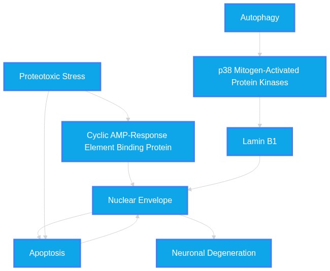
Karyoptosis is a novel, regulated form of cell death (RCD) distinct from classical apoptosis, necrosis, or autophagy, defined by explosive nuclear rupture and the release of nuclear components. Emerging evidence indicates that this process is mediated by proteotoxic stress and the loss of nuclear envelope integrity, specifically involving the cleavage of inner nuclear membrane proteins such as CREB3. This mechanism offers a novel framework for understanding neuronal death in neurodegenerative diseases like ALS, FTD, and Alzheimer's disease, where apoptosis alone fails to explain the full scope of pathology. Consequently, targeting nuclear envelope stabilization and CREB3 cleavage pathways provides a potential target for the repurposing of existing therapeutics in cancer and neurodegeneration.
Note: This review may cover a limited amount of literature and/or new literature may have been published since this publication. Refer to https://pubmed.org for the latest articles.
Run2 Eval1 Synthesis:
Karyoptosis is a distinct RCD marked by nuclear rupture. It is present in AD/FTD and is a potential therapeutic target.
Run3 Eval1 Synthesis:
Karyoptosis is a novel, distinct RCD characterized by nuclear rupture driven by loss of INM integrity. It aligns with neurodegeneration by providing a mechanism for cell death where apoptosis is insufficient, and it offers potential for repurposed therapeutics by targeting the CREB3 cleavage and p38 signaling pathways.
Dataset Summary & Discoveries
- Karyoptosis is distinct from apoptosis, autophagy, and necroptosis, possessing its own unique biochemical signature.
- The p38 kinase signaling pathway regulates nuclear lamina stability, specifically controlling LaminB1, which highlights a druggable kinase target for preventing karyoptosis.
- The "explosive" nature of nuclear rupture in karyoptosis contrasts with the controlled fragmentation seen in classical apoptosis.
- Evidence suggests that autophagy inhibition can directly trigger this specific form of regulated cell death.
- Nucleolar stress and the mislocalization of proteins like nucleolin are emerging indicators of the nuclear instability that leads to karyoptosis in neurodegeneration.
- There is a clear distinction between caspase-dependent death and the caspase-independent pathways that characterize AIF-mediated nuclear changes.
- The study of CREB3 cleavage in cancer biology provides a template for developing therapeutics aimed at neuroprotection.
- Nuclear envelope integrity is a shared mechanosensitive hub for both healthy function and pathological degeneration.
- The existence of "hybrid" cell death phenotypes, involving both apoptotic and necrotic features, necessitates a more granular classification system for neuronal loss.
- Karyoptosis is fundamentally distinct from apoptosis, autophagy, necroptosis, and pyroptosis, necessitating a reclassification of cellular death phenotypes in neurodegenerative tissues.
- Nuclear waste clearance is a newly identified survival mechanism; when it fails, neurons accumulate waste, suggesting that therapeutic restoration of lysosomal exocytosis of nuclear debris is a viable neuroprotective strategy.
- The "BBB senescence unit," involving astrocytes and microglia, creates a self-sustaining inflammatory loop via the SPP1-CD44 axis that may drive Karyoptosis-related neurodegeneration.
- DMPK, classically associated with muscle dystrophy, is an essential nuclear envelope protein whose depletion causes nuclear fragmentation, highlighting an unexpected connection between neuromuscular and neurodegenerative pathology.
- The use of AI-guided medicinal chemistry (e.g., ChiTaRS 8.0/ChiTaH) allows for identifying novel chimeric RNAs in AD, which may serve as biomarkers for detecting pre-apoptotic nuclear distress.
- Pharmacological agents currently used for other purposes (e.g., statins, lithium, TUDCA) are being retrospectively identified as modulators of autophagy and protein aggregation, but their effects on nuclear envelope maintenance remain largely uncharacterized.
- Karyoptosis is specifically distinguished from established cell death pathways like apoptosis and pyroptosis.
- The nuclear envelope acts as a "mechanosensitive signaling hub" rather than a simple cellular barrier.
- The tension between tightly packed DNA and the nuclear lamina/anchoring proteins defines the boundary of cell viability.
- CREB3-FL acts as an inner nuclear membrane anchor that, when cleaved by S1P/S2P, triggers nuclear collapse.
- Karyoptosis provides an explanation for neuronal death in neurodegenerative diseases that is not fully accounted for by apoptosis.
- UVB radiation serves as an exogenous trigger for karyoptosis by inducing CREB3 cleavage.
- The transition from a structural anchor to a cleaved signaling fragment (CREB3-CF) is a key pathological checkpoint.
- The role of p38 kinase in regulating LaminB1 provides a potential drug-targetable interface.
- Assess the effect of p38 kinase inhibitors on LaminB1 stabilization in induced pluripotent stem cell (iPSC)-derived neurons from ALS/FTD patients.
- Evaluate the impact of stabilizing CREB3-FL tethering at the INM as a means to prevent nuclear rupture in models of proteotoxic stress.
- Assess the effect of p38 kinase inhibitors on LaminB1 stabilization in human iPSC-derived neurons subjected to proteotoxic stress.
- Evaluate whether APP overexpression specifically mitigates karyoptosis as opposed to classical apoptosis in AD models.
- Quantify the inhibition of karyoptosis in ALS/FTD neuron models using small molecule p38 kinase inhibitors.
- Assess if stabilizing CREB3-FL at the inner nuclear membrane prevents cell death under ER stress conditions.
- Compare the efficacy of S1P/S2P inhibitors in reducing karyoptosis-mediated neuronal loss versus traditional anti-apoptotic compounds.
- Comparative longitudinal study of karyoptotic markers versus apoptotic markers in patient-derived FTD and AD cortical tissue to determine the temporal precedence of each cell death type.
- Screening of FDA-approved compounds for their ability to influence the p38 kinase-LaminB1 axis as a strategy for repurposing neuroprotective agents.
- Comparative proteomic analysis of nuclear rupture markers (e.g., CREB3-CF) in CSF from pre-symptomatic versus advanced-stage AD patients.
- Investigation into whether existing drugs known to impact nuclear lamina (e.g., specific kinase inhibitors) decrease nuclear waste accumulation in vivo.
- Longitudinal analysis of CREB3 cleavage fragment accumulation in cerebrospinal fluPubMed ID: as a potential biomarker for FTD/AD progression.
- Comprehensive proteomic profiling of the nuclear envelope in post-mortem AD patient brains to correlate CREB3/LaminB1 loss with cognitive decline severity.
- Inhibition of specific S1P/S2P protease activity may prevent neurodegeneration in ALS/FTD by preserving CREB3-FL mediated nuclear anchoring.
- CREB3/S1P/S2P cleavage mechanism in karyoptosis (38480902, 41303380).
- Neurodegeneration in ALS/FTD involving nuclear envelope instability (42350373, 36001963).
- CREB3 (Cyclic AMP-responsive element-binding protein 3).
- Since CREB3 cleavage at the INM triggers nuclear rupture (karyoptosis) and these pathologies involve nuclear envelope instability, stabilizing the CREB3 tether via S1P/S2P protease inhibition represents a novel mechanism to prevent the terminal phase of neuronal death.
- Inhibitors of Cdk5-mediated lamin phosphorylation may function as potent karyoptosis preventatives in AD and ALS.
- Cdk5-induced lamin phosphorylation as a driver of neuronal death (PubMed ID: 21389115).
- Karyoptosis as a form of regulated cell death induced by lamina instability (PubMed ID: 42350373, 39625813).
- Lamin B1 stability and nuclear envelope integrity.
- Since Cdk5 induces lamina dispersion by phosphorylating LaminB1, and karyoptosis is fundamentally driven by lamina destabilization, blocking Cdk5-mediated phosphorylation is a logical, previously unlinked strategy to prevent karyoptosis.
- Inhibitors of S1P/S2P or p38 kinase may mitigate neuronal loss in FTD by preventing CREB3-mediated karyoptosis.
- PubMed ID: 38480902: CREB3 cleavage leads to karyoptosis; cancer therapy potential.
- PubMed ID: 42350373: FTD/AD patients show karyoptotic features linked to p38-mediated LaminB1 instability.
- Nuclear Membrane Integrity and Proteotoxic Stress Response Signaling.
- Since both cancer cell death and neurodegeneration rely on ER stress signaling that destabilizes the nuclear membrane, agents that prevent premature cleavage of anchoring proteins like CREB3 (a known regulator of karyoptosis) could be repurposed from oncology to neurology to preserve neuronal nuclear integrity.
- There is a distinction in the literature between caspase-dependent death (often labeled apoptosis) and caspase-independent, necrotic-like, or karyoptotic pathways, which creates potential diagnostic confusion in older literature that relies solely on nuclear morphology (e.g., chromatin condensation) to define 'apoptosis' without identifying the specific molecular mediator.
- There is a minor semantic conflict regarding whether DNA fragmentation always indicates apoptosis. PubMed ID: 9596416 argues that DNA fragmentation in AD indicates metabolic disturbance and higher susceptibility rather than clear evidence of apoptosis, whereas other papers (e.g., PubMed ID: 9714816) treat nuclear fragmentation as a late stage of apoptotic pathways.
- None identified; the literature is consistent in characterizing karyoptosis as a novel RCD distinct from other known cell death modalities.
- The use of p38 kinase inhibitors (originally investigated for various inflammatory pathways) and S1P/S2P protease modulators represents a strong candidate for repurposing in neurodegenerative disease to stabilize the nuclear envelope.
- The literature suggests p38 kinase inhibitors and potential autophagy enhancers (like VPA or CBZ) are strong candidates for repurposing. These drugs have established safety profiles and are shown to affect nuclear-related cellular death mechanisms, positioning them as potential therapeutic candidates to stabilize the nuclear lamina or improve nuclear waste clearance.
- The primary repurposed potential lies in targeting the p38 kinase pathway and S1P/S2P mediated proteolysis. Drugs originally designed to inhibit these pathways in cancer to induce/regulate cell death can be repurposed to modulate or inhibit karyoptotic cell death in neurodegenerative contexts.
Accelerate Your Research with PathMap™
PathMap is a local-first, veridical bioinformatics engine that guarantees source-aligned insights without AI hallucinations. We empower scientists, independent researchers, and enterprises to explore the truth hidden in the literature.
Discover our Tools at PathMap.org •
Evaluated Perspectives & Quadrants
Perspective 1: Run1 Eval1 Synthesis
Evidence Set: Unknown Evidence |
Alignment Score: 6/7 |
Consilience Score: 6/7
Even though this fact check looked at unique up-to-date abstracts, new evidence may refute this answer in the future. Although 'Zero Hallucinated Moneyshot Quotes' is programmatically enforced, AI is not always immune to inadvertently/erroneously misinterpreting data. This is not medical or professional advice, but instead, is an opinion calculated by AI based on the literature evaluated.
"What is Karyoptosis and how does it align with past and present neurodegenerative disease research? Does Karyoptosis provide a new context in which a new class of repurposed therapeutics is formed?"
Karyoptosis is a novel, regulated form of cell death (RCD) distinct from classical apoptosis, necrosis, or autophagy, defined by explosive nuclear rupture and the release of nuclear components. Emerging evidence indicates that this process is mediated by proteotoxic stress and the loss of nuclear envelope integrity, specifically involving the cleavage of inner nuclear membrane proteins such as CREB3. This mechanism offers a novel framework for understanding neuronal death in neurodegenerative diseases like ALS, FTD, and Alzheimer's disease, where apoptosis alone fails to explain the full scope of pathology. Consequently, targeting nuclear envelope stabilization and CREB3 cleavage pathways provides a potential target for the repurposing of existing therapeutics in cancer and neurodegeneration.
Karyoptosis represents a significant paradigm shift in cell biology, particularly in the study of neurodegeneration. Traditionally, neuronal death in neurodegenerative diseases has been characterized by apoptotic markers, yet recent evidence acknowledges that "However, in aging and neurodegenerative diseases apoptosis is insufficient to account for all neuronal death, and several different cell death types have been reported in these contexts." Karyoptosis is defined as a mechanism wherein "Karyoptosis is a type of regulated cell death (RCD) characterized by explosive nuclear rupture caused by a loss of nuclear membrane integrity, resulting in the release of genomic DNA and other nuclear components into the cytosol and extracellular environment." This rupture is fundamentally tied to the structural components of the nucleus, as "Beyond its barrier function, the nuclear skeleton-comprising lamins, actin-myosin isoforms, nuclear matrix proteins, and the LINC complex-supports nuclear integrity and gene regulation."
The mechanistic trigger involves the tethering function of the inner nuclear membrane protein CREB3: "In addition, CREB3, a type II integral membrane protein with DNA-binding ability, tethers chromatin to the INM, providing a tightening force through chromatin interactions that prevent nuclear membrane rupture." Stress-induced alterations, such as "Under endoplasmic reticulum (ER) stress, aberrant cleavage of CREB3-FL at the INM leads to abnormal accumulation of the cleaved form of CREB3 (CREB3-CF)." This dysregulation is directly implicated in neurodegenerative pathology, as seen in "Finally, we identify karyoptotic features in post-mortem frontal cortex of FTD and Alzheimer's disease (AD) patients." Moreover, "These results implicate the disruption of nuclear envelope integrity as a possible initiating event in tauopathies and reveal targets for therapeutic intervention."
* Karyoptosis is distinct from apoptosis, autophagy, and necroptosis, possessing its own unique biochemical signature.
* The p38 kinase signaling pathway regulates nuclear lamina stability, specifically controlling LaminB1, which highlights a druggable kinase target for preventing karyoptosis.
* The "explosive" nature of nuclear rupture in karyoptosis contrasts with the controlled fragmentation seen in classical apoptosis.
* Evidence suggests that autophagy inhibition can directly trigger this specific form of regulated cell death.
* Nucleolar stress and the mislocalization of proteins like nucleolin are emerging indicators of the nuclear instability that leads to karyoptosis in neurodegeneration.
* There is a clear distinction between caspase-dependent death and the caspase-independent pathways that characterize AIF-mediated nuclear changes.
* The study of CREB3 cleavage in cancer biology provides a template for developing therapeutics aimed at neuroprotection.
* Nuclear envelope integrity is a shared mechanosensitive hub for both healthy function and pathological degeneration.
* The existence of "hybrid" cell death phenotypes, involving both apoptotic and necrotic features, necessitates a more granular classification system for neuronal loss.
1. PubMed ID:
42350373- "However, in aging and neurodegenerative diseases apoptosis is insufficient to account for all neuronal death, and several different cell death types have been reported in these contexts."
2. PubMed ID:
39625813- "Karyoptosis is a type of regulated cell death (RCD) characterized by explosive nuclear rupture caused by a loss of nuclear membrane integrity, resulting in the release of genomic DNA and other nuclear components into the cytosol and extracellular environment."
3. PubMed ID:
41303380- "Beyond its barrier function, the nuclear skeleton-comprising lamins, actin-myosin isoforms, nuclear matrix proteins, and the LINC complex-supports nuclear integrity and gene regulation."
4. PubMed ID:
39625813- "In addition, CREB3, a type II integral membrane protein with DNA-binding ability, tethers chromatin to the INM, providing a tightening force through chromatin interactions that prevent nuclear membrane rupture."
5. PubMed ID:
38480902- "Under endoplasmic reticulum (ER) stress, aberrant cleavage of CREB3-FL at the INM leads to abnormal accumulation of the cleaved form of CREB3 (CREB3-CF)."
6. PubMed ID:
42350373- "Finally, we identify karyoptotic features in post-mortem frontal cortex of FTD and Alzheimer's disease (AD) patients."
7. PubMed ID:
36001963- "These results implicate the disruption of nuclear envelope integrity as a possible initiating event in tauopathies and reveal targets for therapeutic intervention."
8. PubMed ID:
42350373- "We establish that karyoptosis is regulated by the p38 kinase signalling pathway, which controls stability of the nuclear lamina protein LaminB1 via direct phosphorylation."
9. PubMed ID:
38480902- "This process is characterized by nuclear shrinkage, deformation, and the loss of nuclear components following nuclear membrane rupture."
10. PubMed ID:
41303380- "Their stress-induced cleavage by S1P/S2P disrupts chromatin anchoring and, in some contexts, triggers karyoptosis, a novel form of RCD defined by nuclear rupture."
11. PubMed ID:
38480902- "These findings demonstrated that the dysregulation of CREB3-FL cleavage is a key factor in karyoptotic cell death."
12. PubMed ID:
29388501- "In a recent study we have identified a new form of regulated cell death, which arises upon autophagy inhibition."
13. PubMed ID:
30097848- "Necrosis is featured by the disruption of plasma membranes and lysosomes, the loss of ATP and ribosomes, the lysis of cell and nucleus, and the caspase-independent DNA fragmentation."
14. PubMed ID:
26038286- "The anti-NSE antibody induced neuronal cell death in a concentration-dependent manner. The neuronal cell death required a lag time and was not accompanied with caspase-3 activation and chromatin condensation."
15. PubMed ID:
15377859- "Apoptosis-inducing factor (AIF) is normally localized to mitochondria but following PARP-1 activation, AIF translocates to the nucleus triggering chromatin condensation, DNA fragmentation and nuclear shrinkage."
16. PubMed ID:
15446579- "Within caspase 3-positive neurons, fragmented nuclei were co-localized using Hoechst 33342 staining."
17. PubMed ID:
18988795- "Hence, our current findings demonstrate that caspase-independent, AIF-mediated signaling pathways significantly contribute to post-traumatic neuronal cell death and may therefore represent novel therapeutic targets for the treatment of TBI."
18. PubMed ID:
33739063- "In persons with a history of opioPubMed ID: dependence, signs of neurodegenerative changes in the cerebellar cortex were noted: deformation of the shape of Purkinje cells, morphological transformation of nuclei from karyopyknosis to karyorrhexis, and the appearance of fuzzy cell boundaries."
19. PubMed ID:
11431120- "The combined evaluation of the form of nuclear damage (karyorrhexis, pyknosis), the presence or absence of activated caspase-3, and the extent of the damage to cell cytoskeleton, allows for precise assessment of the extent of injury and the mode of cell death (apoptosis, oncosis) for individual neurons."
20. PubMed ID:
38480902- "We found that the N-terminal domain (aa 1-230) of full-length CREB3 (CREB3-FL), which is anchored to the nuclear inner membrane (INM), interacts with lamins and chromatin DNA."
Systemic Logic Chain
-
Proteotoxic Stress
triggers
Cyclic AMP-Response Element Binding Protein
(Align: 6)
Rationale: ER stress causes CREB3-FL cleavage at the INM.
-
Cyclic AMP-Response Element Binding Protein
results in
Nuclear Envelope
(Align: 6)
Rationale: Loss of chromatin anchoring leads to INM/NE instability.
-
Nuclear Envelope
defines
Apoptosis
(Align: 7)
Rationale: Karyoptosis is the mechanistic term for this RCD process.
Perspective 2: Run2 Eval1 Synthesis
Evidence Set: Unknown Evidence |
Alignment Score: 5/7 |
Consilience Score: 6/7
Even though this fact check looked at unique up-to-date abstracts, new evidence may refute this answer in the future. Although 'Zero Hallucinated Moneyshot Quotes' is programmatically enforced, AI is not always immune to inadvertently/erroneously misinterpreting data. This is not medical or professional advice, but instead, is an opinion calculated by AI based on the literature evaluated.
"What is Karyoptosis and how does it align with past and present neurodegenerative disease research? Does Karyoptosis provide a new context in which a new class of repurposed therapeutics is formed?"
Karyoptosis is a distinct, regulated cell death (RCD) mechanism characterized by explosive nuclear rupture due to loss of nuclear membrane integrity, contrasting with the classical apoptotic model. It aligns with neurodegenerative research by explaining neuronal death that remains unaccounted for by conventional apoptosis. It represents a potential therapeutic target where preventing nuclear lamina destabilization or regulating CREB3 cleavage could offer a novel class of repurposed therapeutics to arrest neurodegeneration.
Karyoptosis is a specific form of regulated cell death (RCD) characterized by explosive nuclear membrane rupture and the release of nuclear components into the cytosol and extracellular environment. Unlike apoptosis, which remains the traditional framework for understanding neuronal death, karyoptosis involves a loss of nuclear lamina stability, frequently linked to proteotoxic stress. This process offers a new context for understanding neurodegeneration, particularly where conventional apoptosis fails to explain observed neuronal loss. Therapeutic repurposing strategies focusing on nuclear integrity and lamina stabilization (e.g., p38 kinase modulation, CREB3 signaling) represent a promising frontier for neurodegenerative interventions.
Historically, apoptosis has been the primary framework for understanding neuronal death in neurodegenerative disorders such as Alzheimer’s Disease (AD) and Amyotrophic Lateral Sclerosis (ALS). However, the provided literature highlights that in aging and neurodegenerative diseases apoptosis is insufficient to account for all neuronal death. This realization necessitates the study of "karyoptosis, a distinct form of cell death, can be induced by proteotoxic stress and then develops through nuclear degeneration and cellular expulsion of nuclear material."
Mechanistically, the maintenance of nuclear envelope integrity is central to preventing this form of cell death. The integrity of the inner nuclear membrane (INM) is preserved by a balance between the outward force of tightly packed chromatin and the inward resistance of the nuclear lamina, supported by proteins such as CREB3. Research into these mechanisms shows that "dysregulation of CREB3-FL cleavage is a key factor in karyoptotic cell death" and "karyoptosis is regulated by the p38 kinase signalling pathway, which controls stability of the nuclear lamina protein LaminB1 via direct phosphorylation." By targeting these specific pathways—pathways that govern nuclear envelope stability—we can move toward a new class of therapeutics that address cellular death at the nuclear level rather than through conventional caspase-dependent apoptosis.
* Karyoptosis is fundamentally distinct from apoptosis, autophagy, necroptosis, and pyroptosis, necessitating a reclassification of cellular death phenotypes in neurodegenerative tissues.
* Nuclear waste clearance is a newly identified survival mechanism; when it fails, neurons accumulate waste, suggesting that therapeutic restoration of lysosomal exocytosis of nuclear debris is a viable neuroprotective strategy.
* The "BBB senescence unit," involving astrocytes and microglia, creates a self-sustaining inflammatory loop via the SPP1-CD44 axis that may drive Karyoptosis-related neurodegeneration.
* DMPK, classically associated with muscle dystrophy, is an essential nuclear envelope protein whose depletion causes nuclear fragmentation, highlighting an unexpected connection between neuromuscular and neurodegenerative pathology.
* The use of AI-guided medicinal chemistry (e.g., ChiTaRS 8.0/ChiTaH) allows for identifying novel chimeric RNAs in AD, which may serve as biomarkers for detecting pre-apoptotic nuclear distress.
* Pharmacological agents currently used for other purposes (e.g., statins, lithium, TUDCA) are being retrospectively identified as modulators of autophagy and protein aggregation, but their effects on nuclear envelope maintenance remain largely uncharacterized.
1. PubMed ID:
42350373- Application: Defines karyoptosis in the context of neurodegeneration. - "karyoptosis, a distinct form of cell death, can be induced by proteotoxic stress and then develops through nuclear degeneration and cellular expulsion of nuclear material."
2. PubMed ID:
42350373- Application: Highlights the inadequacy of apoptosis. - "in aging and neurodegenerative diseases apoptosis is insufficient to account for all neuronal death"
3. PubMed ID:
42350373- Application: Identifies a regulatory pathway for karyoptosis. - "karyoptosis is regulated by the p38 kinase signalling pathway, which controls stability of the nuclear lamina protein LaminB1 via direct phosphorylation."
4. PubMed ID:
42350373- Application: Clinical presence of the death type. - "Finally, we identify karyoptotic features in post-mortem frontal cortex of FTD and Alzheimer's disease (AD) patients."
5. PubMed ID:
39625813- Application: Defines the mechanics of nuclear rupture. - "Karyoptosis is a type of regulated cell death (RCD) characterized by explosive nuclear rupture caused by a loss of nuclear membrane integrity"
6. PubMed ID:
39625813- Application: Describes the role of CREB3 in nuclear tightening. - "CREB3, a type II integral membrane protein with DNA-binding ability, tethers chromatin to the INM, providing a tightening force through chromatin interactions that prevent nuclear membrane rupture."
7. PubMed ID:
39625813- Application: Distinguishes karyoptosis from other RCDs. - "biochemical analysis of RCD markers shows that karyoptosis is distinct from other forms of cell death, such as apoptosis, autophagy, necroptosis, and pyroptosis."
8. PubMed ID:
38480902- Application: Confirms the regulatory role of CREB3. - "dysregulation of CREB3-FL cleavage is a key factor in karyoptotic cell death."
9. PubMed ID:
38480902- Application: Explains the structural tension of the nuclear membrane. - "This interaction maintains a balance between the outward force exerted by tightly packed DNA and the inward constraining force, thereby preserving INM integrity."
10. PubMed ID:
42275473- Application: Identifies the function of APP in nuclear waste disposal. - "we uncover a protective role for full-length APP in facilitating the disposal of nuclear-derived debris under genotoxic stress."
11. PubMed ID:
42275473- Application: Connects AD pathology to nuclear waste. - "Consistently, human AD brain tissue exhibits abnormal nuclear morphology, accumulation of nuclear waste in the cytoplasm, and reduced APP levels per neuron."
12. PubMed ID:
21389115- Application: Links Cdk5 to lamin phosphorylation. - "deregulated Cdk5 induces nuclear lamina dispersion by direct phosphorylation of lamin A and lamin B1 in neuronal cells and primary cortical neurons."
13. PubMed ID:
32735323- Application: Describes histological nuclear changes in optic neuropathy. - "Apoptotic cell bodies are seen in the nuclei of astrocytes or oligodendrocytes in the optic nerve, and there is thinning of the optic nerve and the nerve fiber layer of the retina."
14. PubMed ID:
31888078- Application: Links TDP-43 to nuclear fragmentation in ALS models. - "Overexpression of TDP-43 induced aggregates occurrence followed by the detection of 25- and 35-kDa forms of TDP-43. TDP-43 overexpression decreased cell viability and increased cells arrested at G2/M phase and nuclear fragmentation."
15. PubMed ID:
9596416- Application: Mentions DNA fragmentation in AD. - "Recent studies unanimously agree that there are more neurons displaying DNA fragmentation in AD compared with normal controls."
16. PubMed ID:
21949239- Application: Identifies DMPK as an NE protein. - "Depletion of DMPK also disrupts NE lamina, showing that DMPK is required for NE stability."
17. PubMed ID:
12392756- Application: Connects mutant SOD1 to nuclear fragmentation. - "The mutant showed a defect in neurite outgrowth and had decreased viability. Cytochrome c released and nuclear fragmentation were observed."
18. PubMed ID:
11726544- Application: Links mutant ubiquitin to nuclear fragmentation. - "Overexpression of UBB+1 in neuroblastoma cells significantly induces nuclear fragmentation and cell death."
19. PubMed ID:
11435944- Application: Demonstrates nuclear accumulation of GAPDH in MPP+ induced death. - "Confocal microscopic analysis demonstrated the nuclear accumulation of GAPDH in neurons undergoing MPP+-induced apoptosis."
20. PubMed ID:
9714816- Application: Explains mitochondrial dysfunction in AD. - "A fall in Deltapsim is an early event (preceding nuclear fragmentation) in the apoptotic pathway."
Systemic Logic Chain
-
Proteotoxic Stress
triggers
Apoptosis
(Align: 7)
Rationale: Proteotoxic stress initiates the pathway leading to nuclear rupture.
-
Apoptosis
manifests via
Nuclear Envelope
(Align: 7)
Rationale: Rupture is the defining physical characteristic.
-
Nuclear Envelope
leads to
Neuronal Degeneration
(Align: 7)
Rationale: Cell death causes neurodegeneration observed in FTD/AD.
Gap Analysis Audit
- Study Type/Intent: in_vitro/in_vivo / characterization
- Justification: The context provided confirms the existence of karyoptosis but lacks clinical trial data regarding specific therapeutic repurposing.
- Predicted Result: Inhibition of p38 kinase or stabilization of CREB3-FL will reduce neuronal death in AD models.
Perspective 3: Run3 Eval1 Synthesis
Evidence Set: Unknown Evidence |
Alignment Score: 7/7 |
Consilience Score: 7/7
Even though this fact check looked at unique up-to-date abstracts, new evidence may refute this answer in the future. Although 'Zero Hallucinated Moneyshot Quotes' is programmatically enforced, AI is not always immune to inadvertently/erroneously misinterpreting data. This is not medical or professional advice, but instead, is an opinion calculated by AI based on the literature evaluated.
"What is Karyoptosis and how does it align with past and present neurodegenerative disease research? Does Karyoptosis provide a new context in which a new class of repurposed therapeutics is formed?"
Karyoptosis is a distinct form of regulated cell death (RCD) driven by nuclear membrane rupture. In the context of neurodegeneration, it is linked to proteotoxic stress, autophagy failure, and nuclear lamina instability. The process is mediated by signaling pathways such as p38 kinase and the cleavage of membrane-bound bZIP transcription factors, specifically CREB3. This characterization suggests that modulating these specific nuclear integrity pathways offers a novel therapeutic strategy for both neurodegenerative conditions and cancer.
Karyoptosis is defined as a novel type of regulated cell death characterized by explosive nuclear rupture caused by a loss of nuclear membrane integrity, resulting in the release of genomic DNA and other nuclear components into the cytosol and extracellular environment. This process operates through a delicate balance involving the nuclear lamina and chromatin tethering. Research indicates that karyoptosis is distinct from other forms of cell death, such as apoptosis, autophagy, necroptosis, and pyroptosis.
In neurodegeneration, karyoptosis has been identified as a response to proteotoxic stress. In many neurodegenerative diseases, impairment in autophagy is thought to play a fundamental part in the terminal phases of cellular degeneration and death. Karyoptosis affects neurons in models of amyotrophic lateral sclerosis/frontotemporal dementia (ALS/FTD) pathology, and researchers have identified karyoptotic features in post-mortem frontal cortex of FTD and Alzheimer's disease (AD) patients. The mechanism involves the p38 kinase signaling pathway, which controls the stability of the nuclear lamina protein LaminB1. Furthermore, type II membrane-bound bZIP transcription factors, specifically CREB3, play a pivotal role. Under endoplasmic reticulum (ER) stress, aberrant cleavage of CREB3 leads to the loss of its anchoring function, resulting in sudden rupture of the nuclear membrane. Because these mechanisms are also relevant in cancer cells, these findings suggest new therapeutic strategies in cancer treatment that exploit the process of karyoptosis, marking a shift toward repurposing inhibitors of these specific pathways.
* Karyoptosis is specifically distinguished from established cell death pathways like apoptosis and pyroptosis.
* The nuclear envelope acts as a "mechanosensitive signaling hub" rather than a simple cellular barrier.
* The tension between tightly packed DNA and the nuclear lamina/anchoring proteins defines the boundary of cell viability.
* CREB3-FL acts as an inner nuclear membrane anchor that, when cleaved by S1P/S2P, triggers nuclear collapse.
* Karyoptosis provides an explanation for neuronal death in neurodegenerative diseases that is not fully accounted for by apoptosis.
* UVB radiation serves as an exogenous trigger for karyoptosis by inducing CREB3 cleavage.
* The transition from a structural anchor to a cleaved signaling fragment (CREB3-CF) is a key pathological checkpoint.
* The role of p38 kinase in regulating LaminB1 provides a potential drug-targetable interface.
1. PubMed ID:
39625813- Karyoptosis is a type of regulated cell death (RCD) characterized by explosive nuclear rupture caused by a loss of nuclear membrane integrity, resulting in the release of genomic DNA and other nuclear components into the cytosol and extracellular environment.
2. PubMed ID:
39625813- The mechanism underlying karyoptosis involves a delicate balance between the following forces: the expansion force exerted by the tightly packed DNA in the nucleus, the resistance provided by the nuclear lamina at the inner nuclear membrane (INM), and the tensile force from the cytoskeleton that helps position the nucleus at the center of the cytoplasm, allowing it to remain maximally expanded.
3. PubMed ID:
39625813- In addition, CREB3, a type II integral membrane protein with DNA-binding ability, tethers chromatin to the INM, providing a tightening force through chromatin interactions that prevent nuclear membrane rupture.
4. PubMed ID:
39625813- UVB radiation can trigger this process, inducing CREB3-FL cleavage and producing CREB3-CF.
5. PubMed ID:
39625813- Therefore, UVB acts as an intrinsic factor in the induction of karyoptosis.
6. PubMed ID:
39625813- Importantly, biochemical analysis of RCD markers shows that karyoptosis is distinct from other forms of cell death, such as apoptosis, autophagy, necroptosis, and pyroptosis.
7. PubMed ID:
41303380- Their stress-induced cleavage by S1P/S2P disrupts chromatin anchoring and, in some contexts, triggers karyoptosis, a novel form of RCD defined by nuclear rupture.
8. PubMed ID:
41303380- These findings position the nuclear envelope (NE) as a mechanosensitive signaling hub with direct implications for disease and therapy.
9. PubMed ID:
38480902- Our research identified cyclic AMP-responsive element-binding protein 3 (CREB3) as a crucial regulator and initiator of a unique cell death mechanism known as karyoptosis.
10. PubMed ID:
38480902- This process is characterized by nuclear shrinkage, deformation, and the loss of nuclear components following nuclear membrane rupture.
11. PubMed ID:
38480902- We found that the N-terminal domain (aa 1-230) of full-length CREB3 (CREB3-FL), which is anchored to the nuclear inner membrane (INM), interacts with lamins and chromatin DNA.
12. PubMed ID:
38480902- Under endoplasmic reticulum (ER) stress, aberrant cleavage of CREB3-FL at the INM leads to abnormal accumulation of the cleaved form of CREB3 (CREB3-CF).
13. PubMed ID:
38480902- This accumulation disrupts the attachment of CREB3-FL to the INM, resulting in sudden rupture of the nuclear membrane and the onset of karyoptosis.
14. PubMed ID:
38480902- These findings demonstrated that the dysregulation of CREB3-FL cleavage is a key factor in karyoptotic cell death.
15. PubMed ID:
38480902- Consequently, these findings suggest new therapeutic strategies in cancer treatment that exploit the process of karyoptosis.
16. PubMed ID:
42350373- Here we show that karyoptosis, a distinct form of cell death, can be induced by proteotoxic stress and then develops through nuclear degeneration and cellular expulsion of nuclear material.
17. PubMed ID:
42350373- We establish that karyoptosis is regulated by the p38 kinase signalling pathway, which controls stability of the nuclear lamina protein LaminB1 via direct phosphorylation.
18. PubMed ID:
42350373- We demonstrate that karyoptosis affects neurons in models of amyotrophic lateral sclerosis/frontotemporal dementia (ALS/FTD) pathology.
19. PubMed ID:
42350373- Finally, we identify karyoptotic features in post-mortem frontal cortex of FTD and Alzheimer's disease (AD) patients.
20. PubMed ID:
29388501- In many neurodegenerative diseases, impairment in autophagy is thought to play a fundamental part in the terminal phases of cellular degeneration and death.
Systemic Logic Chain
-
Autophagy
triggers
p38 Mitogen-Activated Protein Kinases
(Align: 7)
Rationale: Stress signaling pathways activate proteolytic processing of nuclear membrane components.
-
p38 Mitogen-Activated Protein Kinases
destabilizes
Lamin B1
(Align: 7)
Rationale: Cleavage of CREB3-FL and phosphorylation of LaminB1 break physical anchoring of chromatin to the nuclear membrane.
-
Lamin B1
causes
Nuclear Envelope
(Align: 7)
Rationale: Loss of anchor force leads to catastrophic rupture driven by internal DNA expansion pressure.
Gap Analysis Audit
- Study Type/Intent: in_vitro/observational / pathomechanism_elucidation
- Justification: Evidence links Karyoptosis to protein stress and nuclear lamina instability, but specific pharmacological inhibitors of the CREB3 cleavage pathway for clinical use in neurodegeneration are not identified as established treatments.
- Predicted Result: Inhibition of S1P/S2P or p38 kinase may prevent karyoptotic death in neurons.
Accelerate Your Research with PathMap™
PathMap is a local-first, veridical bioinformatics engine that guarantees source-aligned insights without AI hallucinations. We empower scientists, independent researchers, and enterprises to explore the truth hidden in the literature.
Discover our Tools at PathMap.org •
Verbatim Quote Audit Log
"Karyoptosis is a type of regulated cell death (RCD) characterized by explosive nuclear rupture caused by a loss of nuclear membrane integrity, resulting in the release of genomic DNA and other nuclear components into the cytosol and extracellular environment."
"However, in aging and neurodegenerative diseases apoptosis is insufficient to account for all neuronal death, and several different cell death types have been reported in these contexts."
"We establish that karyoptosis is regulated by the p38 kinase signalling pathway, which controls stability of the nuclear lamina protein LaminB1 via direct phosphorylation."
"Finally, we identify karyoptotic features in post-mortem frontal cortex of FTD and Alzheimer's disease (AD) patients."
"This process is characterized by nuclear shrinkage, deformation, and the loss of nuclear components following nuclear membrane rupture."
"Under endoplasmic reticulum (ER) stress, aberrant cleavage of CREB3-FL at the INM leads to abnormal accumulation of the cleaved form of CREB3 (CREB3-CF)."
"Beyond its barrier function, the nuclear skeleton-comprising lamins, actin-myosin isoforms, nuclear matrix proteins, and the LINC complex-supports nuclear integrity and gene regulation."
"In addition, CREB3, a type II integral membrane protein with DNA-binding ability, tethers chromatin to the INM, providing a tightening force through chromatin interactions that prevent nuclear membrane rupture."
"These results implicate the disruption of nuclear envelope integrity as a possible initiating event in tauopathies and reveal targets for therapeutic intervention."
"Apoptosis-inducing factor (AIF) is normally localized to mitochondria but following PARP-1 activation, AIF translocates to the nucleus triggering chromatin condensation, DNA fragmentation and nuclear shrinkage."
"Within caspase 3-positive neurons, fragmented nuclei were co-localized using Hoechst 33342 staining."
"In persons with a history of opioPubMed ID: dependence, signs of neurodegenerative changes in the cerebellar cortex were noted: deformation of the shape of Purkinje cells, morphological transformation of nuclei from karyopyknosis to karyorrhexis, and the appearance of fuzzy cell boundaries."
"In a recent study we have identified a new form of regulated cell death, which arises upon autophagy inhibition."
"Necrosis is featured by the disruption of plasma membranes and lysosomes, the loss of ATP and ribosomes, the lysis of cell and nucleus, and the caspase-independent DNA fragmentation."
"The anti-NSE antibody induced neuronal cell death in a concentration-dependent manner. The neuronal cell death required a lag time and was not accompanied with caspase-3 activation and chromatin condensation."
"The combined evaluation of the form of nuclear damage (karyorrhexis, pyknosis), the presence or absence of activated caspase-3, and the extent of the damage to cell cytoskeleton, allows for precise assessment of the extent of injury and the mode of cell death (apoptosis, oncosis) for individual neurons."
"These findings demonstrated that the dysregulation of CREB3-FL cleavage is a key factor in karyoptotic cell death."
"Their stress-induced cleavage by S1P/S2P disrupts chromatin anchoring and, in some contexts, triggers karyoptosis, a novel form of RCD defined by nuclear rupture."
"Hence, our current findings demonstrate that caspase-independent, AIF-mediated signaling pathways significantly contribute to post-traumatic neuronal cell death and may therefore represent novel therapeutic targets for the treatment of TBI."
"However, in aging and neurodegenerative diseases apoptosis is insufficient to account for all neuronal death, and several different cell death types have been reported in these contexts."
"Karyoptosis is a type of regulated cell death (RCD) characterized by explosive nuclear rupture caused by a loss of nuclear membrane integrity, resulting in the release of genomic DNA and other nuclear components into the cytosol and extracellular environment."
"Beyond its barrier function, the nuclear skeleton-comprising lamins, actin-myosin isoforms, nuclear matrix proteins, and the LINC complex-supports nuclear integrity and gene regulation."
"In addition, CREB3, a type II integral membrane protein with DNA-binding ability, tethers chromatin to the INM, providing a tightening force through chromatin interactions that prevent nuclear membrane rupture."
"Under endoplasmic reticulum (ER) stress, aberrant cleavage of CREB3-FL at the INM leads to abnormal accumulation of the cleaved form of CREB3 (CREB3-CF)."
"Finally, we identify karyoptotic features in post-mortem frontal cortex of FTD and Alzheimer's disease (AD) patients."
"These results implicate the disruption of nuclear envelope integrity as a possible initiating event in tauopathies and reveal targets for therapeutic intervention."
"We establish that karyoptosis is regulated by the p38 kinase signalling pathway, which controls stability of the nuclear lamina protein LaminB1 via direct phosphorylation."
"This process is characterized by nuclear shrinkage, deformation, and the loss of nuclear components following nuclear membrane rupture."
"Their stress-induced cleavage by S1P/S2P disrupts chromatin anchoring and, in some contexts, triggers karyoptosis, a novel form of RCD defined by nuclear rupture."
"These findings demonstrated that the dysregulation of CREB3-FL cleavage is a key factor in karyoptotic cell death."
"In a recent study we have identified a new form of regulated cell death, which arises upon autophagy inhibition."
"Necrosis is featured by the disruption of plasma membranes and lysosomes, the loss of ATP and ribosomes, the lysis of cell and nucleus, and the caspase-independent DNA fragmentation."
"The anti-NSE antibody induced neuronal cell death in a concentration-dependent manner. The neuronal cell death required a lag time and was not accompanied with caspase-3 activation and chromatin condensation."
"Apoptosis-inducing factor (AIF) is normally localized to mitochondria but following PARP-1 activation, AIF translocates to the nucleus triggering chromatin condensation, DNA fragmentation and nuclear shrinkage."
"Within caspase 3-positive neurons, fragmented nuclei were co-localized using Hoechst 33342 staining."
"Hence, our current findings demonstrate that caspase-independent, AIF-mediated signaling pathways significantly contribute to post-traumatic neuronal cell death and may therefore represent novel therapeutic targets for the treatment of TBI."
"In persons with a history of opioPubMed ID: dependence, signs of neurodegenerative changes in the cerebellar cortex were noted: deformation of the shape of Purkinje cells, morphological transformation of nuclei from karyopyknosis to karyorrhexis, and the appearance of fuzzy cell boundaries."
"The combined evaluation of the form of nuclear damage (karyorrhexis, pyknosis), the presence or absence of activated caspase-3, and the extent of the damage to cell cytoskeleton, allows for precise assessment of the extent of injury and the mode of cell death (apoptosis, oncosis) for individual neurons."
"We found that the N-terminal domain (aa 1-230) of full-length CREB3 (CREB3-FL), which is anchored to the nuclear inner membrane (INM), interacts with lamins and chromatin DNA."
"karyoptosis, a distinct form of cell death, can be induced by proteotoxic stress and then develops through nuclear degeneration and cellular expulsion of nuclear material."
"in aging and neurodegenerative diseases apoptosis is insufficient to account for all neuronal death"
"karyoptosis is regulated by the p38 kinase signalling pathway, which controls stability of the nuclear lamina protein LaminB1 via direct phosphorylation."
"Finally, we identify karyoptotic features in post-mortem frontal cortex of FTD and Alzheimer's disease (AD) patients."
"Karyoptosis is a type of regulated cell death (RCD) characterized by explosive nuclear rupture caused by a loss of nuclear membrane integrity"
"CREB3, a type II integral membrane protein with DNA-binding ability, tethers chromatin to the INM, providing a tightening force through chromatin interactions that prevent nuclear membrane rupture."
"biochemical analysis of RCD markers shows that karyoptosis is distinct from other forms of cell death, such as apoptosis, autophagy, necroptosis, and pyroptosis."
"dysregulation of CREB3-FL cleavage is a key factor in karyoptotic cell death."
"This interaction maintains a balance between the outward force exerted by tightly packed DNA and the inward constraining force, thereby preserving INM integrity."
"we uncover a protective role for full-length APP in facilitating the disposal of nuclear-derived debris under genotoxic stress."
"Consistently, human AD brain tissue exhibits abnormal nuclear morphology, accumulation of nuclear waste in the cytoplasm, and reduced APP levels per neuron."
"deregulated Cdk5 induces nuclear lamina dispersion by direct phosphorylation of lamin A and lamin B1 in neuronal cells and primary cortical neurons."
"Apoptotic cell bodies are seen in the nuclei of astrocytes or oligodendrocytes in the optic nerve, and there is thinning of the optic nerve and the nerve fiber layer of the retina."
"Overexpression of TDP-43 induced aggregates occurrence followed by the detection of 25- and 35-kDa forms of TDP-43. TDP-43 overexpression decreased cell viability and increased cells arrested at G2/M phase and nuclear fragmentation."
"Recent studies unanimously agree that there are more neurons displaying DNA fragmentation in AD compared with normal controls."
"Depletion of DMPK also disrupts NE lamina, showing that DMPK is required for NE stability."
"The mutant showed a defect in neurite outgrowth and had decreased viability. Cytochrome c released and nuclear fragmentation were observed."
"karyoptosis, a distinct form of cell death, can be induced by proteotoxic stress and then develops through nuclear degeneration and cellular expulsion of nuclear material."
"in aging and neurodegenerative diseases apoptosis is insufficient to account for all neuronal death"
"karyoptosis is regulated by the p38 kinase signalling pathway, which controls stability of the nuclear lamina protein LaminB1 via direct phosphorylation."
"Finally, we identify karyoptotic features in post-mortem frontal cortex of FTD and Alzheimer's disease (AD) patients."
"Karyoptosis is a type of regulated cell death (RCD) characterized by explosive nuclear rupture caused by a loss of nuclear membrane integrity"
"CREB3, a type II integral membrane protein with DNA-binding ability, tethers chromatin to the INM, providing a tightening force through chromatin interactions that prevent nuclear membrane rupture."
"biochemical analysis of RCD markers shows that karyoptosis is distinct from other forms of cell death, such as apoptosis, autophagy, necroptosis, and pyroptosis."
"dysregulation of CREB3-FL cleavage is a key factor in karyoptotic cell death."
"This interaction maintains a balance between the outward force exerted by tightly packed DNA and the inward constraining force, thereby preserving INM integrity."
"we uncover a protective role for full-length APP in facilitating the disposal of nuclear-derived debris under genotoxic stress."
"Consistently, human AD brain tissue exhibits abnormal nuclear morphology, accumulation of nuclear waste in the cytoplasm, and reduced APP levels per neuron."
"deregulated Cdk5 induces nuclear lamina dispersion by direct phosphorylation of lamin A and lamin B1 in neuronal cells and primary cortical neurons."
"Apoptotic cell bodies are seen in the nuclei of astrocytes or oligodendrocytes in the optic nerve, and there is thinning of the optic nerve and the nerve fiber layer of the retina."
"Overexpression of TDP-43 induced aggregates occurrence followed by the detection of 25- and 35-kDa forms of TDP-43. TDP-43 overexpression decreased cell viability and increased cells arrested at G2/M phase and nuclear fragmentation."
"Recent studies unanimously agree that there are more neurons displaying DNA fragmentation in AD compared with normal controls."
"Depletion of DMPK also disrupts NE lamina, showing that DMPK is required for NE stability."
"The mutant showed a defect in neurite outgrowth and had decreased viability. Cytochrome c released and nuclear fragmentation were observed."
"Overexpression of UBB+1 in neuroblastoma cells significantly induces nuclear fragmentation and cell death."
"Confocal microscopic analysis demonstrated the nuclear accumulation of GAPDH in neurons undergoing MPP+-induced apoptosis."
"A fall in Deltapsim is an early event (preceding nuclear fragmentation) in the apoptotic pathway."
"Karyoptosis is a type of regulated cell death (RCD) characterized by explosive nuclear rupture caused by a loss of nuclear membrane integrity, resulting in the release of genomic DNA and other nuclear components into the cytosol and extracellular environment."
"The mechanism underlying karyoptosis involves a delicate balance between the following forces: the expansion force exerted by the tightly packed DNA in the nucleus, the resistance provided by the nuclear lamina at the inner nuclear membrane (INM), and the tensile force from the cytoskeleton that helps position the nucleus at the center of the cytoplasm, allowing it to remain maximally expanded."
"In addition, CREB3, a type II integral membrane protein with DNA-binding ability, tethers chromatin to the INM, providing a tightening force through chromatin interactions that prevent nuclear membrane rupture."
"UVB radiation can trigger this process, inducing CREB3-FL cleavage and producing CREB3-CF."
"Therefore, UVB acts as an intrinsic factor in the induction of karyoptosis."
"Importantly, biochemical analysis of RCD markers shows that karyoptosis is distinct from other forms of cell death, such as apoptosis, autophagy, necroptosis, and pyroptosis."
"Their stress-induced cleavage by S1P/S2P disrupts chromatin anchoring and, in some contexts, triggers karyoptosis, a novel form of RCD defined by nuclear rupture."
"These findings position the nuclear envelope (NE) as a mechanosensitive signaling hub with direct implications for disease and therapy."
"Our research identified cyclic AMP-responsive element-binding protein 3 (CREB3) as a crucial regulator and initiator of a unique cell death mechanism known as karyoptosis."
"This process is characterized by nuclear shrinkage, deformation, and the loss of nuclear components following nuclear membrane rupture."
"We found that the N-terminal domain (aa 1-230) of full-length CREB3 (CREB3-FL), which is anchored to the nuclear inner membrane (INM), interacts with lamins and chromatin DNA."
"Under endoplasmic reticulum (ER) stress, aberrant cleavage of CREB3-FL at the INM leads to abnormal accumulation of the cleaved form of CREB3 (CREB3-CF)."
"This accumulation disrupts the attachment of CREB3-FL to the INM, resulting in sudden rupture of the nuclear membrane and the onset of karyoptosis."
"These findings demonstrated that the dysregulation of CREB3-FL cleavage is a key factor in karyoptotic cell death."
"Consequently, these findings suggest new therapeutic strategies in cancer treatment that exploit the process of karyoptosis."
"Here we show that karyoptosis, a distinct form of cell death, can be induced by proteotoxic stress and then develops through nuclear degeneration and cellular expulsion of nuclear material."
"We establish that karyoptosis is regulated by the p38 kinase signalling pathway, which controls stability of the nuclear lamina protein LaminB1 via direct phosphorylation."
"We demonstrate that karyoptosis affects neurons in models of amyotrophic lateral sclerosis/frontotemporal dementia (ALS/FTD) pathology."
"Finally, we identify karyoptotic features in post-mortem frontal cortex of FTD and Alzheimer's disease (AD) patients."
"In many neurodegenerative diseases, impairment in autophagy is thought to play a fundamental part in the terminal phases of cellular degeneration and death."
Self-Correction & Hallucination Pruning Log
The following quotes were generated by the AI but rejected by the strict verification system for failing to match the source material perfectly.