Subchapter 4.1
Perspective: Run1 Eval1 Synthesis
Evidence Sub-Set: Unknown Evidence
Alignment Score: 6/7 |
Consilience Score: 6/7
Even though this fact check looked at unique up-to-date abstracts, new evidence may refute this answer in the future. Although 'Zero Hallucinated Moneyshot Quotes' is programmatically enforced, AI is not always immune to inadvertently/erroneously misinterpreting data. This is not medical or professional advice, but instead, is an opinion calculated by AI based on the literature evaluated.
"What is Karyoptosis and how does it align with past and present neurodegenerative disease research? Does Karyoptosis provide a new context in which a new class of repurposed therapeutics is formed?"
Karyoptosis is a novel, regulated form of cell death (RCD) distinct from classical apoptosis, necrosis, or autophagy, defined by explosive nuclear rupture and the release of nuclear components. Emerging evidence indicates that this process is mediated by proteotoxic stress and the loss of nuclear envelope integrity, specifically involving the cleavage of inner nuclear membrane proteins such as CREB3. This mechanism offers a novel framework for understanding neuronal death in neurodegenerative diseases like ALS, FTD, and Alzheimer's disease, where apoptosis alone fails to explain the full scope of pathology. Consequently, targeting nuclear envelope stabilization and CREB3 cleavage pathways provides a potential target for the repurposing of existing therapeutics in cancer and neurodegeneration.
Karyoptosis represents a significant paradigm shift in cell biology, particularly in the study of neurodegeneration. Traditionally, neuronal death in neurodegenerative diseases has been characterized by apoptotic markers, yet recent evidence acknowledges that "However, in aging and neurodegenerative diseases apoptosis is insufficient to account for all neuronal death, and several different cell death types have been reported in these contexts." Karyoptosis is defined as a mechanism wherein "Karyoptosis is a type of regulated cell death (RCD) characterized by explosive nuclear rupture caused by a loss of nuclear membrane integrity, resulting in the release of genomic DNA and other nuclear components into the cytosol and extracellular environment." This rupture is fundamentally tied to the structural components of the nucleus, as "Beyond its barrier function, the nuclear skeleton-comprising lamins, actin-myosin isoforms, nuclear matrix proteins, and the LINC complex-supports nuclear integrity and gene regulation."
The mechanistic trigger involves the tethering function of the inner nuclear membrane protein CREB3: "In addition, CREB3, a type II integral membrane protein with DNA-binding ability, tethers chromatin to the INM, providing a tightening force through chromatin interactions that prevent nuclear membrane rupture." Stress-induced alterations, such as "Under endoplasmic reticulum (ER) stress, aberrant cleavage of CREB3-FL at the INM leads to abnormal accumulation of the cleaved form of CREB3 (CREB3-CF)." This dysregulation is directly implicated in neurodegenerative pathology, as seen in "Finally, we identify karyoptotic features in post-mortem frontal cortex of FTD and Alzheimer's disease (AD) patients." Moreover, "These results implicate the disruption of nuclear envelope integrity as a possible initiating event in tauopathies and reveal targets for therapeutic intervention."
* Karyoptosis is distinct from apoptosis, autophagy, and necroptosis, possessing its own unique biochemical signature.
* The p38 kinase signaling pathway regulates nuclear lamina stability, specifically controlling LaminB1, which highlights a druggable kinase target for preventing karyoptosis.
* The "explosive" nature of nuclear rupture in karyoptosis contrasts with the controlled fragmentation seen in classical apoptosis.
* Evidence suggests that autophagy inhibition can directly trigger this specific form of regulated cell death.
* Nucleolar stress and the mislocalization of proteins like nucleolin are emerging indicators of the nuclear instability that leads to karyoptosis in neurodegeneration.
* There is a clear distinction between caspase-dependent death and the caspase-independent pathways that characterize AIF-mediated nuclear changes.
* The study of CREB3 cleavage in cancer biology provides a template for developing therapeutics aimed at neuroprotection.
* Nuclear envelope integrity is a shared mechanosensitive hub for both healthy function and pathological degeneration.
* The existence of "hybrid" cell death phenotypes, involving both apoptotic and necrotic features, necessitates a more granular classification system for neuronal loss.
1.
PMID: 42350373- "However, in aging and neurodegenerative diseases apoptosis is insufficient to account for all neuronal death, and several different cell death types have been reported in these contexts."
2.
PMID: 39625813- "Karyoptosis is a type of regulated cell death (RCD) characterized by explosive nuclear rupture caused by a loss of nuclear membrane integrity, resulting in the release of genomic DNA and other nuclear components into the cytosol and extracellular environment."
3.
PMID: 41303380- "Beyond its barrier function, the nuclear skeleton-comprising lamins, actin-myosin isoforms, nuclear matrix proteins, and the LINC complex-supports nuclear integrity and gene regulation."
4.
PMID: 39625813- "In addition, CREB3, a type II integral membrane protein with DNA-binding ability, tethers chromatin to the INM, providing a tightening force through chromatin interactions that prevent nuclear membrane rupture."
5.
PMID: 38480902- "Under endoplasmic reticulum (ER) stress, aberrant cleavage of CREB3-FL at the INM leads to abnormal accumulation of the cleaved form of CREB3 (CREB3-CF)."
6.
PMID: 42350373- "Finally, we identify karyoptotic features in post-mortem frontal cortex of FTD and Alzheimer's disease (AD) patients."
7.
PMID: 36001963- "These results implicate the disruption of nuclear envelope integrity as a possible initiating event in tauopathies and reveal targets for therapeutic intervention."
8.
PMID: 42350373- "We establish that karyoptosis is regulated by the p38 kinase signalling pathway, which controls stability of the nuclear lamina protein LaminB1 via direct phosphorylation."
9.
PMID: 38480902- "This process is characterized by nuclear shrinkage, deformation, and the loss of nuclear components following nuclear membrane rupture."
10.
PMID: 41303380- "Their stress-induced cleavage by S1P/S2P disrupts chromatin anchoring and, in some contexts, triggers karyoptosis, a novel form of RCD defined by nuclear rupture."
11.
PMID: 38480902- "These findings demonstrated that the dysregulation of CREB3-FL cleavage is a key factor in karyoptotic cell death."
12.
PMID: 29388501- "In a recent study we have identified a new form of regulated cell death, which arises upon autophagy inhibition."
13.
PMID: 30097848- "Necrosis is featured by the disruption of plasma membranes and lysosomes, the loss of ATP and ribosomes, the lysis of cell and nucleus, and the caspase-independent DNA fragmentation."
14.
PMID: 26038286- "The anti-NSE antibody induced neuronal cell death in a concentration-dependent manner. The neuronal cell death required a lag time and was not accompanied with caspase-3 activation and chromatin condensation."
15.
PMID: 15377859- "Apoptosis-inducing factor (AIF) is normally localized to mitochondria but following PARP-1 activation, AIF translocates to the nucleus triggering chromatin condensation, DNA fragmentation and nuclear shrinkage."
16.
PMID: 15446579- "Within caspase 3-positive neurons, fragmented nuclei were co-localized using Hoechst 33342 staining."
17.
PMID: 18988795- "Hence, our current findings demonstrate that caspase-independent, AIF-mediated signaling pathways significantly contribute to post-traumatic neuronal cell death and may therefore represent novel therapeutic targets for the treatment of TBI."
18.
PMID: 33739063- "In persons with a history of opiodependence, signs of neurodegenerative changes in the cerebellar cortex were noted: deformation of the shape of Purkinje cells, morphological transformation of nuclei from karyopyknosis to karyorrhexis, and the appearance of fuzzy cell boundaries."
19.
PMID: 11431120- "The combined evaluation of the form of nuclear damage (karyorrhexis, pyknosis), the presence or absence of activated caspase-3, and the extent of the damage to cell cytoskeleton, allows for precise assessment of the extent of injury and the mode of cell death (apoptosis, oncosis) for individual neurons."
20.
PMID: 38480902- "We found that the N-terminal domain (aa 1-230) of full-length CREB3 (CREB3-FL), which is anchored to the nuclear inner membrane (INM), interacts with lamins and chromatin DNA."
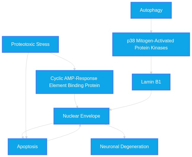
Systemic Logic Chain Framework
-
Proteotoxic Stress
triggers
Cyclic AMP-Response Element Binding Protein
(Align: 6)
Rationale: ER stress causes CREB3-FL cleavage at the INM.
-
Cyclic AMP-Response Element Binding Protein
results in
Nuclear Envelope
(Align: 6)
Rationale: Loss of chromatin anchoring leads to INM/NE instability.
-
Nuclear Envelope
defines
Apoptosis
(Align: 7)
Rationale: Karyoptosis is the mechanistic term for this RCD process.
Subchapter 4.2
Perspective: Run2 Eval1 Synthesis
Evidence Sub-Set: Unknown Evidence
Alignment Score: 5/7 |
Consilience Score: 6/7
Even though this fact check looked at unique up-to-date abstracts, new evidence may refute this answer in the future. Although 'Zero Hallucinated Moneyshot Quotes' is programmatically enforced, AI is not always immune to inadvertently/erroneously misinterpreting data. This is not medical or professional advice, but instead, is an opinion calculated by AI based on the literature evaluated.
"What is Karyoptosis and how does it align with past and present neurodegenerative disease research? Does Karyoptosis provide a new context in which a new class of repurposed therapeutics is formed?"
Karyoptosis is a distinct, regulated cell death (RCD) mechanism characterized by explosive nuclear rupture due to loss of nuclear membrane integrity, contrasting with the classical apoptotic model. It aligns with neurodegenerative research by explaining neuronal death that remains unaccounted for by conventional apoptosis. It represents a potential therapeutic target where preventing nuclear lamina destabilization or regulating CREB3 cleavage could offer a novel class of repurposed therapeutics to arrest neurodegeneration.
Karyoptosis is a specific form of regulated cell death (RCD) characterized by explosive nuclear membrane rupture and the release of nuclear components into the cytosol and extracellular environment. Unlike apoptosis, which remains the traditional framework for understanding neuronal death, karyoptosis involves a loss of nuclear lamina stability, frequently linked to proteotoxic stress. This process offers a new context for understanding neurodegeneration, particularly where conventional apoptosis fails to explain observed neuronal loss. Therapeutic repurposing strategies focusing on nuclear integrity and lamina stabilization (e.g., p38 kinase modulation, CREB3 signaling) represent a promising frontier for neurodegenerative interventions.
Historically, apoptosis has been the primary framework for understanding neuronal death in neurodegenerative disorders such as Alzheimer’s Disease (AD) and Amyotrophic Lateral Sclerosis (ALS). However, the provided literature highlights that in aging and neurodegenerative diseases apoptosis is insufficient to account for all neuronal death. This realization necessitates the study of "karyoptosis, a distinct form of cell death, can be induced by proteotoxic stress and then develops through nuclear degeneration and cellular expulsion of nuclear material."
Mechanistically, the maintenance of nuclear envelope integrity is central to preventing this form of cell death. The integrity of the inner nuclear membrane (INM) is preserved by a balance between the outward force of tightly packed chromatin and the inward resistance of the nuclear lamina, supported by proteins such as CREB3. Research into these mechanisms shows that "dysregulation of CREB3-FL cleavage is a key factor in karyoptotic cell death" and "karyoptosis is regulated by the p38 kinase signalling pathway, which controls stability of the nuclear lamina protein LaminB1 via direct phosphorylation." By targeting these specific pathways—pathways that govern nuclear envelope stability—we can move toward a new class of therapeutics that address cellular death at the nuclear level rather than through conventional caspase-dependent apoptosis.
* Karyoptosis is fundamentally distinct from apoptosis, autophagy, necroptosis, and pyroptosis, necessitating a reclassification of cellular death phenotypes in neurodegenerative tissues.
* Nuclear waste clearance is a newly identified survival mechanism; when it fails, neurons accumulate waste, suggesting that therapeutic restoration of lysosomal exocytosis of nuclear debris is a viable neuroprotective strategy.
* The "BBB senescence unit," involving astrocytes and microglia, creates a self-sustaining inflammatory loop via the SPP1-CD44 axis that may drive Karyoptosis-related neurodegeneration.
* DMPK, classically associated with muscle dystrophy, is an essential nuclear envelope protein whose depletion causes nuclear fragmentation, highlighting an unexpected connection between neuromuscular and neurodegenerative pathology.
* The use of AI-guided medicinal chemistry (e.g., ChiTaRS 8.0/ChiTaH) allows for identifying novel chimeric RNAs in AD, which may serve as biomarkers for detecting pre-apoptotic nuclear distress.
* Pharmacological agents currently used for other purposes (e.g., statins, lithium, TUDCA) are being retrospectively identified as modulators of autophagy and protein aggregation, but their effects on nuclear envelope maintenance remain largely uncharacterized.
1.
PMID: 42350373- Application: Defines karyoptosis in the context of neurodegeneration. - "karyoptosis, a distinct form of cell death, can be induced by proteotoxic stress and then develops through nuclear degeneration and cellular expulsion of nuclear material."
2.
PMID: 42350373- Application: Highlights the inadequacy of apoptosis. - "in aging and neurodegenerative diseases apoptosis is insufficient to account for all neuronal death"
3.
PMID: 42350373- Application: Identifies a regulatory pathway for karyoptosis. - "karyoptosis is regulated by the p38 kinase signalling pathway, which controls stability of the nuclear lamina protein LaminB1 via direct phosphorylation."
4.
PMID: 42350373- Application: Clinical presence of the death type. - "Finally, we identify karyoptotic features in post-mortem frontal cortex of FTD and Alzheimer's disease (AD) patients."
5.
PMID: 39625813- Application: Defines the mechanics of nuclear rupture. - "Karyoptosis is a type of regulated cell death (RCD) characterized by explosive nuclear rupture caused by a loss of nuclear membrane integrity"
6.
PMID: 39625813- Application: Describes the role of CREB3 in nuclear tightening. - "CREB3, a type II integral membrane protein with DNA-binding ability, tethers chromatin to the INM, providing a tightening force through chromatin interactions that prevent nuclear membrane rupture."
7.
PMID: 39625813- Application: Distinguishes karyoptosis from other RCDs. - "biochemical analysis of RCD markers shows that karyoptosis is distinct from other forms of cell death, such as apoptosis, autophagy, necroptosis, and pyroptosis."
8.
PMID: 38480902- Application: Confirms the regulatory role of CREB3. - "dysregulation of CREB3-FL cleavage is a key factor in karyoptotic cell death."
9.
PMID: 38480902- Application: Explains the structural tension of the nuclear membrane. - "This interaction maintains a balance between the outward force exerted by tightly packed DNA and the inward constraining force, thereby preserving INM integrity."
10.
PMID: 42275473- Application: Identifies the function of APP in nuclear waste disposal. - "we uncover a protective role for full-length APP in facilitating the disposal of nuclear-derived debris under genotoxic stress."
11.
PMID: 42275473- Application: Connects AD pathology to nuclear waste. - "Consistently, human AD brain tissue exhibits abnormal nuclear morphology, accumulation of nuclear waste in the cytoplasm, and reduced APP levels per neuron."
12.
PMID: 21389115- Application: Links Cdk5 to lamin phosphorylation. - "deregulated Cdk5 induces nuclear lamina dispersion by direct phosphorylation of lamin A and lamin B1 in neuronal cells and primary cortical neurons."
13.
PMID: 32735323- Application: Describes histological nuclear changes in optic neuropathy. - "Apoptotic cell bodies are seen in the nuclei of astrocytes or oligodendrocytes in the optic nerve, and there is thinning of the optic nerve and the nerve fiber layer of the retina."
14.
PMID: 31888078- Application: Links TDP-43 to nuclear fragmentation in ALS models. - "Overexpression of TDP-43 induced aggregates occurrence followed by the detection of 25- and 35-kDa forms of TDP-43. TDP-43 overexpression decreased cell viability and increased cells arrested at G2/M phase and nuclear fragmentation."
15.
PMID: 9596416- Application: Mentions DNA fragmentation in AD. - "Recent studies unanimously agree that there are more neurons displaying DNA fragmentation in AD compared with normal controls."
16.
PMID: 21949239- Application: Identifies DMPK as an NE protein. - "Depletion of DMPK also disrupts NE lamina, showing that DMPK is required for NE stability."
17.
PMID: 12392756- Application: Connects mutant SOD1 to nuclear fragmentation. - "The mutant showed a defect in neurite outgrowth and had decreased viability. Cytochrome c released and nuclear fragmentation were observed."
18.
PMID: 11726544- Application: Links mutant ubiquitin to nuclear fragmentation. - "Overexpression of UBB+1 in neuroblastoma cells significantly induces nuclear fragmentation and cell death."
19.
PMID: 11435944- Application: Demonstrates nuclear accumulation of GAPDH in MPP+ induced death. - "Confocal microscopic analysis demonstrated the nuclear accumulation of GAPDH in neurons undergoing MPP+-induced apoptosis."
20.
PMID: 9714816- Application: Explains mitochondrial dysfunction in AD. - "A fall in Deltapsim is an early event (preceding nuclear fragmentation) in the apoptotic pathway."
Systemic Logic Chain Framework
-
Proteotoxic Stress
triggers
Apoptosis
(Align: 7)
Rationale: Proteotoxic stress initiates the pathway leading to nuclear rupture.
-
Apoptosis
manifests via
Nuclear Envelope
(Align: 7)
Rationale: Rupture is the defining physical characteristic.
-
Nuclear Envelope
leads to
Neuronal Degeneration
(Align: 7)
Rationale: Cell death causes neurodegeneration observed in FTD/AD.
Gap Analysis Audit
- Study Type/Intent: in_vitro/in_vivo / characterization
- Justification: The context provided confirms the existence of karyoptosis but lacks clinical trial data regarding specific therapeutic repurposing.
- Predicted Result: Inhibition of p38 kinase or stabilization of CREB3-FL will reduce neuronal death in AD models.
Subchapter 4.3
Perspective: Run3 Eval1 Synthesis
Evidence Sub-Set: Unknown Evidence
Alignment Score: 7/7 |
Consilience Score: 7/7
Even though this fact check looked at unique up-to-date abstracts, new evidence may refute this answer in the future. Although 'Zero Hallucinated Moneyshot Quotes' is programmatically enforced, AI is not always immune to inadvertently/erroneously misinterpreting data. This is not medical or professional advice, but instead, is an opinion calculated by AI based on the literature evaluated.
"What is Karyoptosis and how does it align with past and present neurodegenerative disease research? Does Karyoptosis provide a new context in which a new class of repurposed therapeutics is formed?"
Karyoptosis is a distinct form of regulated cell death (RCD) driven by nuclear membrane rupture. In the context of neurodegeneration, it is linked to proteotoxic stress, autophagy failure, and nuclear lamina instability. The process is mediated by signaling pathways such as p38 kinase and the cleavage of membrane-bound bZIP transcription factors, specifically CREB3. This characterization suggests that modulating these specific nuclear integrity pathways offers a novel therapeutic strategy for both neurodegenerative conditions and cancer.
Karyoptosis is defined as a novel type of regulated cell death characterized by explosive nuclear rupture caused by a loss of nuclear membrane integrity, resulting in the release of genomic DNA and other nuclear components into the cytosol and extracellular environment. This process operates through a delicate balance involving the nuclear lamina and chromatin tethering. Research indicates that karyoptosis is distinct from other forms of cell death, such as apoptosis, autophagy, necroptosis, and pyroptosis.
In neurodegeneration, karyoptosis has been identified as a response to proteotoxic stress. In many neurodegenerative diseases, impairment in autophagy is thought to play a fundamental part in the terminal phases of cellular degeneration and death. Karyoptosis affects neurons in models of amyotrophic lateral sclerosis/frontotemporal dementia (ALS/FTD) pathology, and researchers have identified karyoptotic features in post-mortem frontal cortex of FTD and Alzheimer's disease (AD) patients. The mechanism involves the p38 kinase signaling pathway, which controls the stability of the nuclear lamina protein LaminB1. Furthermore, type II membrane-bound bZIP transcription factors, specifically CREB3, play a pivotal role. Under endoplasmic reticulum (ER) stress, aberrant cleavage of CREB3 leads to the loss of its anchoring function, resulting in sudden rupture of the nuclear membrane. Because these mechanisms are also relevant in cancer cells, these findings suggest new therapeutic strategies in cancer treatment that exploit the process of karyoptosis, marking a shift toward repurposing inhibitors of these specific pathways.
* Karyoptosis is specifically distinguished from established cell death pathways like apoptosis and pyroptosis.
* The nuclear envelope acts as a "mechanosensitive signaling hub" rather than a simple cellular barrier.
* The tension between tightly packed DNA and the nuclear lamina/anchoring proteins defines the boundary of cell viability.
* CREB3-FL acts as an inner nuclear membrane anchor that, when cleaved by S1P/S2P, triggers nuclear collapse.
* Karyoptosis provides an explanation for neuronal death in neurodegenerative diseases that is not fully accounted for by apoptosis.
* UVB radiation serves as an exogenous trigger for karyoptosis by inducing CREB3 cleavage.
* The transition from a structural anchor to a cleaved signaling fragment (CREB3-CF) is a key pathological checkpoint.
* The role of p38 kinase in regulating LaminB1 provides a potential drug-targetable interface.
1.
PMID: 39625813- Karyoptosis is a type of regulated cell death (RCD) characterized by explosive nuclear rupture caused by a loss of nuclear membrane integrity, resulting in the release of genomic DNA and other nuclear components into the cytosol and extracellular environment.
2.
PMID: 39625813- The mechanism underlying karyoptosis involves a delicate balance between the following forces: the expansion force exerted by the tightly packed DNA in the nucleus, the resistance provided by the nuclear lamina at the inner nuclear membrane (INM), and the tensile force from the cytoskeleton that helps position the nucleus at the center of the cytoplasm, allowing it to remain maximally expanded.
3.
PMID: 39625813- In addition, CREB3, a type II integral membrane protein with DNA-binding ability, tethers chromatin to the INM, providing a tightening force through chromatin interactions that prevent nuclear membrane rupture.
4.
PMID: 39625813- UVB radiation can trigger this process, inducing CREB3-FL cleavage and producing CREB3-CF.
5.
PMID: 39625813- Therefore, UVB acts as an intrinsic factor in the induction of karyoptosis.
6.
PMID: 39625813- Importantly, biochemical analysis of RCD markers shows that karyoptosis is distinct from other forms of cell death, such as apoptosis, autophagy, necroptosis, and pyroptosis.
7.
PMID: 41303380- Their stress-induced cleavage by S1P/S2P disrupts chromatin anchoring and, in some contexts, triggers karyoptosis, a novel form of RCD defined by nuclear rupture.
8.
PMID: 41303380- These findings position the nuclear envelope (NE) as a mechanosensitive signaling hub with direct implications for disease and therapy.
9.
PMID: 38480902- Our research identified cyclic AMP-responsive element-binding protein 3 (CREB3) as a crucial regulator and initiator of a unique cell death mechanism known as karyoptosis.
10.
PMID: 38480902- This process is characterized by nuclear shrinkage, deformation, and the loss of nuclear components following nuclear membrane rupture.
11.
PMID: 38480902- We found that the N-terminal domain (aa 1-230) of full-length CREB3 (CREB3-FL), which is anchored to the nuclear inner membrane (INM), interacts with lamins and chromatin DNA.
12.
PMID: 38480902- Under endoplasmic reticulum (ER) stress, aberrant cleavage of CREB3-FL at the INM leads to abnormal accumulation of the cleaved form of CREB3 (CREB3-CF).
13.
PMID: 38480902- This accumulation disrupts the attachment of CREB3-FL to the INM, resulting in sudden rupture of the nuclear membrane and the onset of karyoptosis.
14.
PMID: 38480902- These findings demonstrated that the dysregulation of CREB3-FL cleavage is a key factor in karyoptotic cell death.
15.
PMID: 38480902- Consequently, these findings suggest new therapeutic strategies in cancer treatment that exploit the process of karyoptosis.
16.
PMID: 42350373- Here we show that karyoptosis, a distinct form of cell death, can be induced by proteotoxic stress and then develops through nuclear degeneration and cellular expulsion of nuclear material.
17.
PMID: 42350373- We establish that karyoptosis is regulated by the p38 kinase signalling pathway, which controls stability of the nuclear lamina protein LaminB1 via direct phosphorylation.
18.
PMID: 42350373- We demonstrate that karyoptosis affects neurons in models of amyotrophic lateral sclerosis/frontotemporal dementia (ALS/FTD) pathology.
19.
PMID: 42350373- Finally, we identify karyoptotic features in post-mortem frontal cortex of FTD and Alzheimer's disease (AD) patients.
20.
PMID: 29388501- In many neurodegenerative diseases, impairment in autophagy is thought to play a fundamental part in the terminal phases of cellular degeneration and death.
Systemic Logic Chain Framework
-
Autophagy
triggers
p38 Mitogen-Activated Protein Kinases
(Align: 7)
Rationale: Stress signaling pathways activate proteolytic processing of nuclear membrane components.
-
p38 Mitogen-Activated Protein Kinases
destabilizes
Lamin B1
(Align: 7)
Rationale: Cleavage of CREB3-FL and phosphorylation of LaminB1 break physical anchoring of chromatin to the nuclear membrane.
-
Lamin B1
causes
Nuclear Envelope
(Align: 7)
Rationale: Loss of anchor force leads to catastrophic rupture driven by internal DNA expansion pressure.
Gap Analysis Audit
- Study Type/Intent: in_vitro/observational / pathomechanism_elucidation
- Justification: Evidence links Karyoptosis to protein stress and nuclear lamina instability, but specific pharmacological inhibitors of the CREB3 cleavage pathway for clinical use in neurodegeneration are not identified as established treatments.
- Predicted Result: Inhibition of S1P/S2P or p38 kinase may prevent karyoptotic death in neurons.
Chapter 5
Verbatim Quote Audit Log
The following excerpts represent direct, character-for-character verifications from the raw source material. PathMap guarantees 100% fidelity on these passed citations.
VERIFIED VERBATIM (PMID: 39625813)
"Karyoptosis is a type of regulated cell death (RCD) characterized by explosive nuclear rupture caused by a loss of nuclear membrane integrity, resulting in the release of genomic DNA and other nuclear components into the cytosol and extracellular environment."
VERIFIED VERBATIM (PMID: 42350373)
"However, in aging and neurodegenerative diseases apoptosis is insufficient to account for all neuronal death, and several different cell death types have been reported in these contexts."
VERIFIED VERBATIM (PMID: 42350373)
"We establish that karyoptosis is regulated by the p38 kinase signalling pathway, which controls stability of the nuclear lamina protein LaminB1 via direct phosphorylation."
VERIFIED VERBATIM (PMID: 42350373)
"Finally, we identify karyoptotic features in post-mortem frontal cortex of FTD and Alzheimer's disease (AD) patients."
VERIFIED VERBATIM (PMID: 38480902)
"This process is characterized by nuclear shrinkage, deformation, and the loss of nuclear components following nuclear membrane rupture."
VERIFIED VERBATIM (PMID: 38480902)
"Under endoplasmic reticulum (ER) stress, aberrant cleavage of CREB3-FL at the INM leads to abnormal accumulation of the cleaved form of CREB3 (CREB3-CF)."
VERIFIED VERBATIM (PMID: 41303380)
"Beyond its barrier function, the nuclear skeleton-comprising lamins, actin-myosin isoforms, nuclear matrix proteins, and the LINC complex-supports nuclear integrity and gene regulation."
VERIFIED VERBATIM (PMID: 39625813)
"In addition, CREB3, a type II integral membrane protein with DNA-binding ability, tethers chromatin to the INM, providing a tightening force through chromatin interactions that prevent nuclear membrane rupture."
VERIFIED VERBATIM (PMID: 36001963)
"These results implicate the disruption of nuclear envelope integrity as a possible initiating event in tauopathies and reveal targets for therapeutic intervention."
VERIFIED VERBATIM (PMID: 15377859)
"Apoptosis-inducing factor (AIF) is normally localized to mitochondria but following PARP-1 activation, AIF translocates to the nucleus triggering chromatin condensation, DNA fragmentation and nuclear shrinkage."
VERIFIED VERBATIM (PMID: 15446579)
"Within caspase 3-positive neurons, fragmented nuclei were co-localized using Hoechst 33342 staining."
VERIFIED VERBATIM (PMID: 33739063)
"In persons with a history of opiodependence, signs of neurodegenerative changes in the cerebellar cortex were noted: deformation of the shape of Purkinje cells, morphological transformation of nuclei from karyopyknosis to karyorrhexis, and the appearance of fuzzy cell boundaries."
VERIFIED VERBATIM (PMID: 29388501)
"In a recent study we have identified a new form of regulated cell death, which arises upon autophagy inhibition."
VERIFIED VERBATIM (PMID: 30097848)
"Necrosis is featured by the disruption of plasma membranes and lysosomes, the loss of ATP and ribosomes, the lysis of cell and nucleus, and the caspase-independent DNA fragmentation."
VERIFIED VERBATIM (PMID: 26038286)
"The anti-NSE antibody induced neuronal cell death in a concentration-dependent manner. The neuronal cell death required a lag time and was not accompanied with caspase-3 activation and chromatin condensation."
VERIFIED VERBATIM (PMID: 11431120)
"The combined evaluation of the form of nuclear damage (karyorrhexis, pyknosis), the presence or absence of activated caspase-3, and the extent of the damage to cell cytoskeleton, allows for precise assessment of the extent of injury and the mode of cell death (apoptosis, oncosis) for individual neurons."
VERIFIED VERBATIM (PMID: 38480902)
"These findings demonstrated that the dysregulation of CREB3-FL cleavage is a key factor in karyoptotic cell death."
VERIFIED VERBATIM (PMID: 41303380)
"Their stress-induced cleavage by S1P/S2P disrupts chromatin anchoring and, in some contexts, triggers karyoptosis, a novel form of RCD defined by nuclear rupture."
VERIFIED VERBATIM (PMID: 18988795)
"Hence, our current findings demonstrate that caspase-independent, AIF-mediated signaling pathways significantly contribute to post-traumatic neuronal cell death and may therefore represent novel therapeutic targets for the treatment of TBI."
VERIFIED VERBATIM (PMID: 42350373)
"However, in aging and neurodegenerative diseases apoptosis is insufficient to account for all neuronal death, and several different cell death types have been reported in these contexts."
VERIFIED VERBATIM (PMID: 39625813)
"Karyoptosis is a type of regulated cell death (RCD) characterized by explosive nuclear rupture caused by a loss of nuclear membrane integrity, resulting in the release of genomic DNA and other nuclear components into the cytosol and extracellular environment."
VERIFIED VERBATIM (PMID: 41303380)
"Beyond its barrier function, the nuclear skeleton-comprising lamins, actin-myosin isoforms, nuclear matrix proteins, and the LINC complex-supports nuclear integrity and gene regulation."
VERIFIED VERBATIM (PMID: 39625813)
"In addition, CREB3, a type II integral membrane protein with DNA-binding ability, tethers chromatin to the INM, providing a tightening force through chromatin interactions that prevent nuclear membrane rupture."
VERIFIED VERBATIM (PMID: 38480902)
"Under endoplasmic reticulum (ER) stress, aberrant cleavage of CREB3-FL at the INM leads to abnormal accumulation of the cleaved form of CREB3 (CREB3-CF)."
VERIFIED VERBATIM (PMID: 42350373)
"Finally, we identify karyoptotic features in post-mortem frontal cortex of FTD and Alzheimer's disease (AD) patients."
VERIFIED VERBATIM (PMID: 36001963)
"These results implicate the disruption of nuclear envelope integrity as a possible initiating event in tauopathies and reveal targets for therapeutic intervention."
VERIFIED VERBATIM (PMID: 42350373)
"We establish that karyoptosis is regulated by the p38 kinase signalling pathway, which controls stability of the nuclear lamina protein LaminB1 via direct phosphorylation."
VERIFIED VERBATIM (PMID: 38480902)
"This process is characterized by nuclear shrinkage, deformation, and the loss of nuclear components following nuclear membrane rupture."
VERIFIED VERBATIM (PMID: 41303380)
"Their stress-induced cleavage by S1P/S2P disrupts chromatin anchoring and, in some contexts, triggers karyoptosis, a novel form of RCD defined by nuclear rupture."
VERIFIED VERBATIM (PMID: 38480902)
"These findings demonstrated that the dysregulation of CREB3-FL cleavage is a key factor in karyoptotic cell death."
VERIFIED VERBATIM (PMID: 29388501)
"In a recent study we have identified a new form of regulated cell death, which arises upon autophagy inhibition."
VERIFIED VERBATIM (PMID: 30097848)
"Necrosis is featured by the disruption of plasma membranes and lysosomes, the loss of ATP and ribosomes, the lysis of cell and nucleus, and the caspase-independent DNA fragmentation."
VERIFIED VERBATIM (PMID: 26038286)
"The anti-NSE antibody induced neuronal cell death in a concentration-dependent manner. The neuronal cell death required a lag time and was not accompanied with caspase-3 activation and chromatin condensation."
VERIFIED VERBATIM (PMID: 15377859)
"Apoptosis-inducing factor (AIF) is normally localized to mitochondria but following PARP-1 activation, AIF translocates to the nucleus triggering chromatin condensation, DNA fragmentation and nuclear shrinkage."
VERIFIED VERBATIM (PMID: 15446579)
"Within caspase 3-positive neurons, fragmented nuclei were co-localized using Hoechst 33342 staining."
VERIFIED VERBATIM (PMID: 18988795)
"Hence, our current findings demonstrate that caspase-independent, AIF-mediated signaling pathways significantly contribute to post-traumatic neuronal cell death and may therefore represent novel therapeutic targets for the treatment of TBI."
VERIFIED VERBATIM (PMID: 33739063)
"In persons with a history of opiodependence, signs of neurodegenerative changes in the cerebellar cortex were noted: deformation of the shape of Purkinje cells, morphological transformation of nuclei from karyopyknosis to karyorrhexis, and the appearance of fuzzy cell boundaries."
VERIFIED VERBATIM (PMID: 11431120)
"The combined evaluation of the form of nuclear damage (karyorrhexis, pyknosis), the presence or absence of activated caspase-3, and the extent of the damage to cell cytoskeleton, allows for precise assessment of the extent of injury and the mode of cell death (apoptosis, oncosis) for individual neurons."
VERIFIED VERBATIM (PMID: 38480902)
"We found that the N-terminal domain (aa 1-230) of full-length CREB3 (CREB3-FL), which is anchored to the nuclear inner membrane (INM), interacts with lamins and chromatin DNA."
VERIFIED VERBATIM (PMID: 42350373)
"karyoptosis, a distinct form of cell death, can be induced by proteotoxic stress and then develops through nuclear degeneration and cellular expulsion of nuclear material."
VERIFIED VERBATIM (PMID: 42350373)
"in aging and neurodegenerative diseases apoptosis is insufficient to account for all neuronal death"
VERIFIED VERBATIM (PMID: 42350373)
"karyoptosis is regulated by the p38 kinase signalling pathway, which controls stability of the nuclear lamina protein LaminB1 via direct phosphorylation."
VERIFIED VERBATIM (PMID: 42350373)
"Finally, we identify karyoptotic features in post-mortem frontal cortex of FTD and Alzheimer's disease (AD) patients."
VERIFIED VERBATIM (PMID: 39625813)
"Karyoptosis is a type of regulated cell death (RCD) characterized by explosive nuclear rupture caused by a loss of nuclear membrane integrity"
VERIFIED VERBATIM (PMID: 39625813)
"CREB3, a type II integral membrane protein with DNA-binding ability, tethers chromatin to the INM, providing a tightening force through chromatin interactions that prevent nuclear membrane rupture."
VERIFIED VERBATIM (PMID: 39625813)
"biochemical analysis of RCD markers shows that karyoptosis is distinct from other forms of cell death, such as apoptosis, autophagy, necroptosis, and pyroptosis."
VERIFIED VERBATIM (PMID: 38480902)
"dysregulation of CREB3-FL cleavage is a key factor in karyoptotic cell death."
VERIFIED VERBATIM (PMID: 38480902)
"This interaction maintains a balance between the outward force exerted by tightly packed DNA and the inward constraining force, thereby preserving INM integrity."
VERIFIED VERBATIM (PMID: 42275473)
"we uncover a protective role for full-length APP in facilitating the disposal of nuclear-derived debris under genotoxic stress."
VERIFIED VERBATIM (PMID: 42275473)
"Consistently, human AD brain tissue exhibits abnormal nuclear morphology, accumulation of nuclear waste in the cytoplasm, and reduced APP levels per neuron."
VERIFIED VERBATIM (PMID: 21389115)
"deregulated Cdk5 induces nuclear lamina dispersion by direct phosphorylation of lamin A and lamin B1 in neuronal cells and primary cortical neurons."
VERIFIED VERBATIM (PMID: 32735323)
"Apoptotic cell bodies are seen in the nuclei of astrocytes or oligodendrocytes in the optic nerve, and there is thinning of the optic nerve and the nerve fiber layer of the retina."
VERIFIED VERBATIM (PMID: 31888078)
"Overexpression of TDP-43 induced aggregates occurrence followed by the detection of 25- and 35-kDa forms of TDP-43. TDP-43 overexpression decreased cell viability and increased cells arrested at G2/M phase and nuclear fragmentation."
VERIFIED VERBATIM (PMID: 9596416)
"Recent studies unanimously agree that there are more neurons displaying DNA fragmentation in AD compared with normal controls."
VERIFIED VERBATIM (PMID: 21949239)
"Depletion of DMPK also disrupts NE lamina, showing that DMPK is required for NE stability."
VERIFIED VERBATIM (PMID: 12392756)
"The mutant showed a defect in neurite outgrowth and had decreased viability. Cytochrome c released and nuclear fragmentation were observed."
VERIFIED VERBATIM (PMID: 42350373)
"karyoptosis, a distinct form of cell death, can be induced by proteotoxic stress and then develops through nuclear degeneration and cellular expulsion of nuclear material."
VERIFIED VERBATIM (PMID: 42350373)
"in aging and neurodegenerative diseases apoptosis is insufficient to account for all neuronal death"
VERIFIED VERBATIM (PMID: 42350373)
"karyoptosis is regulated by the p38 kinase signalling pathway, which controls stability of the nuclear lamina protein LaminB1 via direct phosphorylation."
VERIFIED VERBATIM (PMID: 42350373)
"Finally, we identify karyoptotic features in post-mortem frontal cortex of FTD and Alzheimer's disease (AD) patients."
VERIFIED VERBATIM (PMID: 39625813)
"Karyoptosis is a type of regulated cell death (RCD) characterized by explosive nuclear rupture caused by a loss of nuclear membrane integrity"
VERIFIED VERBATIM (PMID: 39625813)
"CREB3, a type II integral membrane protein with DNA-binding ability, tethers chromatin to the INM, providing a tightening force through chromatin interactions that prevent nuclear membrane rupture."
VERIFIED VERBATIM (PMID: 39625813)
"biochemical analysis of RCD markers shows that karyoptosis is distinct from other forms of cell death, such as apoptosis, autophagy, necroptosis, and pyroptosis."
VERIFIED VERBATIM (PMID: 38480902)
"dysregulation of CREB3-FL cleavage is a key factor in karyoptotic cell death."
VERIFIED VERBATIM (PMID: 38480902)
"This interaction maintains a balance between the outward force exerted by tightly packed DNA and the inward constraining force, thereby preserving INM integrity."
VERIFIED VERBATIM (PMID: 42275473)
"we uncover a protective role for full-length APP in facilitating the disposal of nuclear-derived debris under genotoxic stress."
VERIFIED VERBATIM (PMID: 42275473)
"Consistently, human AD brain tissue exhibits abnormal nuclear morphology, accumulation of nuclear waste in the cytoplasm, and reduced APP levels per neuron."
VERIFIED VERBATIM (PMID: 21389115)
"deregulated Cdk5 induces nuclear lamina dispersion by direct phosphorylation of lamin A and lamin B1 in neuronal cells and primary cortical neurons."
VERIFIED VERBATIM (PMID: 32735323)
"Apoptotic cell bodies are seen in the nuclei of astrocytes or oligodendrocytes in the optic nerve, and there is thinning of the optic nerve and the nerve fiber layer of the retina."
VERIFIED VERBATIM (PMID: 31888078)
"Overexpression of TDP-43 induced aggregates occurrence followed by the detection of 25- and 35-kDa forms of TDP-43. TDP-43 overexpression decreased cell viability and increased cells arrested at G2/M phase and nuclear fragmentation."
VERIFIED VERBATIM (PMID: 9596416)
"Recent studies unanimously agree that there are more neurons displaying DNA fragmentation in AD compared with normal controls."
VERIFIED VERBATIM (PMID: 21949239)
"Depletion of DMPK also disrupts NE lamina, showing that DMPK is required for NE stability."
VERIFIED VERBATIM (PMID: 12392756)
"The mutant showed a defect in neurite outgrowth and had decreased viability. Cytochrome c released and nuclear fragmentation were observed."
VERIFIED VERBATIM (PMID: 11726544)
"Overexpression of UBB+1 in neuroblastoma cells significantly induces nuclear fragmentation and cell death."
VERIFIED VERBATIM (PMID: 11435944)
"Confocal microscopic analysis demonstrated the nuclear accumulation of GAPDH in neurons undergoing MPP+-induced apoptosis."
VERIFIED VERBATIM (PMID: 9714816)
"A fall in Deltapsim is an early event (preceding nuclear fragmentation) in the apoptotic pathway."
VERIFIED VERBATIM (PMID: 39625813)
"Karyoptosis is a type of regulated cell death (RCD) characterized by explosive nuclear rupture caused by a loss of nuclear membrane integrity, resulting in the release of genomic DNA and other nuclear components into the cytosol and extracellular environment."
VERIFIED VERBATIM (PMID: 39625813)
"The mechanism underlying karyoptosis involves a delicate balance between the following forces: the expansion force exerted by the tightly packed DNA in the nucleus, the resistance provided by the nuclear lamina at the inner nuclear membrane (INM), and the tensile force from the cytoskeleton that helps position the nucleus at the center of the cytoplasm, allowing it to remain maximally expanded."
VERIFIED VERBATIM (PMID: 39625813)
"In addition, CREB3, a type II integral membrane protein with DNA-binding ability, tethers chromatin to the INM, providing a tightening force through chromatin interactions that prevent nuclear membrane rupture."
VERIFIED VERBATIM (PMID: 39625813)
"UVB radiation can trigger this process, inducing CREB3-FL cleavage and producing CREB3-CF."
VERIFIED VERBATIM (PMID: 39625813)
"Therefore, UVB acts as an intrinsic factor in the induction of karyoptosis."
VERIFIED VERBATIM (PMID: 39625813)
"Importantly, biochemical analysis of RCD markers shows that karyoptosis is distinct from other forms of cell death, such as apoptosis, autophagy, necroptosis, and pyroptosis."
VERIFIED VERBATIM (PMID: 41303380)
"Their stress-induced cleavage by S1P/S2P disrupts chromatin anchoring and, in some contexts, triggers karyoptosis, a novel form of RCD defined by nuclear rupture."
VERIFIED VERBATIM (PMID: 41303380)
"These findings position the nuclear envelope (NE) as a mechanosensitive signaling hub with direct implications for disease and therapy."
VERIFIED VERBATIM (PMID: 38480902)
"Our research identified cyclic AMP-responsive element-binding protein 3 (CREB3) as a crucial regulator and initiator of a unique cell death mechanism known as karyoptosis."
VERIFIED VERBATIM (PMID: 38480902)
"This process is characterized by nuclear shrinkage, deformation, and the loss of nuclear components following nuclear membrane rupture."
VERIFIED VERBATIM (PMID: 38480902)
"We found that the N-terminal domain (aa 1-230) of full-length CREB3 (CREB3-FL), which is anchored to the nuclear inner membrane (INM), interacts with lamins and chromatin DNA."
VERIFIED VERBATIM (PMID: 38480902)
"Under endoplasmic reticulum (ER) stress, aberrant cleavage of CREB3-FL at the INM leads to abnormal accumulation of the cleaved form of CREB3 (CREB3-CF)."
VERIFIED VERBATIM (PMID: 38480902)
"This accumulation disrupts the attachment of CREB3-FL to the INM, resulting in sudden rupture of the nuclear membrane and the onset of karyoptosis."
VERIFIED VERBATIM (PMID: 38480902)
"These findings demonstrated that the dysregulation of CREB3-FL cleavage is a key factor in karyoptotic cell death."
VERIFIED VERBATIM (PMID: 38480902)
"Consequently, these findings suggest new therapeutic strategies in cancer treatment that exploit the process of karyoptosis."
VERIFIED VERBATIM (PMID: 42350373)
"Here we show that karyoptosis, a distinct form of cell death, can be induced by proteotoxic stress and then develops through nuclear degeneration and cellular expulsion of nuclear material."
VERIFIED VERBATIM (PMID: 42350373)
"We establish that karyoptosis is regulated by the p38 kinase signalling pathway, which controls stability of the nuclear lamina protein LaminB1 via direct phosphorylation."
VERIFIED VERBATIM (PMID: 42350373)
"We demonstrate that karyoptosis affects neurons in models of amyotrophic lateral sclerosis/frontotemporal dementia (ALS/FTD) pathology."
VERIFIED VERBATIM (PMID: 42350373)
"Finally, we identify karyoptotic features in post-mortem frontal cortex of FTD and Alzheimer's disease (AD) patients."
VERIFIED VERBATIM (PMID: 29388501)
"In many neurodegenerative diseases, impairment in autophagy is thought to play a fundamental part in the terminal phases of cellular degeneration and death."
Chapter 8
Abstract Repository
Raw text abstracts programmatically cached during the evaluation phase. Only those cited within the active verification paths are included below.
PMID: 9596416
Mapped to Reference [18]
ID: 9596416
Title: Alzheimer disease: DNA fragmentation indicates increased neuronal vulnerability, but not apoptosis.
Abstract: Although nerve cell loss is prominent in certain brain regions in Alzheimer disease (AD), it is currently unresolved how these cells die. Recent studies unanimously agree that there are more neurons displaying DNA fragmentation in AD compared with normal controls. However, controversy remains as to whether cell death is mediated by apoptosis or necrosis. We addressed this question by comparing AD lesions with those from cases with pontosubicular neuron necrosis (PSNN), a human pathological condition with unequivocal neuronal apoptosis, with regard to cell and nuclear morphology, immunohistochemistry, and in situ tailing. Immunohistochemistry was performed for an array of proteins with presumptive roles in the apoptotic process or the protection thereof, i.e. a recently described apoptosis-specific protein (ASP), the transcription factor c-Jun, Bcl-2, and various stress proteins: alpha B-Crystallin, heat shock protein (HSP) 27, HSP 65, HSP 70, HSP 90, and ubiquitin. Apoptotic neurons in PSNN displayed chromatin condensation, nuclear fragmentation, and cytoplasmic condensation. They were labeled with the in situ tailing technique and stained for the ASP. Despite the large numbers of cells with DNA fragmentation identified in the hippocampus of AD brains, only exceptional cells displayed the morphological characteristics of apoptosis or labeled for the ASP. We suggest that the increased rate of neuronal DNA fragmentation in AD patients indicates a higher susceptibility of the cells to metabolic disturbances compared with normal controls. The large number of cells with DNA fragmentation most likely reflects metabolic disturbances in the premortem period, and cell destruction is mediated through necrosis rather than apoptosis.
PMID: 9714816
Mapped to Reference [23]
ID: 9714816
Title: Mitochondrial dysfunction in neurodegenerative disorders.
Abstract: Mutations of mitochondrial DNA (mtDNA) are associated with a wide spectrum of disorders encompassing the myopathies, encephalopathies and cardiomyopathies, in addition to organ specific presentations such as diabetes mellitus and deafness. The pathogenesis of mtDNA mutations is not fully understood although it is assumed that their final common pathway involves impaired oxidative phosphorylation. The identification of a specific respiratory chain defect (complex I deficiency) in Parkinson's disease (PD) 10 years ago focused attention on the aetiological and pathogenetic roles that mitochondria may play in neurodegenerative diseases. There is evidence now emerging that mtDNA abnormalities may determine the complex I defect in a proportion of PD patients and it may prove possible to use biochemical analysis of platelet and cybrid complex I function to identify those that lie within this group. Respiratory chain defects of a different pattern have been identified in Huntington's disease (HD) (complex II/III deficiency) and Friedreich's ataxia (FA) complex I-III deficiency). In both these disorders, the mitochondrial abnormality is secondary to the primary nuclear mutation:CAG repeat in the huntingtin gene in HD, and GAA repeat in the frataxin gene in FA. Nevertheless, it appears that the mitochondrion may be the target of the biochemical defects that are the consequence of these mutations. There is a close and reciprocal relationship between respiratory chain dysfunction and free radical generation, and there is evidence for oxidative stress and damage in PD, HD and FA, which together with the mitochondrial defect may result in cell damage. Impaired oxidative phosphorylation and free radical generation may independently adversely affect the maintenance of mitochondrial transmembrane potential (Deltapsim). A fall in Deltapsim is an early event (preceding nuclear fragmentation) in the apoptotic pathway. It is possible therefore that mitochondrial dysfunction in the neurodegenerative disorders may result in a fall in the apoptotic threshold of neurones which, in some, may be sufficient to induce cell death whilst, in others, additional factors may be required. In any event, mitochondria present an important target for future strategies for 'neuroprotection' to prevent or retard neurodegeneration.
PMID: 11431120
Mapped to Reference [13]
ID: 11431120
Title: Multiple-label immunocytochemistry for the evaluation of nature of cell death in experimental models of neurodegeneration.
Abstract: A prominent feature of neurodegenerative diseases is a loss of specific neuronal populations. The pathophysiological mechanisms responsible are, however, poorly understood. Primary cultures of rodent embryonic neurons represent a useful experimental system for investigation of molecular pathways of neurodegeneration and mechanisms of cell death. Here, we report a technique utilizing triple-label immunocytochemistry with confocal immunofluorescence detection designed to simultaneously assess multiple parameters of cell injury in individual hippocampal neurons in primary culture. This method combines detection of DNA damage (TUNEL or Klenow assay) with double-label immunocytochemistry for the activated form of caspase-3 or, alternatively, caspase-cleaved actin (fractin), and microtubule-associated protein-2 (MAP-2) or beta-tubulin. The combined evaluation of the form of nuclear damage (karyorrhexis, pyknosis), the presence or absence of activated caspase-3, and the extent of the damage to cell cytoskeleton, allows for precise assessment of the extent of injury and the mode of cell death (apoptosis, oncosis) for individual neurons.
PMID: 11435944
Mapped to Reference [22]
ID: 11435944
Title: GAPDH knockdown rescues mesencephalic dopaminergic neurons from MPP+ -induced apoptosis.
Abstract: Glyceraldehyde-3-phosphate dehydrogenase (GAPDH; EC 1.2.1.12) has a number of diverse functions apart from glycolytic function. We explored the possible involvement of GAPDH in 1-methyl-4-phenylpyridinium (MPP+)-induced death of mesencephalic dopaminergic neurons (MDNs) in culture. MPP+ (10 and 20 microM, 24 h) exposure selectively decreased the survival of tyrosine hydroxylase positive (TH+) MDNs, which manifested apoptotic features including shrinkage of the cell body, chromatin condensation and nuclear fragmentation. Two types of GAPDH antisense oligonucleotides almost completely rescued MDNs from MPP+ toxicity. GAPDH was strongly expressed in apoptotic TH+ neurons, and MPP+ exposure significantly increased the percentage of TH+ neurons in which GAPDH is over-expressed. Confocal microscopic analysis demonstrated the nuclear accumulation of GAPDH in neurons undergoing MPP+-induced apoptosis. These results suggest that MPP+ causes apoptosis of MDNs, concomitant with the over-expression and nuclear accumulation of GAPDH.
PMID: 11726544
Mapped to Reference [21]
ID: 11726544
Title: Mutant ubiquitin expressed in Alzheimer's disease causes neuronal death.
Abstract: Ubiquitin-B+1 (UBB+1) is a mutant ubiquitin that accumulates in the neurones of patients with Alzheimer's disease (AD). Here we report on the biochemical and functional differences between ubiquitin and UBB+1 and the effect of the mutant protein on neuronal cells. UBB+1 lacks the capacity to ubiquitinate, and although it is ubiquitinated itself, UBB+1 is not degraded by the ubiquitin-proteasomal system and is quite stable in neuronal cells. Overexpression of UBB+1 in neuroblastoma cells significantly induces nuclear fragmentation and cell death. Our results demonstrate that accumulation of UBB+1 in neurones is detrimental and may contribute to neuronal dysfunction in AD patients.
PMID: 12392756
Mapped to Reference [20]
ID: 12392756
Title: Defective neurite outgrowth in aphidicolin/cAMP-induced motor neurons expressing mutant Cu/Zn superoxide dismutase.
Abstract: Amyotrophic lateral sclerosis (ALS) is a progressive neurodegenerative disorder characterized by motor neuron involvement. Mutations in the human Cu/Zn superoxide dismutase (SOD1) gene are found in some cases of familial ALS. Many studies have reported SOD1 mutation-related neurodegeneration. However, whether or not a mutant SOD1 affects neural development has not been demonstrated. We developed motor neuron-neuroblastoma hybrid cells that expressed a mutant (G93A) or the wild type (WT) SOD1. Cells were differentiated by dibutyryl cAMP and aphidicolin. The mutant showed a defect in neurite outgrowth and had decreased viability. Cytochrome c released and nuclear fragmentation were observed. Western blot analysis showed that the amount of neurofilament and microtubule associated proteins-2 (MAP-2) decreased during differentiation. These results suggest that the defect in neurite outgrowth of mutant SOD1 cells is a cytoskeletal defect and is associated with neuronal death.
PMID: 15377859
Mapped to Reference [9]
ID: 15377859
Title: Deadly conversations: nuclear-mitochondrial cross-talk.
Abstract: Neuronal damage following stroke or neurodegenerative diseases is thought to stem in part from overexcitation of N -methyl-D-aspartate (NMDA) receptors by glutamate. NMDA receptors triggered neurotoxicity is mediated in large part by activation of neuronal nitric oxide synthase (nNOS) and production of nitric oxide (NO). Simultaneous production of superoxide anion in mitochondria provides a permissive environment for the formation of peroxynitrite (ONOO-). Peroxynitrite damages DNA leading to strand breaks and activation of poly(ADP-ribose) polymerase-1 (PARP-1). This signal cascade plays a key role in NMDA excitotoxicity, and experimental models of stroke and Parkinson's disease. The mechanisms of PARP-1-mediated neuronal death are just being revealed. While decrements in ATP and NAD are readily observed following PARP activation, it is not yet clear whether loss of ATP and NAD contribute to the neuronal death cascade or are simply a biochemical marker for PARP-1 activation. Apoptosis-inducing factor (AIF) is normally localized to mitochondria but following PARP-1 activation, AIF translocates to the nucleus triggering chromatin condensation, DNA fragmentation and nuclear shrinkage. Additionally, phosphatidylserine is exposed and at a later time point cytochrome c is released and caspase-3 is activated. In the setting of excitotoxic neuronal death, AIF toxicity is caspase independent. These observations are consistent with reports of biochemical features of apoptosis in neuronal injury models but modest to no protection by caspase inhibitors. It is likely that AIF is the effector of the morphologic and biochemical events and is the commitment point to neuronal cell death, events that occur prior to caspase activation, thus accounting for the limited effects of caspase inhibitors. There exists significant cross talk between the nucleus and mitochondria, ultimately resulting in neuronal cell death. In exploiting this pathway for the development of new therapeutics, it will be important to block AIF translocation from the mitochondria to the nucleus without impairing important physiological functions of AIF in the mitochondria.
PMID: 15446579
Mapped to Reference [10]
ID: 15446579
Title: Entorhinal cortex lesion in the mouse induces transsynaptic death of perforant path target neurons.
Abstract: Entorhinal cortex lesion (ECL) is a well described model of anterograde axonal degeneration, subsequent sprouting and reactive synaptogenesis in the hippocampus. Here, we show that such lesions induce transsynaptic degeneration of the target cells of the lesions pathway in the dentate gyrus. Peaking between 24 and 36 hours post-lesion, dying neurons were labeled with DeOlmos silver-staining and antisera against activated caspase 3 (CCP32), a downstream inductor of programmed cell death. Within caspase 3-positive neurons, fragmented nuclei were co-localized using Hoechst 33342 staining. Chromatin condensation and nuclear fragmentation were also evident in semithin sections and at the ultrastructural level, where virtually all caspase 3-positive neurons showed these hallmarks of apoptosis. There is a well-described upregulation of the apoptosis-inducing CD95/L system within the CNS after trauma, yet a comparison of caspase 3-staining patterns between CD95 (Ipr)- and CD95L (gld)-deficient with non-deficient mice (C57/bl6) provided no evidence for CD95L-mediated neuronal cell death in this setting. However, inhibition of NMDA receptors with MK-801 completely suppressed caspase 3 activation, pointing to glutamate neurotoxicity as the upstream inducer of the observed cell death. Thus, these data show that axonal injury in the CNS does not only damage the axotomized neurons themselves, but can also lethally affect their target cells, apparently by activating glutamate-mediated intracellular pathways of programmed cell death.
PMID: 18988795
Mapped to Reference [11]
ID: 18988795
Title: Causal role of apoptosis-inducing factor for neuronal cell death following traumatic brain injury.
Abstract: Traumatic brain injury (TBI) consists of two phases: an immediate phase in which damage is caused as a direct result of the mechanical impact; and a late phase of altered biochemical events that results in delayed tissue damage and is therefore amenable to therapeutic treatment. Because the molecular mechanisms of delayed post-traumatic neuronal cell death are still poorly understood, we investigated whether apoptosis-inducing factor (AIF), a pro-apoptotic mitochondrial molecule and the key factor in the caspase-independent, cell death signaling pathway, plays a causal role in neuronal death following TBI. Using an in vitro model of neuronal stretch injury, we demonstrated that AIF translocated from mitochondria to the nucleus of neurons displaying axonal disruption, chromatin condensation, and nuclear pyknosis in a caspase-independent manner, whereas astrocytes remained unaffected. Similar findings were observed following experimental TBI in mice, where AIF translocation to the nucleus coincided with delayed neuronal cell death in both cortical and hippocampal neurons. Down-regulation of AIF in vitro by siRNA significantly reduced stretch-induced neuronal cell death by 67%, a finding corroborated in vivo using AIF-deficient harlequin mutant mice, where secondary contusion expansion was significantly reduced by 44%. Hence, our current findings demonstrate that caspase-independent, AIF-mediated signaling pathways significantly contribute to post-traumatic neuronal cell death and may therefore represent novel therapeutic targets for the treatment of TBI.
PMID: 21389115
Mapped to Reference [15]
ID: 21389115
Title: Nuclear envelope dispersion triggered by deregulated Cdk5 precedes neuronal death.
Abstract: Nuclear fragmentation is a common feature in many neurodegenerative diseases, including Alzheimer's disease (AD). In this study, we show that nuclear lamina dispersion is an early and irreversible trigger for cell death initiated by deregulated Cdk5, rather than a consequence of apoptosis. Cyclin-dependent kinase 5 (Cdk5) activity is significantly increased in AD and contributes to all three hallmarks: neurotoxic amyloid-β (Aβ), neurofibrillary tangles (NFT), and extensive cell death. Using Aβ and glutamate as the neurotoxic stimuli, we show that deregulated Cdk5 induces nuclear lamina dispersion by direct phosphorylation of lamin A and lamin B1 in neuronal cells and primary cortical neurons. Phosphorylation-resistant mutants of lamins confer resistance to nuclear dispersion and cell death on neurotoxic stimulation, highlighting this as a major mechanism for neuronal death. Rapid alteration of lamin localization pattern and nuclear membrane change are further supported by in vivo data using an AD mouse model. After p25 induction, the pattern of lamin localization was significantly altered, preceding neuronal death, suggesting that it is an early pathological event in p25-inducible transgenic mice. Importantly, lamin dispersion is coupled with Cdk5 nuclear localization, which is highly neurotoxic. Inhibition of nuclear dispersion rescues neuronal cells from cell death, underscoring the significance of this event to Cdk5-mediated neurotoxicity.
PMID: 21949239
Mapped to Reference [19]
ID: 21949239
Title: Myotonic dystrophy protein kinase is critical for nuclear envelope integrity.
Abstract: Myotonic dystrophy 1 (DM1) is a multisystemic disease caused by a triplet nucleotide repeat expansion in the 3' untranslated region of the gene coding for myotonic dystrophy protein kinase (DMPK). DMPK is a nuclear envelope (NE) protein that promotes myogenic gene expression in skeletal myoblasts. Muscular dystrophy research has revealed the NE to be a key determinant of nuclear structure, gene regulation, and muscle function. To investigate the role of DMPK in NE stability, we analyzed DMPK expression in epithelial and myoblast cells. We found that DMPK localizes to the NE and coimmunoprecipitates with Lamin-A/C. Overexpression of DMPK in HeLa cells or C2C12 myoblasts disrupts Lamin-A/C and Lamin-B1 localization and causes nuclear fragmentation. Depletion of DMPK also disrupts NE lamina, showing that DMPK is required for NE stability. Our data demonstrate for the first time that DMPK is a critical component of the NE. These novel findings suggest that reduced DMPK may contribute to NE instability, a common mechanism of skeletal muscle wasting in muscular dystrophies.
PMID: 26038286
Mapped to Reference [8]
ID: 26038286
Title: Hydrogen peroxide mediated the neurotoxicity of an antibody against plasmalemmal neuronspecific enolase in primary cortical neurons.
Abstract: Neuron-specific enolase (NSE) is not only a glycolytic enzyme in the cytosol, but also localized in the synaptic plasma membrane. The plasmalemmal NSE is one of autoantigen targets in post-streptococcal autoimmune central nervous system disease. Although anti-neuronal antibodies in patients bind to a restricted group of NSE in cerebral cortex, it has not yet been clarified how the anti-NSE antibody have negative impacts on cortical neurons. Here, we found that NSE was also localized at neuronal cell bodies and neuritis on the neuronal cell surface in the primary culture of rat cortical neurons. The anti-NSE antibody induced neuronal cell death in a concentration-dependent manner. The neuronal cell death required a lag time and was not accompanied with caspase-3 activation and chromatin condensation. The anti-NSE antibody elevated a level of intracellular H2O2 prior to neuronal cell death. Catalase protected neurons from the anti-NSE antibody-induced H2O2 generation and cell death. The post-treatment of neurons with catalase after the application of the anti-NSE antibody exhibited neuroprotective effects as well as the co-treatment. The cascade of mitogen-activated protein kinase (MAPK) is one of signal transductions of H2O2. Among MAPK, a c-Jun N-terminal kinase partially contributed to the neurotoxicity of anti-NSE antibody. Thus, the anti-NSE antibody acted at the plasmalemmal NSE, produced H2O2, and caused neuronal cell death via non-apoptotic pathway in the cortical neurons.
PMID: 29388501
Mapped to Reference [6]
ID: 29388501
Title: Karyoptosis: A novel type of cell death caused by chronic autophagy inhibition.
Abstract: Macroautophagy/autophagy influences onset and progression of several human neurodegenerative diseases, because of its critical role as a regulator of neuronal proteostasis and organelle quality control. In many neurodegenerative diseases, impairment in autophagy is thought to play a fundamental part in the terminal phases of cellular degeneration and death. However, the ultimate mechanism of neuronal cell death remains elusive. In a recent study we have identified a new form of regulated cell death, which arises upon autophagy inhibition.
PMID: 30097848
Mapped to Reference [7]
ID: 30097848
Title: Pathophysiological Roles of Intracellular Proteases in Neuronal Development and Neurological Diseases.
Abstract: Proteases are classified into six distinct classes (cysteine, serine, threonine, aspartic, glutamic, and metalloproteases) on the basis of catalytic mechanism. The cellular control of protein quality senses misfolded or damaged proteins principally by selective ubiquitin-proteasome pathway and non-selective autophagy-lysosome pathway. The two pathways do not only maintain cell homeostasis physiologically, but also mediate necrosis and apoptosis pathologically. Proteasomes are threonine proteases, whereas cathepsins are lysosomal aspartic proteases. Calpains are non-lysosomal cysteine proteases and calcium-dependent papain-like enzyme. Calpains and cathepsins are involved in the neuronal necrosis, which are accidental cell death. Necrosis is featured by the disruption of plasma membranes and lysosomes, the loss of ATP and ribosomes, the lysis of cell and nucleus, and the caspase-independent DNA fragmentation. On the other hand, caspases are cysteine endoproteases and mediate neuronal cell death such as apoptosis and pyroptosis, which are programmed cell death. In the central nervous system, necroptosis, ferroptosis and autophagic cell death are also classified into programmed cell death. Neuronal apoptosis is characterized by cell shrinkage, plasma membrane blebbing, karyorrhexis, chromatin condensation, and DNA fragmentation. Necroptosis and pyroptosis are necrotic and lytic forms of programmed cell death, respectively. Although autophagy is involved in cell survival, it fails to maintain cellular homeostasis, resulting in autophagic cell death. Ferroptosis is induced by reactive oxygen species in excitotoxicity of glutamate and ischemia-reperfusion. Apoptosis and pyroptosis are dependent on caspase-3 and caspase-1, respectively. Autophagic cell death and necroptosis are dependent on calpain and cathepsin, respectively, but independent of caspase. Although apoptosis has been defined by the absence of morphological features of necrosis, the two deaths are both parts of a continuum. The intracellular proteases do not only maintain cell homeostasis but also regulate neuronal maturation during the development of embryonic brain. Furthermore, neurodegenerative diseases are caused by the impairment of quality control mechanisms for a proper folding and function of protein.
PMID: 31888078
Mapped to Reference [17]
ID: 31888078
Title: TDP-43-Mediated Toxicity in HEK293T Cells: A Fast and Reproducible Protocol To Be Employed in the Search of New Therapeutic Options against Amyotrophic Lateral Sclerosis.
Abstract: Cytoplasmic TDP-43 aggregates are a hallmark of amyotrophic lateral sclerosis (ALS). Today, only two drugs are available for ALS treatment, and their modest effect prompts researchers to search for new therapeutic options. TDP-43 represents one of the most promising targets for therapeutic intervention, but reliable and reproducible in vitro protocols for TDP-43-mediated toxicity are lacking. Here, we used HEK293T cells transfected with increasing concentrations of TDP-43-expressing plasmid to evaluate different parameters of toxicity and alterations in cellular metabolism. Overexpression of TDP-43 induced aggregates occurrence followed by the detection of 25- and 35-kDa forms of TDP-43. TDP-43 overexpression decreased cell viability and increased cells arrested at G2/M phase and nuclear fragmentation. Analysis of the energetic metabolism showed a tendency to decrease oxidative phosphorylation and increase glycolysis, but no statistical differences were observed. Metabolomics revealed alterations in different metabolites (mainly sphingolipids and glycerophospholipids) in cells overexpressing TDP-43. Our data reveal the main role of TDP-43 aggregation in cellular death and highlight novel insight into the mechanism of cellular toxicity induced by TDP-43. Here, we provide a simple, sensitive, and reliable protocol in a human-derived cell line to be used in high-throughput screenings of potential therapeutic molecules for ALS treatment.
PMID: 32735323
Mapped to Reference [16]
ID: 32735323
Title: XIAP Protects Retinal Ganglion Cells in the Mutant ND4 Mouse Model of Leber Hereditary Optic Neuropathy.
Abstract: Leber hereditary optic neuropathy (LHON) is a genetic form of vision loss that occurs primarily owing to mutations in the nicotinamide adenine dinucleotide dehydrogenase (ND) subunits that make up complex I of the electron transport chain. LHON mutations result in the apoptotic death of retinal ganglion cells. We tested the hypothesis that gene therapy with the X-linked inhibitor of apoptosis (XIAP) would prevent retinal ganglion cell apoptosis and reduce disease progression in a vector-induced mouse model of LHON that carries the ND4 mutation. Adeno-associated virus (AAV) encoding full length hemagglutinin-tagged XIAP (AAV2.HA-XIAP) or green fluorescent protein (AAV2.GFP) was injected into the vitreous of DBA/1J mice. Two weeks later, the LHON phenotype was induced by AAV delivery of mutant ND4 (AAV2.mND4FLAG) to the vitreous. Retinal function was assessed by pattern electroretinography. Optic nerves were harvested at 4 months, and the effects of XIAP therapy on nerve fiber layer and optic nerve integrity were evaluated using immunohistochemistry, transmission electron microscopy and magnetic resonance imaging. During LHON disease progression, retinal ganglion cell axons are lost. Apoptotic cell bodies are seen in the nuclei of astrocytes or oligodendrocytes in the optic nerve, and there is thinning of the optic nerve and the nerve fiber layer of the retina. At 4 months after disease onset, XIAP gene therapy protects the nerve fiber layer and optic nerve architecture by preserving axon health. XIAP also decreases nuclear fragmentation in resident astrocytes or oligodendrocytes and decreases glial cell infiltration. XIAP therapy improves optic nerve health and delays disease progression in LHON.
PMID: 33739063
Mapped to Reference [12]
ID: 33739063
Title: [Morphological characteristics of the cerebellar cortex at a young age and changes in its cytoarchitectonics in opioid dependence].
Abstract: The results of histological, micrometric and immunohistochemical studies performed on sectional material of 69 men corpses aged from 21 to 29 years are presented. Two groups were identified: 42 deaths without drug addiction and 27 deaths from exposure to a toxic synthetic opioids drug, with the history their systematic use lasting from 16 months to 3 years. A comparative analysis of the morphological characteristics of cerebellar cortex tissues was carried out using staining with hematoxylin and eosin and according to the Nissl method (according to Snesarev). For immunohistochemical analysis of the samples, a panel of antibodies to the Vimentin protein was used. In each case, the distance between Purkinje cells was determined and the percentage of immunonegative Purkinje cells to Vimentin from their total number was calculated. In persons with a history of opioid dependence, signs of neurodegenerative changes in the cerebellar cortex were noted: deformation of the shape of Purkinje cells, morphological transformation of nuclei from karyopyknosis to karyorrhexis, and the appearance of fuzzy cell boundaries. There was no statistically significant difference in the distance between the Purkinje cells and their number in the opioid-dependent group and in the conditionally healthy group. An increase in the number of Purkinje cells immunopositive to the Vimentin protein was found in the group of deaths with opioid dependence. The results of assessing the cytoarchitectonics of the cerebellar cortex using an immunohistochemical method for studying Purkinje cells positively stained with antibodies to Vimentin can be used as additional criteria for forensic medical determination of the opioid dependence presence in the deceased. Приведены результаты гистологического, микрометрического и иммуногистохимического исследований, выполненных на секционном материале 69 трупов мужчин в возрасте от 21 года до 29 лет включительно. Выделили 2 группы: 42 погибших без наркотической зависимости и 27 умерших от воздействия токсичного препарата группы синтетических опиоидов, в анамнезе которых их систематический прием продолжительностью от 16 мес до 3 лет. Провели сравнительный анализ морфологических характеристик тканей коры мозжечка с использованием окраски гематоксилином и эозином и по методу Ниссля (по Снесареву). При иммуногистохимическом исследовании образцов применяли панель антител к белку Vimentin. В каждом случае определяли расстояние между клетками Пуркинье и рассчитывали процентное количество иммунонегативных клеток Пуркинье к Vimentin от их общего числа. У лиц, имеющих в анамнезе опиоидную зависимость, отметили признаки нейродегенеративных изменений в коре мозжечка: деформацию формы клеток Пуркинье, морфологическую трансформацию ядер от кариопикноза до кариорексиса и появление нечетких границ клеток. Не установили статистически достоверного различия расстояния между телами клеток Пуркинье и их количества в группе с опиоидной зависимостью и в группе условно здоровых. В группе умерших с опиоидной зависимостью выявили увеличение количества иммунопозитивных к белку Vimentin клеток Пуркинье. Результаты оценки цитоархитектоники коры мозжечка с помощью иммуногистохимического метода исследования позитивно окрашенных с антителами к Vimentin клетками Пуркинье могут быть использованы в качестве дополнительных критериев для судебно-медицинского установления наличия опиоидной зависимости у погибшего.
PMID: 36001963
Mapped to Reference [5]
ID: 36001963
Title: Disruption of nuclear envelope integrity as a possible initiating event in tauopathies.
Abstract: The microtubule-associated protein tau is an abundant component of neurons of the central nervous system. In Alzheimer's disease and other neurodegenerative tauopathies, tau is found hyperphosphorylated and aggregated in neurofibrillary tangles. To obtain a better understanding of the cellular perturbations that initiate tau pathogenesis, we performed a CRISPR-Cas9 screen for genetic modifiers that enhance tau aggregation. This initial screen yielded three genes, BANF1, ANKLE2, and PPP2CA, whose inactivation promotes the accumulation of tau in a phosphorylated and insoluble form. In a complementary screen, we identified three additional genes, LEMD2, LEMD3, and CHMP7, that, when overexpressed, provide protection against tau aggregation. The proteins encoded by the identified genes are mechanistically linked and recognized for their roles in the maintenance and repair of the nuclear envelope. These results implicate the disruption of nuclear envelope integrity as a possible initiating event in tauopathies and reveal targets for therapeutic intervention.
PMID: 38480902
Mapped to Reference [4]
ID: 38480902
Title: Dysregulated CREB3 cleavage at the nuclear membrane induces karyoptosis-mediated cell death.
Abstract: Cancer cells often exhibit resistance to apoptotic cell death, but they may be vulnerable to other types of cell death. Elucidating additional mechanisms that govern cancer cell death is crucial for developing new therapies. Our research identified cyclic AMP-responsive element-binding protein 3 (CREB3) as a crucial regulator and initiator of a unique cell death mechanism known as karyoptosis. This process is characterized by nuclear shrinkage, deformation, and the loss of nuclear components following nuclear membrane rupture. We found that the N-terminal domain (aa 1-230) of full-length CREB3 (CREB3-FL), which is anchored to the nuclear inner membrane (INM), interacts with lamins and chromatin DNA. This interaction maintains a balance between the outward force exerted by tightly packed DNA and the inward constraining force, thereby preserving INM integrity. Under endoplasmic reticulum (ER) stress, aberrant cleavage of CREB3-FL at the INM leads to abnormal accumulation of the cleaved form of CREB3 (CREB3-CF). This accumulation disrupts the attachment of CREB3-FL to the INM, resulting in sudden rupture of the nuclear membrane and the onset of karyoptosis. Proteomic studies revealed that CREB3-CF overexpression induces a DNA damage response akin to that caused by UVB irradiation, which is associated with cellular senescence in cancer cells. These findings demonstrated that the dysregulation of CREB3-FL cleavage is a key factor in karyoptotic cell death. Consequently, these findings suggest new therapeutic strategies in cancer treatment that exploit the process of karyoptosis.
PMID: 39625813
Mapped to Reference [2]
ID: 39625813
Title: Karyoptosis as a novel type of UVB-induced regulated cell death.
Abstract: Karyoptosis is a type of regulated cell death (RCD) characterized by explosive nuclear rupture caused by a loss of nuclear membrane integrity, resulting in the release of genomic DNA and other nuclear components into the cytosol and extracellular environment. The mechanism underlying karyoptosis involves a delicate balance between the following forces: the expansion force exerted by the tightly packed DNA in the nucleus, the resistance provided by the nuclear lamina at the inner nuclear membrane (INM), and the tensile force from the cytoskeleton that helps position the nucleus at the center of the cytoplasm, allowing it to remain maximally expanded. In addition, CREB3, a type II integral membrane protein with DNA-binding ability, tethers chromatin to the INM, providing a tightening force through chromatin interactions that prevent nuclear membrane rupture. UVB radiation can trigger this process, inducing CREB3-FL cleavage and producing CREB3-CF. Therefore, UVB acts as an intrinsic factor in the induction of karyoptosis. Importantly, biochemical analysis of RCD markers shows that karyoptosis is distinct from other forms of cell death, such as apoptosis, autophagy, necroptosis, and pyroptosis. This review explores the mechanisms involved in maintaining nuclear membrane integrity and the role of CREB3 in triggering karyoptosis and provides brief suggestions on the potential implications for targeting cancer cells.
PMID: 41303380
Mapped to Reference [3]
ID: 41303380
Title: New Roles of bZIP-Containing Membrane-Bound Transcription Factors in Chromatin Tethering and Karyoptosis.
Abstract: The nuclear membrane has emerged as a dynamic regulatory platform coordinating genome organization, mechanotransduction, and regulated cell death (RCD). Beyond its barrier function, the nuclear skeleton-comprising lamins, actin-myosin isoforms, nuclear matrix proteins, and the LINC complex-supports nuclear integrity and gene regulation. Recent evidence shows that type II membrane-bound bZIP transcription factors such as cAMP-responsive element-binding protein 3 (CREB3) and CREB3L1 localize to the inner nuclear membrane (INM), linking chromatin tethering with stress signaling. Their stress-induced cleavage by S1P/S2P disrupts chromatin anchoring and, in some contexts, triggers karyoptosis, a novel form of RCD defined by nuclear rupture. These findings position the nuclear envelope (NE) as a mechanosensitive signaling hub with direct implications for disease and therapy. In this review, we provide a comprehensive discussion on how type II membrane-bound bZIP transcription factors and chromatin acting as a nucleoskeleton cooperate to regulate nuclear membrane integrity.
PMID: 42275473
Mapped to Reference [14]
ID: 42275473
Title: A protective role for APP in nuclear waste clearance via lysosomal exocytosis.
Abstract: Amyloid precursor protein (APP) is widely known for its role in Alzheimer's disease (AD) pathogenesis through its proteolytic processing into amyloid-β peptides. However, its physiological functions remain incompletely understood. Here, we uncover a protective role for full-length APP in facilitating the disposal of nuclear-derived debris under genotoxic stress. In both cultured cells and in vivo mouse models, loss of APP leads to nuclear waste accumulation, increased inflammation, and cell death, whereas APP overexpression mitigates these effects. Mechanistically, we show that APP supports the extracellular release of nuclear waste material through lysosomal exocytosis. APP mutants associated with familial AD fail to mediate this process. Consistently, human AD brain tissue exhibits abnormal nuclear morphology, accumulation of nuclear waste in the cytoplasm, and reduced APP levels per neuron. These findings highlight a conserved cellular mechanism by which APP contributes to nuclear and cellular homeostasis, and suggest that impaired nuclear waste clearance may represent an underappreciated contributor to neurodegeneration.
PMID: 42350373
Mapped to Reference [1]
ID: 42350373
Title: Karyoptosis mediates cell death and neurodegeneration upon proteotoxic stress.
Abstract: Neurodegenerative diseases are frequently associated with proteotoxic stress linked to disease specific proteins. The autophagy-lysosome system provides essential control of proteotoxic stress and its failure can lead to initiation of apoptosis. However, in aging and neurodegenerative diseases apoptosis is insufficient to account for all neuronal death, and several different cell death types have been reported in these contexts. Here we show that karyoptosis, a distinct form of cell death, can be induced by proteotoxic stress and then develops through nuclear degeneration and cellular expulsion of nuclear material. We establish that karyoptosis is regulated by the p38 kinase signalling pathway, which controls stability of the nuclear lamina protein LaminB1 via direct phosphorylation. We demonstrate that karyoptosis affects neurons in models of amyotrophic lateral sclerosis/frontotemporal dementia (ALS/FTD) pathology. Finally, we identify karyoptotic features in post-mortem frontal cortex of FTD and Alzheimer's disease (AD) patients. Together these findings characterise a form of cell death directly linked to proteotoxic stress and nuclear lamina stability that is associated with neurodegeneration.