Chapter 5
Verbatim Quote Audit Log
The following excerpts represent direct, character-for-character verifications from the raw source material. PathMap guarantees 100% fidelity on these passed citations.
VERIFIED VERBATIM (PMID: 42350373)
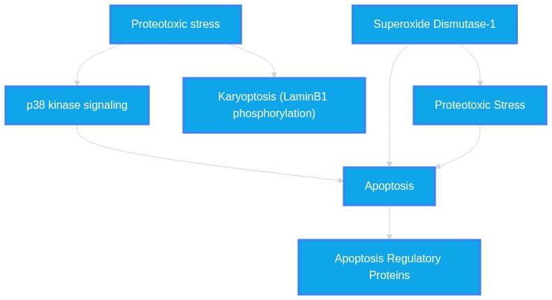
"Here we show that karyoptosis, a distinct form of cell death, can be induced by proteotoxic stress and then develops through nuclear degeneration and cellular expulsion of nuclear material."
VERIFIED VERBATIM (PMID: 42350373)
"We establish that karyoptosis is regulated by the p38 kinase signalling pathway, which controls stability of the nuclear lamina protein LaminB1 via direct phosphorylation."
VERIFIED VERBATIM (PMID: 42350373)
"Finally, we identify karyoptotic features in post-mortem frontal cortex of FTD and Alzheimer's disease (AD) patients."
VERIFIED VERBATIM (PMID: 42350373)
"We demonstrate that karyoptosis affects neurons in models of amyotrophic lateral sclerosis/frontotemporal dementia (ALS/FTD) pathology."
VERIFIED VERBATIM (PMID: 42165865)
"Fluoride exposure caused a dose-dependent downregulation of antioxidant and ER stress-related genes and concurrent upregulation of the pro-apoptotic genes."
VERIFIED VERBATIM (PMID: 42184491)
"Brazilin reduced HepG2 viability in a concentration-dependent manner and suppressed pro-survival signaling (Akt phosphorylation and Bcl-2), accompanied by caspase-3 cleavage and increased Annexin V positivity, indicating apoptosis-associated cytotoxicity."
VERIFIED VERBATIM (PMID: 42194024)
"Nrf2 knockdown via lentiviral shRNA or pharmacological inhibition with brusatol significantly exacerbated BDE-47-induced apoptosis and immune dysfunction"
VERIFIED VERBATIM (PMID: 42190857)
"Acridine orange (AO) staining showed increased apoptosis-related fluorescence signals in the head region, with AO-positive puncta density differing significantly among groups."
VERIFIED VERBATIM (PMID: 42165865)
"Histopathological analysis revealed structural damage in hippocampus and cerebral cortex, including neuronal shrinkage, vacuolization, and apoptotic features."
VERIFIED VERBATIM (PMID: 42008072)
"Mechanistically, RS-PP-059 triggered endoplasmic reticulum (ER) stress and unfolded protein response (UPR), upregulating key markers including GRP78, IRE1α, CHOP, and spliced XBP1 (XBP1s) at both mRNA and protein levels."
VERIFIED VERBATIM (PMID: 41924369)
"Immunohistochemistry confirmed that EGCG significantly reduced microglial activation, glial fibrillary acidic protein (GFAP) expression, and cleaved caspase-3-positive cells (P<0.01 to P<0.001)."
VERIFIED VERBATIM (PMID: 41917198)
"Lisinopril upregulated BI1 protein expression, stabilizing mitochondrial membrane potential and protecting against SOD1G93A-induced apoptosis in NSC34 cells."
VERIFIED VERBATIM (PMID: 41905503)
"Repeated ISO exposure impaired learning and memory, increased anxiety-like behaviors, and caused hippocampal damage, along with elevated pro-apoptotic markers (Bax/Bcl-2 ratio, cleaved caspase-3, PARP1 fragments)"
VERIFIED VERBATIM (PMID: 41903067)
"Thioquercetins (thioQ, thioQ(OAc)4, and thioQ(OAc)5), when used at low micromolar range, induced apoptotic cell death in melanoma cells compared to normal cells."
VERIFIED VERBATIM (PMID: 42156174)
"Mutations in superoxide dismutase 1 (SOD1) compromise its metal-binding capacity, resulting in protein misfolding and aggregation, which ultimately induces cellular apoptosis in amyotrophic lateral sclerosis (ALS)."
VERIFIED VERBATIM (PMID: 41983194)
"The apoptotic rate of sALS (Day 30: 61.37% ± 9.63%; Day 60: 78.41% ± 6.63%) and SOD1 (Day 30: 73.69% ± 8.81%; Day 60: 60.37% ± 11.53%) -derived MNs was significantly higher than those of HCs at both Day 30 (30.72% ± 7.57%) and Day 60 (50.85% ± 19.36%) (p < 0.001)."
VERIFIED VERBATIM (PMID: 42351313)
"The p.N598S variant induced pathological phenotypes consistent with NEK1 haploinsufficiency, including increased susceptibility to DNA damage, increased apoptosis, ciliary dysmorphia, and nucleocytoplasmic translocation of TDP-43."
VERIFIED VERBATIM (PMID: 42243993)
"Hyperoside also reduced cleaved caspase-3 and corrected the Bax/Bcl-2 ratio, improving cell viability under basal and oxidative stress conditions."
VERIFIED VERBATIM (PMID: 42194024)
"These results demonstrate that Nrf2/ARE pathway activation represents an adaptive antioxidant response and contributes to limiting BDE-47-induced cytotoxicity and immune impairment in macrophages."
VERIFIED VERBATIM (PMID: 42350373)
"Here we show that karyoptosis, a distinct form of cell death, can be induced by proteotoxic stress and then develops through nuclear degeneration and cellular expulsion of nuclear material."
VERIFIED VERBATIM (PMID: 42350373)
"We establish that karyoptosis is regulated by the p38 kinase signalling pathway, which controls stability of the nuclear lamina protein LaminB1 via direct phosphorylation."
VERIFIED VERBATIM (PMID: 42350373)
"We demonstrate that karyoptosis affects neurons in models of amyotrophic lateral sclerosis/frontotemporal dementia (ALS/FTD) pathology."
VERIFIED VERBATIM (PMID: 42350373)
"Finally, we identify karyoptotic features in post-mortem frontal cortex of FTD and Alzheimer's disease (AD) patients."
VERIFIED VERBATIM (PMID: 42156174)
"Mutations in superoxide dismutase 1 (SOD1) compromise its metal-binding capacity, resulting in protein misfolding and aggregation, which ultimately induces cellular apoptosis in amyotrophic lateral sclerosis (ALS)."
VERIFIED VERBATIM (PMID: 41983194)
"The apoptotic rate of sALS (Day 30: 61.37% ± 9.63%; Day 60: 78.41% ± 6.63%) and SOD1 (Day 30: 73.69% ± 8.81%; Day 60: 60.37% ± 11.53%) -derived MNs was significantly higher than those of HCs at both Day 30 (30.72% ± 7.57%) and Day 60 (50.85% ± 19.36%) (p < 0.001)."
VERIFIED VERBATIM (PMID: 42190857)
"Acridine orange (AO) staining showed increased apoptosis-related fluorescence signals in the head region, with AO-positive puncta density differing significantly among groups."
VERIFIED VERBATIM (PMID: 42165865)
"Histopathological analysis revealed structural damage in hippocampus and cerebral cortex, including neuronal shrinkage, vacuolization, and apoptotic features."
VERIFIED VERBATIM (PMID: 42165865)
"Fluoride exposure caused a dose-dependent downregulation of antioxidant and ER stress-related genes and concurrent upregulation of the pro-apoptotic genes."
VERIFIED VERBATIM (PMID: 42008072)
"Mechanistically, RS-PP-059 triggered endoplasmic reticulum (ER) stress and unfolded protein response (UPR), upregulating key markers including GRP78, IRE1α, CHOP, and spliced XBP1 (XBP1s) at both mRNA and protein levels."
VERIFIED VERBATIM (PMID: 41924369)
"Immunohistochemistry confirmed that EGCG significantly reduced microglial activation, glial fibrillary acidic protein (GFAP) expression, and cleaved caspase-3-positive cells (P<0.01 to P<0.001)."
VERIFIED VERBATIM (PMID: 41917198)
"Lisinopril upregulated BI1 protein expression, stabilizing mitochondrial membrane potential and protecting against SOD1G93A-induced apoptosis in NSC34 cells."
VERIFIED VERBATIM (PMID: 41905503)
"Repeated ISO exposure impaired learning and memory, increased anxiety-like behaviors, and caused hippocampal damage, along with elevated pro-apoptotic markers (Bax/Bcl-2 ratio, cleaved caspase-3, PARP1 fragments)"
VERIFIED VERBATIM (PMID: 41903067)
"Thioquercetins (thioQ, thioQ(OAc)4, and thioQ(OAc)5), when used at low micromolar range, induced apoptotic cell death in melanoma cells compared to normal cells."
VERIFIED VERBATIM (PMID: 42184491)
"Brazilin reduced HepG2 viability in a concentration-dependent manner and suppressed pro-survival signaling (Akt phosphorylation and Bcl-2), accompanied by caspase-3 cleavage and increased Annexin V positivity, indicating apoptosis-associated cytotoxicity."
VERIFIED VERBATIM (PMID: 42194024)
"These results demonstrate that Nrf2/ARE pathway activation represents an adaptive antioxidant response and contributes to limiting BDE-47-induced cytotoxicity and immune impairment in macrophages."
VERIFIED VERBATIM (PMID: 42194024)
"Nrf2 knockdown via lentiviral shRNA or pharmacological inhibition with brusatol significantly exacerbated BDE-47-induced apoptosis and immune dysfunction"
VERIFIED VERBATIM (PMID: 42243993)
"Hyperoside also reduced cleaved caspase-3 and corrected the Bax/Bcl-2 ratio, improving cell viability under basal and oxidative stress conditions."
VERIFIED VERBATIM (PMID: 42351313)
"The p.N598S variant induced pathological phenotypes consistent with NEK1 haploinsufficiency, including increased susceptibility to DNA damage, increased apoptosis, ciliary dysmorphia, and nucleocytoplasmic translocation of TDP-43."
VERIFIED VERBATIM (PMID: 42389275)
"The best-established histopathological sequence involves AL-related epithelial apoptosis, phagocytosis of apoptotic bodies by macrophages, and subsequent lipofuscin deposition."
VERIFIED VERBATIM (PMID: 42350373)
"We establish that karyoptosis is regulated by the p38 kinase signalling pathway, which controls stability of the nuclear lamina protein LaminB1 via direct phosphorylation."
VERIFIED VERBATIM (PMID: 42350373)
"We demonstrate that karyoptosis affects neurons in models of amyotrophic lateral sclerosis/frontotemporal dementia (ALS/FTD) pathology."
VERIFIED VERBATIM (PMID: 42156174)
"Mutations in superoxide dismutase 1 (SOD1) compromise its metal-binding capacity, resulting in protein misfolding and aggregation, which ultimately induces cellular apoptosis in amyotrophic lateral sclerosis (ALS)."
VERIFIED VERBATIM (PMID: 42171198)
"Research indicates evidence of ferroptosis in ALS, and the natural compound kaempferol has been demonstrated to inhibit neuronal ferroptosis."
VERIFIED VERBATIM (PMID: 42054746)
"Among these, necroptosis has recently emerged as a potential mediator linking inflammaging to neurodegeneration."
VERIFIED VERBATIM (PMID: 41789732)
"biological results reveal an increase of TAR DNA-binding protein 43 aggregates, NOD-like receptor pyrin domain containing protein 3, interleukin (IL)-18, IL-6, and nitrites in 3D skin of ALS patients, thus indicating pyroptosis activation linked to neurodegeneration."
VERIFIED VERBATIM (PMID: 41807703)
"Pharmacological inhibition of RIPK3 with GSK872 markedly attenuated these pathological effects in vitro."
VERIFIED VERBATIM (PMID: 42278291)
"mitigated apoptosis by modulating Bax/Bcl-2 balance and reducing TUNEL-positive cells."
VERIFIED VERBATIM (PMID: 42390647)
"At 2.5 Gy, cell numbers declined to near zero by 6 h (Mean normalized adherent nuclear count decreased to approximately 5% of baseline.), associated with marked nuclear abnormalities, including multinucleation and nuclear fragmentation."
VERIFIED VERBATIM (PMID: 42105621)
"SiNPs significantly increased reactive oxygen species (ROS) levels and malondialdehyde (MDA) content, and decreased the mRNA levels of antioxidant-related genes, including SOD1, SOD2, CAT, GPX1, PRDX2, and NRF2."
VERIFIED VERBATIM (PMID: 42085907)
"ALAN increased hippocampal apoptosis, which was exacerbated by Bmal1 knockdown and mitigated by its overexpression."
VERIFIED VERBATIM (PMID: 42186564)
"During sexual reproduction, the MICs undergo meiosis and the MAC deforms and fragments, providing a unique model to study the regulation of diverse nuclear events."
VERIFIED VERBATIM (PMID: 42353187)
"Compared with control cells, STC2-overexpressing cells exhibited higher cell viability, reduced ROS accumulation, and decreased cell death."
VERIFIED VERBATIM (PMID: 42148083)
"Ferroptosis is a form of regulated cell death driven by iron-dependent lipperoxidation, which plays a pivotal role in regulating the inflammatory-immune microenvironment of central nervous system (CNS) diseases."
VERIFIED VERBATIM (PMID: 42031063)
"Disulfidptosis is a recently identified form of regulated cell death driven by disulfide stress and cytoskeletal collapse under conditions of impaired reducing capacity."
VERIFIED VERBATIM (PMID: 42350373)
"We establish that karyoptosis is regulated by the p38 kinase signalling pathway, which controls stability of the nuclear lamina protein LaminB1 via direct phosphorylation."
VERIFIED VERBATIM (PMID: 42350373)
"We demonstrate that karyoptosis affects neurons in models of amyotrophic lateral sclerosis/frontotemporal dementia (ALS/FTD) pathology."
VERIFIED VERBATIM (PMID: 42156174)
"Mutations in superoxide dismutase 1 (SOD1) compromise its metal-binding capacity, resulting in protein misfolding and aggregation, which ultimately induces cellular apoptosis in amyotrophic lateral sclerosis (ALS)."
VERIFIED VERBATIM (PMID: 42171198)
"Research indicates evidence of ferroptosis in ALS, and the natural compound kaempferol has been demonstrated to inhibit neuronal ferroptosis."
VERIFIED VERBATIM (PMID: 42054746)
"Among these, necroptosis has recently emerged as a potential mediator linking inflammaging to neurodegeneration."
VERIFIED VERBATIM (PMID: 41789732)
"biological results reveal an increase of TAR DNA-binding protein 43 aggregates, NOD-like receptor pyrin domain containing protein 3, interleukin (IL)-18, IL-6, and nitrites in 3D skin of ALS patients, thus indicating pyroptosis activation linked to neurodegeneration."
VERIFIED VERBATIM (PMID: 41807703)
"Pharmacological inhibition of RIPK3 with GSK872 markedly attenuated these pathological effects in vitro."
VERIFIED VERBATIM (PMID: 42278291)
"mitigated apoptosis by modulating Bax/Bcl-2 balance and reducing TUNEL-positive cells."
VERIFIED VERBATIM (PMID: 42390647)
"At 2.5 Gy, cell numbers declined to near zero by 6 h (Mean normalized adherent nuclear count decreased to approximately 5% of baseline.), associated with marked nuclear abnormalities, including multinucleation and nuclear fragmentation."
VERIFIED VERBATIM (PMID: 42105621)
"SiNPs significantly increased reactive oxygen species (ROS) levels and malondialdehyde (MDA) content, and decreased the mRNA levels of antioxidant-related genes, including SOD1, SOD2, CAT, GPX1, PRDX2, and NRF2."
VERIFIED VERBATIM (PMID: 42085907)
"ALAN increased hippocampal apoptosis, which was exacerbated by Bmal1 knockdown and mitigated by its overexpression."
VERIFIED VERBATIM (PMID: 42186564)
"During sexual reproduction, the MICs undergo meiosis and the MAC deforms and fragments, providing a unique model to study the regulation of diverse nuclear events."
VERIFIED VERBATIM (PMID: 42353187)
"Compared with control cells, STC2-overexpressing cells exhibited higher cell viability, reduced ROS accumulation, and decreased cell death."
VERIFIED VERBATIM (PMID: 42148083)
"Ferroptosis is a form of regulated cell death driven by iron-dependent lipperoxidation, which plays a pivotal role in regulating the inflammatory-immune microenvironment of central nervous system (CNS) diseases."
VERIFIED VERBATIM (PMID: 42031063)
"Disulfidptosis is a recently identified form of regulated cell death driven by disulfide stress and cytoskeletal collapse under conditions of impaired reducing capacity."
VERIFIED VERBATIM (PMID: 42212756)
"5-HT synapses in the spinal cord and 5-HT neurons in the brainstem gradually increased following the progression of disease and presented a significantly negative correlation between the increased distribution of 5-HT synapses and neurons and the reduction of neural cell number (positively correlated with the increase in neural cell death) at the onset and/or progression stage of TG mice."
VERIFIED VERBATIM (PMID: 41968900)
"GS1XR efficiently enters normal cells in low matrix metalloproteinases (MMP)-2/9 environments, scavenges radiation-induced reactive oxygen species (ROS), maintains the Nrf2 antioxidant pathway, suppresses apoptosis, and increases clonogenic survival."
VERIFIED VERBATIM (PMID: 41823531)
"Paradoxically, ferritin can be degraded via ferritinophagy, a selective autophagic process that releases toxic ferrous iron and directly triggers ferroptosis."
VERIFIED VERBATIM (PMID: 41807703)
"In this study, we demonstrated that cytoplasmically mis-localized TDP-43 exacerbated neuroinflammation, induced cell death, and impaired phagocytic function in microglial cells, primarily through receptor interacting serine/threonine kinase 3 (RIPK3)-dependent necroptosis."
VERIFIED VERBATIM (PMID: 42074133)
"Exposure of ALS NPCs to the ER stressor tunicamycin increased the ER stress markers binding immunoglobulin protein (BiP) and C/EBP homologous protein (CHOP), disrupted mitochondrial membrane potential, upregulated expression of the mitochondrial apoptotic marker, BAX, increased caspase-3 activation, and reduced cell viability."
VERIFIED VERBATIM (PMID: 42350373)
"karyoptosis, a distinct form of cell death, can be induced by proteotoxic stress and then develops through nuclear degeneration and cellular expulsion of nuclear material."
VERIFIED VERBATIM (PMID: 42350373)
"We establish that karyoptosis is regulated by the p38 kinase signalling pathway, which controls stability of the nuclear lamina protein LaminB1 via direct phosphorylation."
VERIFIED VERBATIM (PMID: 42350373)
"We demonstrate that karyoptosis affects neurons in models of amyotrophic lateral sclerosis/frontotemporal dementia (ALS/FTD) pathology."
VERIFIED VERBATIM (PMID: 42156174)
"Mutations in superoxide dismutase 1 (SOD1) compromise its metal-binding capacity, resulting in protein misfolding and aggregation, which ultimately induces cellular apoptosis in amyotrophic lateral sclerosis (ALS)."
VERIFIED VERBATIM (PMID: 42250707)
"The P66R mutation in SOD1 destabilizes local structure and promotes β-sheet-driven fibrillation, which makes it a suitable model for exploring approaches for reducing pathogenic aggregation."
VERIFIED VERBATIM (PMID: 41997149)
"The nicotinamide adenine dinucleotide (NAD+) hydrolase sterile alpha and Toll/interleukin-1 receptor motif-containing 1 (SARM1) is the central executioner of pathological axon degeneration and is allosterically activated by an increased nicotinamide mononucleotide (NMN)/NAD+ ratio."
VERIFIED VERBATIM (PMID: 42086533)
"Our study demonstrates that proteasomal-dependent CHK1 and ASF1A downregulation contributes to accumulation of DNA damage in cells affected by ALS-linked protein aggregates."
VERIFIED VERBATIM (PMID: 42165865)
"These findings indicate that prolonged NaF exposure impairs antioxidant defenses, induces ER stress, and activates apoptotic pathways, thereby contributing to neuronal damage."
VERIFIED VERBATIM (PMID: 42045773)
"Furthermore, CAPE treatment restored these parameters, reduced oxidative stress, and enhanced antioxidant defenses (SOD, CAT, r-GSH)."
VERIFIED VERBATIM (PMID: 42243993)
"Hyperoside also reduced cleaved caspase-3 and corrected the Bax/Bcl-2 ratio, improving cell viability under basal and oxidative stress conditions."
VERIFIED VERBATIM (PMID: 42062868)
"Removal of TMED9 selectively impairs ATF6 activation without altering IRE1 or PERK signaling, resulting in increased sensitivity to ER stress-induced apoptosis."
VERIFIED VERBATIM (PMID: 42358374)
"Nevertheless, both probiotics and IF markedly reduced serum MDA, hippocampal CLOCK gene expression, gliosis, and apoptosis and enhanced memory performance."
VERIFIED VERBATIM (PMID: 42177227)
"ATX reduces oxidative stress and ameliorates liver and brain damage in aged rats via activation of the Nrf2/Bach1-ARE pathway."
VERIFIED VERBATIM (PMID: 42334525)
"Compounds significantly down-regulated anti-apoptotic genes, whereas mRNA levels of pro-apoptotic genes were up-regulated in MCF-7 cells."
VERIFIED VERBATIM (PMID: 42353197)
"Aβ also decreased the level of GTP-bound Ras and phosphorylation of the downstream mitogen-activated protein kinase/extracellular signal-regulated kinase (MAPK/ERK)."
VERIFIED VERBATIM (PMID: 42400730)
"Resveratrol activates SIRT1 and AMPK signaling in preclinical models, enhancing mitochondrial biogenesis, lowering apoptosis, and restoring cellular resilience."
VERIFIED VERBATIM (PMID: 42144025)
"Hepatic levels of malonaldehyde and caspase-3 were increased, while the levels of superoxide dismutase activity and glutathione and B cell lymphoma-2 were decreased."
VERIFIED VERBATIM (PMID: 42343420)
"LAG-3 expression was progressively upregulated in spinal cord microglia during disease progression, and LAG-3-high microglia exhibited a disease-associated microglia (DAM) transcriptional signature."
VERIFIED VERBATIM (PMID: 42327312)
"MERFISH revealed upregulation of microglial, astrocytic, oligodendrocytic, and T cell markers post-ICI treatment, revealing unique pathways driving neuroinflammation, synaptic function, and cellular signaling."
VERIFIED VERBATIM (PMID: 42327318)
"Here, using a mutant superoxide dismutase 1 (SOD1-G37R) mouse model of familial ALS, Cre-mediated excision of the mutant SOD1 gene within the oligodendrocyte lineage prior to myelin compaction is shown to slow disease onset, improve motor performance, and prolong survival."
Chapter 8
Abstract Repository
Raw text abstracts programmatically cached during the evaluation phase. Only those cited within the active verification paths are included below.
PMID: 41789732
Mapped to Reference [18]
ID: 41789732
Title: 3D Artificial Skin Model As a Novel Strategy for the Detection of Pyroptosis-Cascade Activation in Amyotrophic Lateral Sclerosis.
Abstract: Amyotrophic lateral sclerosis (ALS) is a severe adult-onset neurodegenerative disease with limited treatment approaches. Evidence has shown that degeneration of cutaneous nerves may reflect neurodegenerative processes occurring within the central nervous system. Although skin biopsy is widely adopted in clinical practice, the procedure is invasive and requires multiple patients' tissue removals. Therefore, we developed a 3D innervated skin model by combining 3D printing of methacrylated hyaluronic acid as an innovative tool for better reproducing the dermis and epidermis and electrospinning of polylactic acid for mimicking skin innervation. Later, 3D artificial skin was colonized with a preneuronal cell line (SH-SY5Y) and fibroblasts isolated from skin biopsy of ALS patients at different disease stages. 3D skin possesses a porosity suitable for cell colonization and a high stability. Importantly, biological results reveal an increase of TAR DNA-binding protein 43 aggregates, NOD-like receptor pyrin domain containing protein 3, interleukin (IL)-18, IL-6, and nitrites in 3D skin of ALS patients, thus indicating pyroptosis activation linked to neurodegeneration. This physiologically relevant 3D skin model reduces the need for repeated biopsies, allows standardized experimental conditions, and supports biomarker research and preclinical drug testing in ALS.
PMID: 41807703
Mapped to Reference [19]
ID: 41807703
Title: TDP-43 pathology triggers neuroinflammation and cognitive impairment by inducing microglial necroptosis.
Abstract: Pathological TAR DNA-binding protein-43 (TDP-43) is a defining feature of several neurodegenerative diseases, including amyotrophic lateral sclerosis (ALS), frontotemporal dementia (FTD) and Alzheimer's disease (AD). However, the mechanism by which TDP-43 pathology disrupts microglial function and drives neuroinflammation remains unclear. In this study, we demonstrated that cytoplasmically mis-localized TDP-43 exacerbated neuroinflammation, induced cell death, and impaired phagocytic function in microglial cells, primarily through receptor interacting serine/threonine kinase 3 (RIPK3)-dependent necroptosis. Pharmacological inhibition of RIPK3 with GSK872 markedly attenuated these pathological effects in vitro. These findings were further corroborated in a murine model with cytoplasmic TDP-43 mis-localization, where GSK872 treatment remarkably alleviated neuroinflammation and restored cognitive deficits. Mechanistically, our findings indicate that the nuclear depletion of TDP-43, resulted from its cytoplasmic mis-localization, impairs its ability to transcriptionally repress the Ripk3 gene, subsequently leading to RIPK3 upregulation and activation of RIPK3-dependent necroptosis. Collectively, our findings establish RIPK3-dependent necroptosis as a critical driver of TDP-43 pathology-mediated neuroinflammation and identified necroptosis as a promising therapeutic target in TDP-43-associated neurodegenerative disorders.
PMID: 41823531
Mapped to Reference [30]
ID: 41823531
Title: Ferritin in ferroptosis: Implications for neurodegenerative diseases (Review).
Abstract: Neurodegenerative diseases, including Alzheimer's disease, Parkinson's disease and amyotrophic lateral sclerosis, are characterized by progressive loss of neurons. Although the precise pathogenesis of such diseases is complex and multifactorial, several molecular pathways have been implicated, including the aggregation of misfolded proteins, mitochondrial dysfunction, oxidative stress, neuroinflammation and disrupted iron homeostasis. Emerging evidence has underscored the pivotal role of ferroptosis, an iron‑dependent, non‑apoptotic form of cell death, in neurodegenerative disease progression. Ferritin, characterized by a 24‑subunit hollow sphere structure composed of heavy and light chains, plays a key role in the network regulating cerebral iron homeostasis. In response to cellular iron overload, ferritin expression is upregulated to sequester labile iron and mitigate Fenton reaction‑mediated toxicity, thus exerting a cytoprotective function. Paradoxically, ferritin can be degraded via ferritinophagy, a selective autophagic process that releases toxic ferrous iron and directly triggers ferroptosis. This review systematically reviews the role of ferritin within the iron homeostasis network to elucidate the connection between the dysregulation of iron metabolism and the pathological mechanisms of neurodegenerative diseases. The study focused on the potential role of ferritin as a biomarker for early diagnosis, therapeutic strategies targeting ferritin pathways to restore iron homeostasis and the clinical translational value of magnetic resonance imaging‑based non‑invasive quantification of cerebral iron deposition. It is crucial to elucidate the multidimensional roles of ferritin in neurodegeneration to provide a theoretical foundation for precision diagnostic and therapeutic approaches.
PMID: 41903067
Mapped to Reference [10]
ID: 41903067
Title: HSP90 inhibition potentiates oxidant-based antimelanoma action of novel thioquercetin derivatives by compromising AhR/CYP1A1 pathway.
Abstract: Quercetin, a plant-derived dietary flavonoid, has multifunctional biological activities, including anticancer action; however, its applications may be restricted due to limited bioavailability. Thus, novel synthetic quercetin derivatives (QDs) with improved properties and/or drug combinations should be designed and tested. In the present study, anticancer activity of fourteen newly synthesized QDs was investigated using four cellular models of melanoma, namely A375, MM370, G-361, and SH-4 cells. Thioquercetins (thioQ, thioQ(OAc)4, and thioQ(OAc)5), when used at low micromolar range, induced apoptotic cell death in melanoma cells compared to normal cells. Thioquercetins also reduced the population of spheroid-forming cells and suppressed the growth of A375 cells in 3D spheroid models. Thioquercetin-mediated antimelanoma action was potentiated upon heat shock protein 90 (HSP90) inhibition. Co-treatment with the HSP90 inhibitor 17-DMAG and thioquercetins augmented oxidative stress (increased superoxide production, decreased levels of antioxidant proteins SOD1, and PRDX1-2), and impaired the aryl hydrocarbon receptor (AhR)/cytochrome P450 1A1 (CYP1A1) signaling pathway-based detoxification of thioquercetins by the inhibition of AhR translocation to the nucleus and AhR-mediated stimulation of CYP1A1 expression leading to enhanced cytotoxic effects against melanoma cells. The senolytic activity of thioQ(OAc)4 with four acetylated hydroxy groups against cisplatin-induced senescent melanoma cells was also revealed in selected experimental settings. We suggest that the use of novel thioquercetin-based derivatives along with HSP90 inhibitors should be further validated in vivo and considered for the design of more effective antimelanoma strategies in the future.
PMID: 41905503
Mapped to Reference [9]
ID: 41905503
Title: Intranasal rosmarinic acid reduces cognitive and hippocampal damage from repeated neonatal isoflurane exposure by regulating apoptotic/oxidative/inflammatory responses, and heat-shock and 14-3-3 proteins.
Abstract: Repeated or prolonged exposure to general anesthetics like isoflurane (ISO) during neurodevelopment can lead to long-term neurocognitive and behavioral deficits, particularly because pediatric brains lack adequate antioxidant defenses, and no preventive therapies currently exist. Rosmarinic acid (RA), a polyphenolic compound with antioxidant and neuroprotective properties, has not yet been evaluated for mitigating ISO-induced toxicity. In this study, Wistar albino rat pups were exposed to ISO (1.5% in 30% oxygen/air, 3-h) on postnatal days (P)7 +P9 + P11, and the protective effects of intranasal RA (25 mg/kg) pretreatment (1-h before anesthesia) were investigated for the first time. Control groups received either oxygen alone or RA before oxygen exposure. On P12, hippocampal tissue was examined for detecting acute neuronal apoptosis, oxidative stress, inflammation, and stress-related proteins using histopathology and immunoblotting. Cognitive performance was assessed using Morris Water Maze tests that evaluated spatial learning (P28-P32) and both short- and long-term memory (P33, P60, P90). Repeated ISO exposure impaired learning and memory, increased anxiety-like behaviors, and caused hippocampal damage, along with elevated pro-apoptotic markers (Bax/Bcl-2 ratio, cleaved caspase-3, PARP1 fragments), redox imbalance and inflammation (reduced SOD1; increased GPX1, 4HNE, NF-κB-p65, TNF-α). ISO also disrupted stress signaling by reducing p-HSF1, Hsp90, and Hsp60 levels, while raising Hsp70 and decreasing 14-3-3 isoforms. RA pretreatment countered these effects by restoring antioxidant and stress-response proteins, reducing inflammation and apoptosis, and maintaining neuronal integrity and cognitive function. No harmful effects were observed in the RA-only group. These findings suggest that intranasal RA pretreatment may be a preventive strategy against anesthesia-related neurotoxicity in pediatric patients.
PMID: 41917198
Mapped to Reference [8]
ID: 41917198
Title: Lisinopril activates BI1 to reprogram lipid metabolism and restore autophagy in ALS.
Abstract: Amyotrophic lateral sclerosis (ALS) involves disrupted lipid metabolism. Bax inhibitor 1 (BI1), an endoplasmic reticulum protein downregulated in ALS neuroprotective, represents a therapeutic target, but its metabolic regulatory mechanisms are incompletely understood. Using transcriptomics in skeletal muscle of ALS mice pre- and post-BI1 treatment, we identified BI1-regulated pathways. Structure-based virtual screening of FDA-approved compounds nominated lisinopril as a BI1 activator. Lisinopril upregulated BI1 protein expression, stabilizing mitochondrial membrane potential and protecting against SOD1G93A-induced apoptosis in NSC34 cells. Concurrently, it regulated TGF-β1/mTOR-dependent autophagy, maintained NMJ integrity, and reshaped triglyceride/sphingolipid/glycerophospholipid metabolism to attenuate spinal cord pathology in ALS mice, promoting energy metabolism shift toward glucose oxidation. Additionally, lisinopril inhibited the TGF-β1/Smad2/3 pathway to alleviate muscle fibrosis, downregulate Acp5/FN expression, and reduce type I collagen deposition. In conclusion, this study provides evidence that pharmacological activation of BI1 by lisinopril suppresses TGF-β1, modulates lipid metabolism, and ameliorates ALS pathology, demonstrating promising therapeutic repurposing potential.
PMID: 41924369
Mapped to Reference [7]
ID: 41924369
Title: Epigallocatechin gallate in N-methyl-N-nitrosourea-induced retinitis pigmentosa mouse model.
Abstract: To investigate the therapeutic efficacy and underlying mechanisms of epigallocatechin gallate (EGCG), a major green tea catechin with potent antioxidant properties, in an N-methyl-N-nitrosourea (MNU)-induced mouse model of retinitis pigmentosa (RP). C57BL/6 mice were randomly divided into control (PBS), MNU-induced RP, and MNU+EGCG pretreatment groups. EGCG (50 mg/kg, intraperitoneal) was administered daily for 3 consecutive days prior to a single MNU injection (50 mg/kg). Retinal function was evaluated by scotopic electroretinography (ERG). Retinal structure was assessed using optical coherence tomography (OCT) and hematoxylin-eosin staining, with outer nuclear layer (ONL) thickness measurement. Mechanisms were explored via RNA sequencing, reverse transcription quantitative real-time polymerase chain reaction (RT-qPCR) validation of oxidative stress- and inflammation-related genes, and immunohistochemistry for microglial activation, astrocytic gliosis, and apoptosis markers. Compared with the MNU group, EGCG pretreatment significantly preserved scotopic ERG a-wave and b-wave amplitudes (P<0.001). OCT and histological analysis showed that EGCG markedly attenuated MNU-induced thinning of total retina and ONL (P<0.005, P<0.001, respectively). RNA sequencing identified 1147 differentially expressed genes modulated by EGCG, with significant upregulation of antioxidant genes (Nrf2, Sod1, Gpx4, Cat1, Ho-1) and downregulation of pro-inflammatory genes. Immunohistochemistry confirmed that EGCG significantly reduced microglial activation, glial fibrillary acidic protein (GFAP) expression, and cleaved caspase-3-positive cells (P<0.01 to P<0.001). EGCG exerts robust neuroprotective effects in MNU-induced RP through enhancement of antioxidant defenses, suppression of neuroinflammation, and preservation of retinal structure and function. These findings suggest EGCG as a promising candidate for adjuvant antioxidant therapy in RP.
PMID: 41968900
Mapped to Reference [29]
ID: 41968900
Title: Selective Radioprotection by the Fusion Antioxidant Enzyme GS1XR Via an MMP-2/9-Cleavable Cell-Penetrating Switch.
Abstract: Radiotherapy is central to cancer treatment but radiation-induced oxidative stress also damages normal tissues. To achieve selective radioprotection of normal tissues, we systematically evaluated, in vitro and in vivo, the antioxidant and radioprotective performance of a previously engineered fusion antioxidant enzyme, GS1XR (GST-SOD1-X-R9). GS1XR efficiently enters normal cells in low matrix metalloproteinases (MMP)-2/9 environments, scavenges radiation-induced reactive oxygen species (ROS), maintains the Nrf2 antioxidant pathway, suppresses apoptosis, and increases clonogenic survival. In contrast, in 3D tumor microenvironments with high MMP-2/9, cleavage of the X peptide removes R9, resulting in a loss of transmembrane capacity and a pronounced reduction in intracellular ROS scavenging. Animal studies further showed that GS1XR significantly alleviates whole-body irradiation-induced hematopoietic injury and, in tumor-bearing models receiving radiotherapy, does not compromise radiotherapy-mediated tumor control. Collectively, GS1XR couples microenvironment-responsive cell entry with enzymatic antioxidation to achieve selective radioprotection while preserving radiotherapy-mediated tumor control.
PMID: 41983194
Mapped to Reference [3]
ID: 41983194
Title: Dynamic changes in excitability and viability of sporadic and SOD1-related amyotrophic lateral sclerosis iPSC-derived motor neurons.
Abstract: To explore the dynamic changes in excitability and viability of induced pluripotent stem cells (iPSC)-derived motor neurons from sporadic amyotrophic lateral sclerosis (ALS) and compare them with SOD1-related ALS patients and healthy control. Peripheral blood samples were collected from ALS patients and healthy controls (HC) to establish the iPSC-derived motor neurons (MNs). Whole-cell patch-clamp recordings at different culture stages was made using an Axopatch 700B amplifier in combination with pClamp 11 software (Molecular Devices). The frequency of action potentials (APs) was recorded. Additionally, Terminal deoxynucleotidyl transferase (TdT)-mediated deoxyuridine triphosphate (dUTP) Nick-End Labeling (TUNEL) was used to assess the apoptosis of MNs. ALS patient-derived MNs exhibited significantly higher firing rates compared to HCs at both 4-7 weeks (p = 0.004) and 7-9 weeks (p = 0.009). Further analysis revealed that SOD1-derived MNs showed significantly higher firing frequencies than sALS (p = 0.009) and HCs (p < 0.001) in 4-7 weeks. In 7-9 weeks, it remained significant between SOD1 and HC-derived MNs (p = 0.015), but became insignificant between SOD1 and sALS (p = 0.855). The apoptotic rate of sALS (Day 30: 61.37% ± 9.63%; Day 60: 78.41% ± 6.63%) and SOD1 (Day 30: 73.69% ± 8.81%; Day 60: 60.37% ± 11.53%) -derived MNs was significantly higher than those of HCs at both Day 30 (30.72% ± 7.57%) and Day 60 (50.85% ± 19.36%) (p < 0.001). MNs derived from both patients with mutant SOD1 and sporadic ALS exhibited increased excitability compared to HCs. The increased excitability of MNs derived from ALS patients with mutant SOD1 occurred earlier, and over time, became consistent with the excitability observed in MNs derived from sporadic ALS. The apoptosis rates of MNs showed similar trends. iPSC-derived MNs from both sporadic and mutant ALS may serve as useful cell models for ALS in future studies.
PMID: 41997149
Mapped to Reference [33]
ID: 41997149
Title: SARM1 executes neuronal parthanatos and promotes excitotoxic cell death.
Abstract: The nicotinamide adenine dinucleotide (NAD+) hydrolase sterile alpha and Toll/interleukin-1 receptor motif-containing 1 (SARM1) is the central executioner of pathological axon degeneration and is allosterically activated by an increased nicotinamide mononucleotide (NMN)/NAD+ ratio. DNA damage induces NAD+ loss and an increased NMN/NAD+ ratio by hyperactivating poly(ADP-ribose) polymerase 1 (PARP1), which triggers the parthanatos cell death pathway. Multiple mechanistically distinct DNA-damaging agents activate SARM1 and induce axon degeneration following PARP1 activation. Remarkably, SARM1 is required for key steps downstream of hyperactivated PARP1, which are pathognomonic of parthanatos, including mitochondrial depolarization, nuclear translocation of apoptosis-inducing factor (AIF), and cell death. Hence, SARM1 is an essential component of neuronal parthanatos. Moreover, complex neurodegenerative stimuli whose mechanisms include activation of parthanatos, such as 1-methyl-4-phenyl-pyridinium (MPP+) dopaminergic neuron toxicity and N-methyl-D-aspartate (NMDA) excitotoxicity, are potently protected by SARM1 inhibition. These findings place SARM1 at the nexus of multiple mechanisms driving neuronal cell death, thereby greatly expanding the potential clinical utility of SARM1 inhibitors beyond diseases of axon loss.
PMID: 42008072
Mapped to Reference [6]
ID: 42008072
Title: A novel 14-deoxy-12-hydroxyandrographolide analogue promotes apoptosis in colorectal cancer through ROS-dependent endoplasmic reticulum stress activation.
Abstract: Colorectal cancer is the second leading cause of cancer-related mortality worldwide, highlighting the critical need for novel therapeutic strategies. In this study, we investigated the anticancer activity and molecular mechanisms of RS-PP-059, a derivative of 14-deoxy-12-hydroxyandrographolide, in colorectal cancer cells. RS-PP-059 exhibited potent cytotoxicity and selectivity toward HT-29 cells, suppressing viability and clonogenic growth. The compound induced apoptotic cell death, as shown by increased Annexin V-positive cells, PARP-1 cleavage, p53 activation, and γ-H2AX accumulation, indicating DNA damage, and was accompanied by a reduction in total caspase-3 protein levels. Mechanistically, RS-PP-059 triggered endoplasmic reticulum (ER) stress and unfolded protein response (UPR), upregulating key markers including GRP78, IRE1α, CHOP, and spliced XBP1 (XBP1s) at both mRNA and protein levels. Co-treatment with the ER stress inhibitor 4-phenylbutyrate (4-PBA) only partially reversed these effects, suggesting robust ER stress activation by RS-PP-059. In parallel, RS-PP-059 increased intracellular reactive oxygen species (ROS) in a time-dependent manner, accompanied by differential regulation of antioxidant genes with strong induction of HO-1 and suppression of CAT, SOD1, and GPX-1. Importantly, pretreatment with N-acetyl-L-cysteine (NAC) abolished ROS accumulation, ER stress activation, apoptosis, and loss of viability, confirming the ROS-dependent mechanism. In conclusion, our findings demonstrate that RS-PP-059 exerts potent anticancer effects in colorectal cancer cells by promoting ROS-mediated ER stress, leading to DNA damage and apoptosis.
PMID: 42031063
Mapped to Reference [27]
ID: 42031063
Title: Disulfidptosis in neurodegenerative diseases: From redox imbalance to neuronal dysfunction.
Abstract: Disulfidptosis is a recently identified form of regulated cell death driven by disulfide stress and cytoskeletal collapse under conditions of impaired reducing capacity. Neurodegenerative diseases (NDs), including Parkinson's disease, Alzheimer's disease, and amyotrophic lateral sclerosis, are characterized by oxidative stress, mitochondrial dysfunction, metabolic impairment, protein aggregation, and cytoskeletal instability-features that may provide a permissive intracellular context for disulfidptosis. However, its occurrence and pathological relevance in these disorders remain incompletely understood. In this review, we examine the potential involvement of disulfidptosis in neurodegenerative diseases from a disease-centered perspective. We emphasize that current evidence is largely indirect and based on mechanistic overlap rather than direct experimental validation in neural systems. Accordingly, we distinguish between direct evidence, indirect mechanistic support, and pathophysiological plausibility. We further discuss cell-type-specific susceptibility across neurons and glial cells, analyze its relationship with other cell death pathways, and consider potential therapeutic implications. Overall, disulfidptosis is best regarded as a context-dependent and emerging mechanism that may contribute to neuronal vulnerability under specific metabolic and redox constraints. Clarifying its disease relevance will be essential for determining its significance in neurodegeneration and its potential as a therapeutic target.
PMID: 42045773
Mapped to Reference [35]
ID: 42045773
Title: Caffeic Acid Phenethyl Ester Enhanced the Klotho/SIRT1/Nrf2/HO-1 Axis to Protect Against Methylmercury-Induced ALS-Like Neurodegeneration.
Abstract: Amyotrophic lateral sclerosis (ALS) is a neurodegenerative disorder characterized by motor neuron degeneration, oxidative stress, and neuroinflammation. This study evaluated the neuroprotective potential of caffeic acid phenethyl ester (CAPE) against MTME + 5-induced neurotoxicity in an ALS-like pathology model. CAPE (50 and 100 mg/kg., p.o.) demonstrated significant therapeutic efficacy by improving motor and cognitive deficits, restoring oxidative balance, and mitigating neuroinflammatory and apoptotic pathways. Behavioral assessments, including the open field, grip strength, forced swim, and Morris water maze, highlighted CAPE's ability to restore neuromuscular coordination and cognitive function in a dose-dependent manner. Cellular and Molecular analyses revealed that MTME+5 exposure significantly disrupted Klotho/SIRT-1/Nrf2/HO-1 antioxidant signaling, increased pro-inflammatory cytokines (TNF-α, IL-1β), and elevated apoptotic markers (Bax, caspase-3) while depleting anti-inflammatory cytokines (IL-10) and neuroprotective proteins. Furthermore, CAPE treatment restored these parameters, reduced oxidative stress, and enhanced antioxidant defenses (SOD, CAT, r-GSH). Furthermore, CAPE normalized neurotransmitter imbalances, including acetylcholine, dopamine, GABA, serotonin, and glutamate, alleviating excitotoxicity. Histopathological and gross morphological analyses confirmed CAPE50 and CAPE100 ability to preserve neuronal and myelin integrity across key brain regions, including the cerebral cortex, hippocampus, striatum, midbrain, and cerebellum. CAPE also reduced methylmercury accumulation in the brain and cerebrospinal fluid, indicating detoxifying effects. Co-administration of vitamin B1 (VTB1(200)) further amplified CAPE's therapeutic efficacy. Complete blood count (CBC) analysis demonstrated MTME+5-induced hematological abnormalities, including reduced RBCs, hemoglobin, WBCs, and platelets, alongside elevated eosinophils and basophils. CAPE treatment normalized these parameters, indicating systemic recovery. These findings establish CAPE as a promising neuroprotective agent for ALS, capable of targeting neurocomplications.
PMID: 42054746
Mapped to Reference [17]
ID: 42054746
Title: From necroptosis to neuroinflammation: Unraveling mechanisms and therapeutic targets in age-related cognitive decline.
Abstract: Aging is the major risk factor for several chronic conditions, including cognitive decline and dementia. It is accompanied by profound immune alterations characterized by a progressive decline in immune competence, a process known as immunosenescence. The resulting dysregulation of immune function leads to the overproduction of proinflammatory cytokines and fuels a persistent, low-grade inflammatory state termed inflammaging. This chronic inflammation contributes to dysfunction across the central and peripheral nervous systems, promoting neuronal damage and accelerating neurodegenerative processes such as Alzheimer's disease, Parkinson's disease, amyotrophic lateral sclerosis, and other age-related cognitive disorders. Within this framework, prolonged activation of inflammatory pathways can trigger regulated forms of cell death. Among these, necroptosis has recently emerged as a potential mediator linking inflammaging to neurodegeneration. Its core molecular effectors, including the receptor-interacting protein kinases RIPK1 and RIPK3 and the mixed-lineage kinase domain-like protein (MLKL), are increasingly expressed in aged neural tissues, promoting the release of damage-associated molecular patterns (DAMPs) that amplify glial activation, oxidative stress, and blood-brain barrier disruption. Growing evidence suggests that necroptotic signaling may be upregulated in the aging brain and in neurodegenerative disorders, where it could contribute to neuronal loss and cognitive impairment. This review discusses the potential role of necroptosis in the continuum between inflammation and neurodegeneration, highlighting emerging diagnostic and therapeutic perspectives. Epigenetic and circulating biomarkers, such as phosphorylated MLKL and specific microRNAs, may support early detection, while pharmacological and nutraceutical strategies targeting necroptosis show promising neuroprotective effects in preclinical studies.
PMID: 42062868
Mapped to Reference [36]
ID: 42062868
Title: IRE1-XBP1driven induction of TMED9 stabilizes ATF6 during ER stress to promote cell survival.
Abstract: The endoplasmic reticulum (ER) plays a central role in protein homeostasis by facilitating the folding, modification, and quality control of secretory and membrane proteins. Disruption of ER function results in protein misfolding and ER stress, which activate the unfolded protein response (UPR). While the three canonical UPR branches, inositol-requiring enzyme 1 (IRE1), protein kinase RNA-like endoplasmic reticulum kinase (PERK), and activating transcription factor 6 (ATF6), have been extensively studied, the mechanisms that coordinate their activities and ultimately dictate survival or death remain poorly understood. Transmembrane P24 trafficking protein 9 (TMED9), a cargo receptor that cycles between the ER and Golgi, has been implicated in protein quality control under pathological conditions, but its physiological role in ER proteostasis and UPR signaling is unclear. The ER stress response was studied in cellular human models including normal epithelial cells and patient-derived pediatric glioma cultures. To define the regulatory mechanisms dictating TMED9 expression, quantitative Reverse Transcription polymerase chain reaction (qRT-PCR), luciferase reporter assay, and western blotting were employed. To elucidate TMED9 function, loss-of-function approaches, including clustered regularly interspaced short palindromic repeats (CRISPR)-Cas9-mediated knockout and small interfering RNA knockdown were used in combination with RNA-seq and live imaging. Protein stability was tested by pulse-chase experiments, ubiquitination, and degradation analyses. To study the implications of TMED9 activation, we screened curated gene expression datasets from the European Molecular Biology Laboratory- European Bioinformatics Institute (EMBL-EBI) Expression Atlas and employed live-cell imaging-based assays and functional assays (cell viability, apoptosis, migration, and self-renewal). Our study uncovers a physiological role for TMED9 in ER proteostasis and UPR signaling. We show that, under ER stress, TMED9 expression is transcriptionally induced by the IRE1-spliced X-box binding protein 1 (XBP1s) pathway via a conserved unfolded protein response element (UPRE)-like element in its promoter. Removal of TMED9 selectively impairs ATF6 activation without altering IRE1 or PERK signaling, resulting in increased sensitivity to ER stress-induced apoptosis. Mechanistically, we identify TMED9 as a stress-induced stabilizer of ATF6 that prevents its ubiquitin-dependent proteasomal degradation. Functionally, TMED9 regulation is exploited by tumor cells, which sustain IRE1-XBP1s activity to upregulate TMED9, thereby enhancing survival under ER stress conditions. Collectively, our findings establish TMED9 as a critical regulator of ER stress adaptation. TMED9 emerges as a molecular mediator that links IRE1-dependent transcriptional response to ATF6 stabilization, ultimately supporting increased secretory demand under stress conditions and in cancer development.
PMID: 42074133
Mapped to Reference [31]
ID: 42074133
Title: Pridopidine Protects ALS Patient-Derived Neural Progenitor Cells via Sigma-1 Receptor Activation.
Abstract: The sigma-1 receptor (S1R) is an endoplasmic reticulum (ER)-resident protein enriched at the mitochondria-associated ER membranes (MAMs) that supports ER homeostasis, preserves mitochondrial function, and enhances cell survival under stress. Disruptions of MAM integrity and prolonged ER stress are well-recognized pathological features of amyotrophic lateral sclerosis (ALS), contributing to motor neuron dysfunction and degeneration. In this study, we evaluated the protective effects of pridopidine, a highly selective and potent S1R agonist currently in clinical development for Huntington's disease (HD) and ALS, using neural progenitor cells (NPCs) derived from induced pluripotent stem cells (iPSCs) from a patient with sporadic ALS. Exposure of ALS NPCs to the ER stressor tunicamycin increased the ER stress markers binding immunoglobulin protein (BiP) and C/EBP homologous protein (CHOP), disrupted mitochondrial membrane potential, upregulated expression of the mitochondrial apoptotic marker, BAX, increased caspase-3 activation, and reduced cell viability. Pridopidine significantly attenuated tunicamycin-induced BiP and CHOP expression in a biphasic, dose-dependent manner (with maximal efficacy at 1 µM), consistent with the typical pharmacology of S1R agonists. Pridopidine restored mitochondrial membrane potential, reduced mitochondrial apoptotic signaling, shown by decreased BAX expression and caspase-3 activation, and improved survival of ALS-NPCs under ER stress. Co-treatment with the selective S1R antagonist, NE-100, attenuated these effects, supporting an S1R-mediated mechanism of action for pridopidine. Together, these results demonstrate that S1R activation by pridopidine mitigates ER-stress-induced mitochondrial dysfunction and cell loss in ALS-NPCs, resulting in enhanced survival of NPCs supporting the therapeutic potential of pridopidine in ALS.
PMID: 42085907
Mapped to Reference [23]
ID: 42085907
Title: Bmal1 regulates hippocampal oxidative damage in cognitive memory impairment induced by artificial light at night in mice.
Abstract: Artificial light at night (ALAN) has emerged as a significant public health concern, yet its effects on cognitive impairment remain poorly understood. This study investigated the impact of 28 consecutive days of 5-lx ALAN exposure on hippocampal function in C57BL/6 J mice. We evaluated locomotor behavior, neuronal morphology, neurogenesis, oxidative stress, and circadian rhythms, revealing that ALAN induces cognitive impairment. ALAN exposure reduced Bmal1 expression, increased reactive oxygen species (ROS) and malondialdehyde accumulation, and disrupted the time-of-day-dependent differences expression of NRF2, SOD1, and GPX1. These alterations suppressed SOD and GPX enzymatic activity, leading to hippocampal oxidative damage. To clarify BMAL1's role, we used adeno-associated virus (AAV) to modulate Bmal1 expression in the hippocampus. ALAN increased hippocampal apoptosis, which was exacerbated by Bmal1 knockdown and mitigated by its overexpression. These findings suggest that ALAN can contribute to memory impairment by disrupting hippocampal damage and impairing neurogenesis through its effect on Bmal1. This study identifies potential molecular targets for preventing and treating cognitive impairment and neurodegenerative disorders.
PMID: 42086533
Mapped to Reference [34]
ID: 42086533
Title: Proteasomal-dependent CHK1 degradation leads to DNA damage accumulation in ALS cellular model systems.
Abstract: Amyotrophic lateral sclerosis (ALS) is characterised by the aggregation of TDP-43 and mutant FUS in the cytoplasm of affected motor neurons. Accumulation of DNA damage is emerging as a novel correlative trait of ALS. We recently showed that formation of TDP-43 and FUS cytoplasmic inclusions (CIs) lead to DNA damage accumulation through dysregulation of the DNA damage response (DDR). However, the multiple molecular mechanisms contributing to DNA damage accumulation in affected motor neurons in ALS have not been fully elucidated. In recent years, chemical inhibition of the serine/threonine kinase CHK1 was shown to lead to accumulation of DNA breaks as well as increased apoptosis, in differentiated cortical neurons. Notably, CHK1 has been involved in DNA double-strand break repair in non-dividing cells, by acting through the histone chaperone ASF1A. In this article, we show that cells bearing FUS and TDP-43 CIs show downregulation of the protein levels of CHK1 and ASF1A. We observe CHK1 protein downregulation in neuronal cell lines, as well as in patient-derived motor neurons progenitors and in the spinal cord of a FUS-ALS mouse model. Restoration of the nuclear levels of CHK1 and ASF1A via transient overexpression, is sufficient to reduce DNA damage signal accumulation and rescues DDR defects. Importantly, we show that the ubiquitin-proteasome pathway is responsible for CHK1 degradation in cells bearing FUS CI, since its inhibition restores CHK1 and ASF1A protein levels. Our study demonstrates that proteasomal-dependent CHK1 and ASF1A downregulation contributes to accumulation of DNA damage in cells affected by ALS-linked protein aggregates.
PMID: 42105621
Mapped to Reference [22]
ID: 42105621
Title: Silica nanoparticles suppress porcine oocyte maturation via oxidative stress, metabolic dysfunction, and impaired cholesterol trafficking.
Abstract: Silica nanoparticles (SiNPs), as common feed additives, are widely applied in livestock diets and pose potential risks to reproductive health owing to their tissue accumulation. In the present study, we explored the effects and underlying mechanisms of SiNPs exposure during in vitro maturation (IVM) of porcine oocytes. The results showed that SiNPs significantly suppress porcine oocyte maturation as evidenced by decreased first polar body (PB1) release rate. Notably, SiNPs significantly induced abnormal expansion of cumulus cells and impaired gap junction intercellular communication (GJIC), accompanied by decreased Connexin 43 (CX43) expression and aberrant F-actin structure. Furthermore, DCFH-DA staining showed that SiNPs significantly increased reactive oxygen species (ROS) levels and malondialdehyde (MDA) content, and decreased the mRNA levels of antioxidant-related genes, including SOD1, SOD2, CAT, GPX1, PRDX2, and NRF2. JC-1 staining showed that SiNPs significantly induced mitochondrial dysfunction via diminished mitochondrial membrane potential (ΔΨm) and aberrant distribution, and decreased the mRNA levels of energy metabolism-related genes, such as NOX4 and COX2. Additionally, SiNPs significantly disrupted lysosomal function and cholesterol trafficking and decreased the mRNA levels of LDLR, NPC1, NPC2, and LAMP2, leading to reduced free cholesterol levels and the mRNA levels of estrogen synthesis-related genes, including STAR, CYP19A1, and HSD-3β. Collectively, SiNPs suppress porcine oocyte maturation, at least partly, through oxidative stress, metabolic disruption, and impaired cholesterol trafficking.
PMID: 42144025
Mapped to Reference [42]
ID: 42144025
Title: Lycopene, quercetin, and silymarin alleviate tartrazine-induced liver injury via modulating Nrf2 signaling and endoplasmic reticulum stress pathways.
Abstract: Tartrazine is a synthetic lemon-yellow azo dye that is widely used as a coloring agent in food products, drugs, and cosmetics. Tartrazine was reported to induce hepatotoxicity, however, the effects of tartrazine on nuclear factor erythroid two-related factor two (Nrf2) signaling and endoplasmic reticulum (ER) stress pathways in rat liver have not been investigated. Therefore, this study aimed to investigate the effects of tartrazine on Nrf2 signaling and ER stress pathways in rat liver, to examine the potential therapeutic effects of lycopene, quercetin, and silymarin, and to investigate the mechanisms through which they may mitigate tartrazine-induced liver injury. Rats were allocated into five experimental groups including a normal control group and a tartrazine group that received tartrazine (10 mg/kg) orally for 13 weeks. The lycopene, quercetin, and silymarin groups received tartrazine (10 mg/kg) for 13 weeks and were treated with lycopene (10 mg/kg), quercetin (50 mg/kg), and silymarin (150 mg/kg), respectively, for the last 8 weeks. Tartrazine-induced liver injury was characterized by increased alanine aminotransferase, aspartate aminotransferase, and alkaline phosphatase serum levels and histopathological changes in the liver. Furthermore, tartrazine suppressed hepatic Nrf2 signaling and induced ER stress. Hepatic levels of malonaldehyde and caspase-3 were increased, while the levels of superoxide dismutase activity and glutathione and B cell lymphoma-2 were decreased. Moreover, hepatic transforming growth factor-beta one levels and collagen deposition were increased. Treatment with lycopene, quercetin, and silymarin ameliorated the tartrazine-induced changes, upregulated Nrf2 signaling and alleviated ER stress. Our findings suggest that lycopene, quercetin, and silymarin may provide a promising therapeutic approach against tartrazine-induced liver injury.
PMID: 42148083
Mapped to Reference [26]
ID: 42148083
Title: Ferroptosis-immune crosstalk in CNS diseases: mechanisms and translational insights.
Abstract: Ferroptosis is a form of regulated cell death driven by iron-dependent lipid peroxidation, which plays a pivotal role in regulating the inflammatory-immune microenvironment of central nervous system (CNS) diseases. Mounting evidence indicates that dysregulated iron metabolism and an imbalance in antioxidant defenses can induce ferroptosis in neurons and glial cells while simultaneously remodeling immune cell function, thereby establishing a bidirectional feedback loop that amplifies neuroinflammation and tissue damage. In neurodegenerative diseases, including Alzheimer's disease (AD), Parkinson's disease (PD), and amyotrophic lateral sclerosis (ALS), pro-inflammatory cytokines such as TNF-α and IL-1β released by activated microglia upregulate neuronal iron transporters (e.g., DMT1 and TfR1), promoting iron accumulation and ferroptotic cell death. In turn, damage-associated molecular patterns released from ferroptotic cells further potentiate immune activation, forming a self-amplifying cycle. In contrast, within the glioma microenvironment, CD8+ T cell-derived IFN-γ suppresses SLC7A11 expression in tumor cells, leading to glutathione depletion and glutathione peroxidase 4 inactivation, thereby triggering ferroptosis and modulating anti-tumor immunity. Although targeting ferroptosis or neuroimmune pathways has shown therapeutic promise in mitigating neurological deficits and enhancing anti-tumor responses, the underlying mechanisms governing ferroptosis-immune crosstalk remain inadequately characterized. Herein, this review systematically summarizes the key biological characteristics of ferroptosis and immune responses, with particular emphasis on their interplay across major CNS disorders (i.e., AD, PD, ALS, multiple sclerosis, stroke, and glioma). Furthermore, we discuss emerging therapeutic strategies encompassing small molecules, immunomodulatory approaches, and nanotechnology-based interventions, highlighting the ferroptosis-immune axis as a promising therapeutic target for CNS diseases.
PMID: 42156174
Mapped to Reference [2]
ID: 42156174
Title: COMMD1 Induces Copper Deficiency of SOD1 by Inhibiting the Palmitoylation of CCS in ALS.
Abstract: Mutations in superoxide dismutase 1 (SOD1) compromise its metal-binding capacity, resulting in protein misfolding and aggregation, which ultimately induces cellular apoptosis in amyotrophic lateral sclerosis (ALS). Copper metabolism domain containing 1 (COMMD1), a gene implicated in copper homeostasis, has not been thoroughly characterized in the context of ALS pathogenesis. In this study, we identified elevated COMMD1 expression in ALS, potentially contributing to diminished copper incorporation into SOD1. Knockdown of COMMD1 enhanced palmitoylation of the copper chaperone for SOD1 (CCS), facilitating its membrane translocation and promoting copper loading into SOD1, thereby conferring neuroprotection in ALS. Mechanistically, we established that COMMD1 knockdown augments CCS palmitoylation via activation of the hypoxia-inducible factor 1 subunit alpha (HIF-1α)/fatty acid synthase (FASN) signaling axis. In vivo investigations utilizing male hSOD1G93A transgenic mice demonstrated that COMMD1 deficiency markedly ameliorated the deterioration of motor function and prolonged survival duration. These findings collectively suggest that COMMD1 represents a potential therapeutic target for ALS intervention.
PMID: 42165865
Mapped to Reference [5]
ID: 42165865
Title: Molecular mechanisms underlaying fluoride-induced neurotoxicity: interplay of antioxidants and endoplasmic reticulum stress-mediated apoptotic pathways in rats.
Abstract: Fluoride is a naturally occurring compound widely present in soil, water, rocks and is essential to maintain the physiological function and structure of bones and teeth. However, chronic exposure to elevated fluoride levels has been linked to adverse neurological effects. Despite its widespread environmental presence, the molecular mechanisms underlying fluoride-induced neurotoxicity remain incompletely understood. This study aimed to elucidate the effects of fluoride on oxidative stress, endoplasmic reticulum (ER) stress, apoptosis, and associated histopathological alterations in brain tissue. Forty Sprague-Dawley rats were randomly assigned to four groups (n = 10 per group; 5 male + 5 female) and administered sodium fluoride (NaF) in drinking water at concentrations of < 0.5 ppm (control), 50 ppm, 150 ppm, and 300 ppm for 90 consecutive days. The expression of antioxidant genes (SOD1 and GCLC), ER stress, and apoptosis-related genes (XBP1, GRP78, BCL-2, and BAX) was quantified using real-time quantitative PCR (RT-qPCR), and histopathological analysis of the brain tissues was performed. Fluoride exposure caused a dose-dependent downregulation of antioxidant and ER stress-related genes and concurrent upregulation of the pro-apoptotic genes. Histopathological analysis revealed structural damage in hippocampus and cerebral cortex, including neuronal shrinkage, vacuolization, and apoptotic features. These findings indicate that prolonged NaF exposure impairs antioxidant defenses, induces ER stress, and activates apoptotic pathways, thereby contributing to neuronal damage. This study provides mechanistic insights into fluoride-induced neurotoxicity and highlights the need for further research on potential therapeutic strategies targeting oxidative and ER stress pathways.
PMID: 42171198
Mapped to Reference [16]
ID: 42171198
Title: Targeting lipid nanoparticle mediated co-delivery of edaravone and kaempferol for amyotrophic lateral sclerosis therapy.
Abstract: Amyotrophic lateral sclerosis (ALS) is characterized by a progressive and selective loss of motor neurons in the central nervous system, particularly in the brain and spinal cord. However, the main cellular mechanisms and cell death pathways leading to motor neuron degeneration have not yet been clarified. Research indicates evidence of ferroptosis in ALS, and the natural compound kaempferol has been demonstrated to inhibit neuronal ferroptosis. However, damage to the blood-brain barrier (BBB) prevents the drug from penetrating the central nervous system, which significantly reduces its therapeutic efficacy. Here, we developed a targeted delivery system named Eda/Kae@Lip-RGD (EKLR), which consisted of liposome-grafted RGD peptides for the co-delivery of the drugs kaempferol and edaravone, capable of crossing the BBB to provide co-delivery of kaempferol and edaravone for combined treatment of ALS. As expected, treatment with EKLR for one month significantly slowed down weight loss and improved athletic performance in SOD1G93A transgenic mice. Mechanistically, this nanomedicine suppressed ferroptosis by upregulating the antioxidant proteins GPX4 and SLC7A11, alongside the downregulation of Nrf2 and ACSL4 levels, thus collectively preserving neuronal integrity. Meanwhile, EKLR restored the normal morphology and the survival rate of neurons and maintained the mitochondrial structure and morphological integrity. Accordingly, this nanoplatform may represent a distinctive and potentially effective strategy for achieving neuroprotection in ALS as well as in other disorders of the central nervous system.
PMID: 42177227
Mapped to Reference [38]
ID: 42177227
Title: Nrf2/Bach1-ARE pathway are involved in the ameliorative effects of astaxanthin on D-galactose-induced liver and brain aging and injury.
Abstract: Astaxanthin (ATX), a natural antioxidant whose benefits in age-related liver and kidney damage remain unclear. We established a D-galactose-induced ageing model in rats and observed the daily behaviour of the rats. Using staining methods to detect ROS, apoptosis and histopathological changes in liver and brain tissue. Determination of antioxidant levels of IL-2, IL-6 and AGES in rats. Assessment of cognitive function using the Morris water maze and ChAT. The mRNA and protein expression levels of Nrf2, Bach1, SOD1, SOD2, HO-1 were determined by real-time PCR and Western blotting. To investigate the role of the Nrf2/Bach1-ARE pathway, we used ML385, a specific inhibitor of the Nrf2 pathway, to treat rats in the inhibitor group. Aging rats showed impaired learning and memory, along with decreased levels of neurotransmitters and antioxidant enzymes. ATX and vitamin E (VE) interventions significantly alleviated these symptoms and activated the Nrf2/Bach1-ARE pathway in liver and brain tissues of aged SD rats. Furthermore, the protective effects of ATX were attenuated by the addition of the Nrf2 inhibitor ML385. ATX reduces oxidative stress and ameliorates liver and brain damage in aged rats via activation of the Nrf2/Bach1-ARE pathway.
PMID: 42184491
Mapped to Reference [11]
ID: 42184491
Title: Real-time profiling of brazilin-induced cytotoxic responses in HepG2 cells and exosome-associated metabolic signaling under lipid accumulation.
Abstract: Hepatocellular carcinoma (HCC) frequently arises in metabolically altered livers and often exhibits limited responses to systemic therapies, highlighting the need for agents that target both tumor cell survival and metabolic vulnerabilities. Brazilin, a bioactive compound from Caesalpinia sappan, has been reported to exert anticancer activities, yet its effects on intercellular communication under lipid-enriched conditions remain unclear. Here, we profiled brazilin-induced cytotoxic responses in HepG2 cells using real-time imaging-based monitoring of cell morphology, number, and area, together with viability assays. Brazilin reduced HepG2 viability in a concentration-dependent manner and suppressed pro-survival signaling (Akt phosphorylation and Bcl-2), accompanied by caspase-3 cleavage and increased Annexin V positivity, indicating apoptosis-associated cytotoxicity. We then isolated extracellular vesicles released from brazilin-treated HepG2 cells and confirmed exosome-like characteristics by TEM, exosomal marker expression (CD9/CD63/CD81), and nanoparticle tracking analysis. Notably, in palmitate/oleate-loaded HepG2 cells, exosomes derived from brazilin-treated cells modulated metabolic and stress-associated proteins, including reduced CPT1A and SOD1 levels and increased p27 expression, consistent with altered fatty-acid oxidation-related signaling under lipid accumulation. Collectively, these findings suggest that brazilin exerts direct cytotoxic effects in HepG2 cells and that exosomes released upon brazilin exposure may contribute to metabolic signaling changes in lipid-loaded cancer cells.
PMID: 42186564
Mapped to Reference [24]
ID: 42186564
Title: Dual roles of two Tfdps in germline nuclear meiosis and somatic nuclear fragmentation during sexual reproduction in the unicellular organism Paramecium tetraurelia.
Abstract: Meiosis is regulated by phase-specific genes to orchestrate nuclear and cytoskeletal dynamics essential for sexual reproduction. The ciliate Paramecium tetraurelia exhibits nuclear dimorphism, harboring two germline micronuclei (MICs) and one somatic macronucleus (MAC) within a single cell during vegetative growth. During sexual reproduction, the MICs undergo meiosis and the MAC deforms and fragments, providing a unique model to study the regulation of diverse nuclear events. Transcription factor DP (TFDP), which heterodimerizes with E2Fs, can bind to specific DNA motifs in promoters of cell cycle-regulated genes to activate or repress their expression. Here, we identified 16 TFDP homologs in P. tetraurelia, representing an exceptional gene family expansion accompanied by functional domain diversification. The functions of Tfdp1a and Tfdp1b, which are specifically expressed during sexual reproduction and localize in the old and new MACs, were further investigated. Their depletion resulted in meiotic arrest at metaphase in the MIC, failure of old MAC fragmentation, and abortive cytokinesis. Transcriptomic analysis revealed that TFDP1A/1B knockdown primarily causes gene downregulation, with > 60% of downregulated genes being specifically highly expressed during sexual reproduction. Functional annotation and enrichment analyses demonstrated significant downregulation of proteins involved in meiosis, DNA replication, and DNA repair. Critically, multiple downregulated meiotic regulators are essential for proper homologous chromosome segregation and sister chromatid separation, providing a mechanistic basis for the observed MIC meiotic arrest. This study uncovers Tfdp1a/1b as essential regulators of the distinctive meiotic process in P. tetraurelia, providing crucial insights into nuclear dynamics and the regulation of sexual reproduction in binucleate systems. The online version contains supplementary material available at 10.1007/s42995-026-00378-1.
PMID: 42190857
Mapped to Reference [4]
ID: 42190857
Title: Neurodevelopmental and behavioral effects of early-life esketamine hydrochloride exposure in zebrafish (Danio rerio).
Abstract: Esketamine hydrochloride is increasingly used as a rapid-acting antidepressant, and its expanding clinical and non-medical use has raised concerns regarding its release into aquatic systems via wastewater treatment plant effluents as an emerging psychoactive contaminant. However, its potential neurodevelopmental toxicity in aquatic organisms remains insufficiently characterized. In this study, zebrafish embryos were exposed to esketamine hydrochloride during early development, and its toxic effects were evaluated using an integrated framework combining developmental, behavioral, histological, transcriptomic, oxidative stress-related, and apoptosis-related endpoints. Early-life esketamine exposure altered multiple developmental indicators, including head length, eye depth, interocular distance, and body length, and disrupted locomotor regulation at later stages, particularly light-dark responsiveness and spatial preference. Histological examination further revealed exposure-related alterations in brain tissue organization. Transcriptomic profiling identified coordinated changes in pathways associated with redox homeostasis, protein synthesis, and phototransduction-related signaling. Targeted validation demonstrated significant upregulation of oxidative stress-related genes, including sod1 and sod2, while ELISA-based assays showed exposure-dependent alterations in SOD, CAT, GSH, and MDA levels. Acridine orange (AO) staining showed increased apoptosis-related fluorescence signals in the head region, with AO-positive puncta density differing significantly among groups. Integrative correlation analysis further linked developmental, behavioral, oxidative stress-related, and apoptosis-related endpoints. Notably, these effects occurred in the absence of overt lethality. Collectively, these findings demonstrate that esketamine interferes with neurodevelopmental and behavioral processes in zebrafish larvae and support the incorporation of early-life neurobehavioral endpoints into risk assessment frameworks for neuroactive pharmaceuticals.
PMID: 42194024
Mapped to Reference [12]
ID: 42194024
Title: Activation of the Nrf2/ARE Pathway Attenuates BDE-47-Induced Immunotoxicity in RAW264.7 Macrophages.
Abstract: Polybrominated diphenyl ethers (PBDEs), widely used as brominated flame retardants, are known to exert persistent adverse effects on the immune systems of humans and other organisms. Previous studies have demonstrated that 2,2',4,4'-tetrabromodiphenyl ether (BDE-47), a prevalent congener, induces apoptosis, impairs phagocytic function, and triggers aberrant immune-inflammatory reactions in RAW264.7 macrophages via the induction of elevated intracellular reactive oxygen species (ROS). However, the underlying regulatory mechanism remains unclear. The nuclear factor erythroid 2-related factor 2/antioxidant response element (Nrf2/ARE) signaling pathway is a key cellular defense system against oxidative stress. In this study, we investigated the role of the Nrf2/ARE pathway in BDE-47-induced macrophage immunotoxicity. Network toxicology analysis identified Nrf2 as a hub gene within the BDE-47-associated immunotoxicity network. Molecular docking and molecular dynamics simulations suggested a potential interaction between BDE-47 and the Keap1-Nrf2 complex, with moderate binding affinity. Experimental studies in RAW264.7 cells showed that BDE-47 exposure activated the Nrf2/ARE pathway, as evidenced by Nrf2 nuclear translocation and the differential upregulation of downstream genes (GCLC, GCLM, HO-1, NQO1, SOD1, and CAT). Importantly, Nrf2 knockdown via lentiviral shRNA or pharmacological inhibition with brusatol significantly exacerbated BDE-47-induced apoptosis and immune dysfunction, including enhanced pro-inflammatory cytokine production and impaired phagocytosis. These results demonstrate that Nrf2/ARE pathway activation represents an adaptive antioxidant response and contributes to limiting BDE-47-induced cytotoxicity and immune impairment in macrophages.
PMID: 42212756
Mapped to Reference [28]
ID: 42212756
Title: 5-Hydroxytryptamine Distribution Alteration in Both Neuron and Synapse of Tg(SOD1*G93A)1gur Mice: A Potential Intervention Candidate Strategy for Amyotrophic Lateral Sclerosis.
Abstract: Amyotrophic lateral sclerosis (ALS) is a fatal neurodegenerative disease; the precise pathogenesis of sporadic ALS (sALS) has not yet been elucidated up to now. Previous studies revealed that the abnormal alterations of some non-motor neurons (non-MN) were a potential pathogenesis of sALS. Therefore, this study aims to search the potential evidences of non-MN in the pathogenesis of ALS via exploring potential relationships between 5-hydroxytryptamine (5-HT) neurons and the development of ALS. We employed fluorescent immunohistochemistry to investigate the altered distribution patterns of 5-HT and tryptophan hydroxylase 2 in the spinal cord and brainstem of Tg(SOD1*G93A)1Gur (TG) and wild-type (WT) mice. Additionally, we used western blot to analyze the expression levels of 5-hydroxytryptamine receptor 1A (5-HTR1A) and 5-HTR2A. Our findings revealed that 5-HT synapses were primarily distributed in the funiculus lateralis, anterior horn, posterior horn, central lateral column, and the area around the central canal of cervical, thoracic, and lumbar segments, and raphe nucleus as well as lateral paragigantocellular nucleus, and gradually reduced following age increase in WT mice. However, 5-HT synapses in the spinal cord and 5-HT neurons in the brainstem gradually increased following the progression of disease and presented a significantly negative correlation between the increased distribution of 5-HT synapses and neurons and the reduction of neural cell number (positively correlated with the increase in neural cell death) at the onset and/or progression stage of TG mice. 5-HTR1A significantly increased, while 5-HTR2A significantly decreased at the onset stage of TG mice. Our study speculated that the distribution changes of 5-HT synapses in the spinal cord and 5-HT neurons in the brainstem play a potential protective role in the pathogenesis of sALS through a compensatory 5-HT increase.
PMID: 42243993
Mapped to Reference [13]
ID: 42243993
Title: Hyperoside protects against poly-GR-mediated neurodegeneration via regulation of mitochondrial fission and oxidative stress in C9orf72-associated ALS.
Abstract: Arginine-rich poly-glycine-arginine (poly-GR), a toxic dipeptide repeat protein generated from C9orf72 hexanucleotide repeat expansion, drives mitochondrial dysfunction, oxidative stress, and neuronal loss in amyotrophic lateral sclerosis (ALS). Hyperoside, a bioactive flavonoid, exhibits antioxidant and cytoprotective properties, but its therapeutic relevance to C9orf72-associated ALS remains unclear. To determine whether hyperoside attenuates poly-GR-induced mitochondrial and oxidative injury and improves neuronal survival in cellular and animal models of C9orf72-ALS. A combined in vitro and in vivo experimental study using motor neuron-like cells and an AAV-mediated neonatal mouse model of poly-GR toxicity. NSC34 cells expressing EGFP-GR50 were analyzed for mitochondrial morphology, membrane potential, ROS generation, antioxidant signaling, and apoptosis using confocal microscopy, CellROX/MitoTracker assays, Western blot analysis, and viability testing. For in vivo assessment, neonatal mice received intracerebroventricular AAV9-EGFP-GR50 followed by intraperitoneal hyperoside (10 mg/kg). Survival, cerebral hemisphere length, and cortical NeuN⁺ neuron numbers were quantified. Poly-GR expression induced pronounced mitochondrial fragmentation, reduced membrane potential, elevated ROS, and suppressed Nrf2/HO-1/GPx4 signaling, accompanied by increased Drp1 and reduced Opa1 expression. Hyperoside reversed these abnormalities by restoring mitochondrial integrity, normalizing the Drp1/Opa1 balance, enhancing Nrf2 nuclear accumulation, and increasing the expression of HO-1 and GPx4. Hyperoside also reduced cleaved caspase-3 and corrected the Bax/Bcl-2 ratio, improving cell viability under basal and oxidative stress conditions. In vivo, hyperoside modestly prolonged survival, increased cerebral hemisphere length, and significantly preserved cortical neuronal numbers in AAV9-EGFP-GR50 mice. Hyperoside mitigates poly-GR-induced neurotoxicity by alleviating excessive mitochondrial fission, strengthening Nrf2-dependent antioxidant defenses, and suppressing apoptosis. These findings support hyperoside as a promising multi-target therapeutic candidate for C9orf72-associated ALS.
PMID: 42250707
Mapped to Reference [32]
ID: 42250707
Title: Inhibitory effect of silymarin on amyloid formation in ALS-associated hSOD1 P66R mutant.
Abstract: The aberrant aggregation of human superoxide dismutase 1 (hSOD1) into β-sheet-rich amyloid fibrils is a crucial process in the pathogenesis of amyotrophic lateral sclerosis (ALS), enhancing motor neuron degeneration and disease progression. The P66R mutation in SOD1 destabilizes local structure and promotes β-sheet-driven fibrillation, which makes it a suitable model for exploring approaches for reducing pathogenic aggregation. Here, we evaluate silymarin, a polyphenolic compound with known antioxidant and neuroprotective properties, for its potential to inhibit P66R-hSOD1 aggregation. ThT fluorescence and transmission electron microscopy analyses demonstrate a significant decrease in amyloid fibril formation in the presence of silymarin; in addition, FTIR spectroscopy confirms the suppression of β-sheet formation. Fluorescence quenching and ANS binding assays indicate a moderate-affinity binding between silymarin and the mutant protein, along with a reduction in surface hydrophobicity. Hemolysis assays confirm its protective effect against membrane damage induced by aggregates, while molecular docking and dynamic simulations indicate that silymarin stabilizes aggregation-prone areas with hydrogen bonding and hydrophobic interactions, thereby promoting compact conformations and reducing solvent-exposed surfaces. The findings identified silymarin as an effective anti-amyloidogenic agent that reduces β-sheet accumulation and fibril formation while also decreasing cytotoxicity, highlighting its potential as a therapeutic candidate for ALS.
PMID: 42278291
Mapped to Reference [20]
ID: 42278291
Title: Targeting Autoimmune Myocarditis with Lemon Balm Extract: In Vivo Molecular Approach.
Abstract: Due to the complex pathophysiology and serious outcomes of autoimmune myocarditis, we sought to determine whether ethanolic lemon balm extract (LBE) could attenuate disease progression and development of dilative cardiomyopathy (DCM). EAM was induced in Dark Agouti rats by immunization with porcine myosin. Fifty animals were allocated to five groups: healthy controls, untreated EAM, and EAM treated with LBE (50, 100, or 200 mg/kg) for six weeks. Hemodynamic parameters were monitored, and echocardiography assessed cardiac structure and function. Inflammatory, oxidative, fibrotic, and apoptotic markers were analyzed. Immunological profiling revealed that LBE significantly decreased proinflammatory cytokines (IL-1, IL-6, TNF-α, IL-4, IL-17) while restoring anti-inflammatory IL-10 levels (p < 0.05). Antioxidant activity was confirmed by reduced levels of O2-, H2O2, and TBARS, accompanied by significant increases in SOD, CAT, and GSH activity (p < 0.05), and upregulation of SOD1 and SOD2 gene expression. Additionally, LBE (200 mg/kg) markedly reversed fibrotic remodeling through suppression of TGF-β expression and collagen deposition, as shown by Sirius Red staining, and mitigated apoptosis by modulating Bax/Bcl-2 balance and reducing TUNEL-positive cells. Collectively, these findings suggest that LBE exerts strong cardioprotective effects in EAM by regulating inflammatory, oxidative, fibrotic, and apoptotic pathways, thereby preventing myocarditis progression toward DCM.
PMID: 42327312
Mapped to Reference [44]
ID: 42327312
Title: Spatial Transcriptomics reveals a T cell-mediated microglial activation axis of neurodegeneration following immune checkpoint inhibition.
Abstract: Immune checkpoint inhibitor (ICI) combinations that block cytotoxic T-lymphocyte-associated protein 4 (CTLA-4) and programmed cell death protein 1 (PD-1) signaling have revolutionized cancer care but also exert a range of immune-related adverse events (irAE) in various tissues, including the brain. Our understanding of the mechanisms of irAE in the brain is still evolving, and we recently demonstrated that ICI (blockade of CTLA-4 and PD-1) perturbs hippocampal-dependent memory function by derailing neuro-immune homeostasis and compromising synaptic integrity. However, the spatial patterns and the cell-type-specific molecular mechanisms underlying ICI-related brain dysfunction remain not well-defined. To address this gap, we performed spatial transcriptomic profiling of the hippocampal region using multiplexed error-robust fluorescence in situ hybridization (MERFISH) to map gene expression at single-cell resolution. By integrating spatial single-cell data with bulk RNA-seq, we define the distribution of microglia, astrocytes, synaptic, and neuroinflammatory markers, and determine how ICI reshapes hippocampal cellular composition in a syngeneic murine melanoma model. MERFISH revealed upregulation of microglial, astrocytic, oligodendrocytic, and T cell markers post-ICI treatment, revealing unique pathways driving neuroinflammation, synaptic function, and cellular signaling. Furthermore, immunofluorescence analysis of postmortem brains from patients treated with ICI corroborates our findings of ICI-related immune activation of microglia. Finally, using a conditional deletion model, we show that T cells are indispensable for ICI-driven microglial activation. Altogether, our study provides a high-resolution spatial framework for understanding irAEs in brain function and a T cell-microglia crosstalk axis as a driving mechanism of dysregulated neuro-immune homeostasis during ICI.
PMID: 42327318
Mapped to Reference [45]
ID: 42327318
Title: Mutant SOD1 expressed by oligodendrocytes aggregates in myelinic nanochannels and accelerates disease progression in familial ALS mice.
Abstract: Amyotrophic lateral sclerosis (ALS) is a highly debilitating and fatal disease characterized by the progressive loss of motor neurons. Reduced oligodendroglial support has been implicated in ALS progression but remains mechanistically unexplained. Here, using a mutant superoxide dismutase 1 (SOD1-G37R) mouse model of familial ALS, Cre-mediated excision of the mutant SOD1 gene within the oligodendrocyte lineage prior to myelin compaction is shown to slow disease onset, improve motor performance, and prolong survival. In contrast, silencing mutant SOD1 expression within oligodendrocytes after myelin compaction failed to ameliorate disease phenotype. Electron microscopy is used to identify aggregation of mutant SOD1 within paranodal loops and the inner periaxonal tongue of 'myelinic nanochannels', narrow cytosolic compartments for the diffusion of metabolites and motor-driven transport processes. In a second mouse model (SOD1-G93A) of familial, SOD1 mutant-mediated ALS, we show that induction of excessive myelin compaction and myelinic channel collapse (by depletion of CNP from myelin) accelerates disease and diminishes survival. Our data support loss of myelinic channel integrity as a contributor to familial ALS disease initiation and progression, findings likely relevant to neurodegenerative disease involving other aggregation prone proteins that are expressed in myelinating oligodendrocytes. Oligodendrocytes have been implicated in the progression of amyotrophic lateral sclerosis (ALS) but the underlying mechanisms have remained obscure. Here we show in genetic mouse models that the familial ALS causing isoform of a ubiquitously expressed mutant enzyme (SOD1) aggregates in cytosolic channels within myelin that are responsible for delivery of transporters and nutrients necessary to support the axonal compartment. ALS disease progression was accelerated in mice when myelinic channels were collapsed by deleting CNP, a structural protein necessary for myelinic channel maintenance. Disruption of transport through myelinic channels by aggregation of mutant SOD1 may perturb oligodendrocyte support of motor axons and contribute to disease in this form of ALS.
PMID: 42334525
Mapped to Reference [39]
ID: 42334525
Title: Synthesis and multilevel evaluation of benzimidazole-based anticancer compounds: DNA/serum albumin interaction, and in-depth theoretical insights.
Abstract: Within the scope of this investigation, two novel compounds (3a and 3b) were designed and synthesized in two steps. Compounds 3a and 3b were tested utilizing the MTT study to evaluate their in vitro cytotoxic activity against healthy human embryonic kidney, lung cancer, breast cancer, and human liver cancer cell lines. It was determined that compound 3b exhibited high levels of cytotoxic activity against both liver and breast cancer cell lines, with IC50 values of 10.83 and 11.55 µM, respectively. Also, to elucidate the anticancer mechanism of compounds, pro-apoptotic BAX and BiD, anti-apoptotic BCL2 and BCL-xl, oxidant enzymes PRDX1 and SOD1 levels were examined by RT-qPCR. Moreover, cellular oxidative stress levels were spectrophotometrically measured, and cellular senescence was evaluated via the senescence-associated β-galactosidase test. DFT calculations and RDG, ELF, and LOL analyses were performed to demonstrate the reactivity of these compounds. Compounds significantly down-regulated anti-apoptotic genes, whereas mRNA levels of pro-apoptotic genes were up-regulated in MCF-7 cells. Moreover, oxidative stress status was significantly increased depending on the compound treatment. Consistently, PRDX1 and SOD1 levels were also significantly up-regulated. Furthermore, cellular senescence was significantly induced by the compounds. Fluorescence spectroscopy demonstrated strong binding of both compounds with CT-DNA, characterized by static quenching mechanism and binding constants of 6.02 × 10⁶M- 1(for 3a) and 8.69 × 10⁶M- 1(for 3b), suggesting an intercalative binding mode. In contrast, moderate affinity toward BSA indicated suitable transport characteristics with reduced nonspecific protein binding. Molecular docking studies supported the experimental findings. These combined results verify the potential of the synthesized derivatives as promising candidates for further investigation as biologically active anticancer agents.
PMID: 42343420
Mapped to Reference [43]
ID: 42343420
Title: Immune checkpoint LAG-3 governs stage-dependent and disease-associated microglial modules in ALS model mice.
Abstract: Immune checkpoint molecules, inhibitory receptors originally characterized in T cell biology, have recently emerged as regulators of microglial function in neurodegeneration, yet their roles in amyotrophic lateral sclerosis (ALS) remain unexplored. Here, we investigated LAG-3, an inhibitory immune checkpoint receptor, in microglial regulation during ALS pathogenesis using SOD1G93A mice. LAG-3 expression was progressively upregulated in spinal cord microglia during disease progression, and LAG-3-high microglia exhibited a disease-associated microglia (DAM) transcriptional signature. Genetic deletion of LAG-3 produced a biphasic phenotype, with accelerated disease onset but significantly prolonged disease duration. LAG-3 deficiency enhanced inflammatory microglial responses at the early disease stage, whereas at the late stage it suppressed inflammatory signaling while selectively preserving phagocytic effector gene expression, demonstrating that LAG-3 dissociates the inflammatory and phagocytic modules within the DAM program in a stage-dependent manner. These transcriptional changes translated into enhanced phagocytic capacity in primary microglia and amelioration of the spinal cord environment through suppression of inflammatory pathways and restoration of oxidative phosphorylation. Our findings identify LAG-3 as a stage-dependent regulator of microglial functional states in ALS and support the concept that immune checkpoint molecules constitute a class of module-level regulators of microglial function in neurodegeneration.
PMID: 42350373
Mapped to Reference [1]
ID: 42350373
Title: Karyoptosis mediates cell death and neurodegeneration upon proteotoxic stress.
Abstract: Neurodegenerative diseases are frequently associated with proteotoxic stress linked to disease specific proteins. The autophagy-lysosome system provides essential control of proteotoxic stress and its failure can lead to initiation of apoptosis. However, in aging and neurodegenerative diseases apoptosis is insufficient to account for all neuronal death, and several different cell death types have been reported in these contexts. Here we show that karyoptosis, a distinct form of cell death, can be induced by proteotoxic stress and then develops through nuclear degeneration and cellular expulsion of nuclear material. We establish that karyoptosis is regulated by the p38 kinase signalling pathway, which controls stability of the nuclear lamina protein LaminB1 via direct phosphorylation. We demonstrate that karyoptosis affects neurons in models of amyotrophic lateral sclerosis/frontotemporal dementia (ALS/FTD) pathology. Finally, we identify karyoptotic features in post-mortem frontal cortex of FTD and Alzheimer's disease (AD) patients. Together these findings characterise a form of cell death directly linked to proteotoxic stress and nuclear lamina stability that is associated with neurodegeneration.
PMID: 42351313
Mapped to Reference [14]
ID: 42351313
Title: A rare missense variant impacting NEK1 kinase function is associated with ALS.
Abstract: Heterozygous truncating loss-of-function (LoF) variants in NEK1 are a known cause of amyotrophic lateral sclerosis (ALS). NEK1 encodes the pleiotropic serine/threonine kinase NIMA-related kinase 1, and prior in vitro studies have implicated kinase dysfunction as the principal pathogenic mechanism underlying NEK1-associated ALS. However, bona fide pathogenic missense variants causally linked to ALS have not previously been reported, leaving this hypothesis unconfirmed. Here, we identify a rare NEK1 missense variant, p.N598S, that co-segregates with disease in a familial ALS pedigree and is enriched in European ALS cohorts. This variant exhibits normal protein expression levels, indicating a functional rather than quantitative defect. Using isogenic human motor neurons, we directly compared the effects of p.N598S with those of the ALS-associated truncating variant p.R812* to delineate disease mechanisms. The p.N598S variant induced pathological phenotypes consistent with NEK1 haploinsufficiency, including increased susceptibility to DNA damage, increased apoptosis, ciliary dysmorphia, and nucleocytoplasmic translocation of TDP-43. Importantly, p.N598S impaired NEK1 kinase activity, and pharmacological inhibition of NEK1 recapitulated the cellular phenotypes observed in both p.N598S- and p.R812*-mutant motor neurons. Collectively, these findings provide strong genetic and functional evidence for a disease-causing role of NEK1 kinase disruption in NEK1-ALS. Our findings provide immediate diagnostic and therapeutic implications, particularly for the functional interpretation of missense variants of uncertain significance and the development of targeted treatment strategies.
PMID: 42353187
Mapped to Reference [25]
ID: 42353187
Title: Overexpression of Stanniocalcin 2 Protects Differentiated QM7 Cells from H2O2-Induced Oxidative Damage.
Abstract: Oxidative stress, caused by excessive reactive oxygen species (ROS) accumulation, is a major factor in muscle cell damage and muscle atrophy-related disorders. Although Stanniocalcin 2 (STC2) is involved in cellular stress and exhibits cytoprotective effects in various cell types, its role in skeletal muscle cells during oxidative stress is unclear. This study investigated the effects of STC2 overexpression in quail muscle (QM7) cells exposed to H2O2-induced oxidative stress. STC2 expression was upregulated in non-transfected QM7 cells following H2O2 treatment. Stable STC2-overexpressing cells were differentiated for 4 days, and then assessed for cell viability, ROS accumulation, cell death, and myotube morphology following H2O2 treatment. Compared with control cells, STC2-overexpressing cells exhibited higher cell viability, reduced ROS accumulation, and decreased cell death. STC2 overexpression also attenuated the H2O2-induced reduction in MyHC protein expression. Antioxidant-related genes, including Superoxide Dismutase 1, Glutathione Peroxidase 1, Heme Oxygenase 1, and NAD(P)H Quinone Dehydrogenase 1, were significantly upregulated in STC2-overexpressing cells. Compared with the control cells, nuclear factor erythroid 2-related factor 2 protein levels were not increased in STC2-overexpressing cells under oxidative stress conditions. These findings suggest that STC2 overexpression alleviates oxidative stress-induced cellular damage and may contribute to protective antioxidant responses in muscle cells.
PMID: 42353197
Mapped to Reference [40]
ID: 42353197
Title: Hesperetin Rescues Amyloid Beta-Induced Defects in Neurite Outgrowth Under In Vitro Mild Cognitive Impairment-like Cellular Conditions.
Abstract: Accumulation of aggregated amyloid beta (Aβ) species is a defining pathological hallmark of Alzheimer's disease and is associated with extensive neuronal structural abnormalities. Mild cognitive impairment (MCI), a transitional stage between normal aging and the onset of dementia, is thought to represent an early phase of this pathological continuum. Studies at the cellular level suggest that the conditions impair the maintenance of established neuronal processes/networks and restrict their capacity for elongation or re-elongation. They may also attenuate the activation and process extension of quiescent neural progenitor or stem-like cells. These early cellular changes precede overt neurodegeneration in neural tissue and are likely to contribute to cognitive decline. They highlight the importance of in vitro models for identifying molecular targets involved in recovery from disease. In this study, we investigated the effects of aggregated Aβ (25-35) on neuronal process elongation and associated intracellular events in the N1E-115 cell line, a widely used model of neuronal differentiation. Addition of aggregated Aβ to cultured N1E-115 cells attenuated process elongation in a concentration-dependent manner. This morphological impairment was accompanied by decreased expression of neuronal differentiation markers. In contrast, at the half-maximal inhibitory concentration for process elongation, long-term cultured cells did not exhibit apparent process retraction or degenerative morphology. This mild but progressive impairment, without extensive cell death, is consistent with the cellular features of early-stage conditions rather than advanced Alzheimer's pathologies. Similar results were observed in primary cortical neurons. Aβ also decreased the level of GTP-bound Ras and phosphorylation of the downstream mitogen-activated protein kinase/extracellular signal-regulated kinase (MAPK/ERK). Furthermore, treatment with hesperetin, a bioactive flavonoid compound, recovered the Aβ-induced inhibition of neuronal process elongation. Hesperetin also restored Ras and MAPK/ERK states, suggesting that its effects are associated, at least in part, with modulation of signaling through Ras and MAPK/ERK. Our findings suggest that hesperetin may serve as a useful molecular probe for modulating early cellular responses associated with Alzheimer's disease-related pathology. This in vitro model might serve as a useful platform for investigating the molecular target candidates involved in recovery from nervous system disorders.
PMID: 42358374
Mapped to Reference [37]
ID: 42358374
Title: Comparative neuroprotective and exercise capacity effects of prophylactic intermittent fasting and probiotics in sleep-deprived rats: insights into anti-inflammatory marker modulation and CLOCK gene regulation.
Abstract: Prophylactic probiotics and intermittent fasting (IF) substantially modulate the neuropsychological functions and exercise capacity in rats subjected to sleep deprivation (SD). A comparative study was conducted to analyze the effects of probiotics and IF on SD-induced neuropsychological disturbances and compromised muscle endurance. Forty albino Wistar rats were randomly assigned to four groups. The NSD group was maintained on a standard chow diet for 12 weeks. The SD group followed an SD regimen for 72 h per week over 8 weeks, starting from the fifth week. The SDP group received probiotics at a dose of colony-forming units (CFUs)/100 g/day for 4 weeks prior to SD, followed by 8 weeks of concurrent probiotic administration with SD. The SDIF group underwent an alternate-day fasting regimen for 4 weeks before SD, followed by 8 weeks of simultaneous SD combined with IF. Neuropsychological functions and exercise capacity were tested, and then the brains were carefully dissected, sectioned, and processed for hematoxylin and eosin, cresyl violet, and immunohistochemical staining. Inflammatory markers, including interleukin-6 (IL-6), tumor necrosis factor-α (TNF-α), and hippocampal expression of the circadian locomotor output cycles kaput (CLOCK) gene, were significantly elevated in the SD group. Conversely, it showed significant decreases in endurance, exploratory behavior, hippocampal superoxide dismutase (SOD) activity, and fecal short-chain fatty acids (SCFAs). Histological analysis also revealed hippocampal gliosis, apoptosis, CA1 pyramidal cell degeneration, layer disorganization, and upregulation of glial fibrillary acidic protein (GFAP), NF-κB, and cleaved caspase-3. Nevertheless, both probiotics and IF markedly reduced serum MDA, hippocampal CLOCK gene expression, gliosis, and apoptosis and enhanced memory performance. In addition, they significantly increased hippocampal SOD activity and SCFAs. These findings indicate that prophylactic probiotics decrease cognitive disruption and impaired muscle endurance caused by SD through CLOCK gene regulation compared to that with IF. This highlights the need for further research to elucidate these mechanisms. Histological findings also supported these results, showing improved neuronal structure in the hippocampus following probiotic treatment.
PMID: 42389275
Mapped to Reference [15]
ID: 42389275
Title: Role of gut microbiota in melanosis coli: from anthraquinone biotransformation to mucosal homeostasis dysbiosis.
Abstract: Melanosis coli (MC) is a benign and usually reversible condition characterized by brownish-black pigmentation of the colonic mucosa and is commonly associated with chronic exposure to anthraquinone laxatives (ALs). The best-established histopathological sequence involves AL-related epithelial apoptosis, phagocytosis of apoptotic bodies by macrophages, and subsequent lipofuscin deposition. Emerging evidence suggests that the gut microbiota (GM) may contribute to this process by converting pharmacologically inactive anthraquinone glycosides into active anthrone metabolites, including rhein anthrone. This narrative review summarizes available MC-specific findings and clearly distinguishes them from mechanistic hypotheses extrapolated from constipation, intestinal barrier, and microbiome literature. We discuss microbial β-glucosidases and reductases involved in AL biotransformation, reported changes in microbial diversity and SCFA-producing taxa in MC or constipation-associated cohorts, and plausible links with barrier dysfunction, bile-acid metabolism, tryptophan-derived metabolites, and LPS-TLR4 signaling. We therefore present the "Microbiota-Apoptosis Axis" as a proposed framework rather than a validated causal pathway. Finally, we review GM-targeted strategies, including probiotics, synbiotics, and fecal microbiota transplantation, while emphasizing that direct clinical evidence in MC remains limited and that cessation of anthraquinone laxatives remains the primary management strategy.
PMID: 42390647
Mapped to Reference [21]
ID: 42390647
Title: Real-time confocal imaging for evaluation of dose-dependent effects of gamma knife radiosurgery on U87 glioblastoma cell line.
Abstract: To characterize, in real time, how single-fraction low-dose Gamma Knife stereotactic radiosurgery (GKRS) affects proliferation and cell-cycle dynamics in glioblastoma (GBM) cells across a range of doses. We hypothesized that a 2.5 Gy dose would induce mitotic failure and cell death, whereas low doses (<1 Gy) might elicit adaptive or hormetic responses. Human U-87 MG glioblastoma cells were cultured on coated glass-bottom dishes, stained with Hoechst 33,342, and maintained at 37°C/5% CO₂. A custom agarose phantom with an embedded dish and metal grid enabled precise delivery of graded SRS doses. CT-based planning localized the dish in the Leksell Gamma Knife Perfexion®. A single 50% isodose shot delivered 2.5 Gy at the central grid cell (C9), generating adjacent compartments with 2.0, 1.5, 1.0, 0.8, and 0.5 Gy. Live confocal imaging was performed at baseline and at 0, 3, 6, and 24 h post-irradiation. Total cell number and mitotic cells (condensed chromosomes) were quantified per field. Three independent experiments were analyzed using two-way ANOVA with Tukey's post hoc test (α = 0.05). At 2.5 Gy, cell numbers declined to near zero by 6 h (Mean normalized adherent nuclear count decreased to approximately 5% of baseline.), associated with marked nuclear abnormalities, including multinucleation and nuclear fragmentation. Mitotic figures were absent at all post-SRS time points. Intermediate doses (1.0-1.5 Gy) caused a transient delay: cell counts increased (up to ~150% at 3-6 h, p < 0.05) before declining by 24 h. In contrast, low doses (0.5-0.8 Gy) resulted in net proliferation. The 0.8 Gy group showed a 182% increase in cell count at 24 h (p = 0.01), with a peak mitotic fraction (~12%) at 6 h versus ~2% in controls (p < 0.01). These findings are consistent with radiation hormesis. Two-way ANOVA confirmed significant effects of dose and time (p < 0.001). To our knowledge, this is the first study integrating live-cell imaging with GKRS isodose mapping to assess temporal cellular responses to stereotactic radiosurgery. Gamma Knife SRS induces a dose-dependent response in GBM cells: high doses abolish proliferation, whereas sub-therapeutic doses paradoxically stimulate cell-cycle progression. These findings highlight a potential clinical concern, as low-dose regions may transiently promote tumor growth, but may also be exploited to enhance radiosensitivity.
PMID: 42400730
Mapped to Reference [41]
ID: 42400730
Title: Neuroprotective potential of resveratrol in Parkinson, Huntington, amyotrophic lateral sclerosis, and multiple sclerosis: a comprehensive review.
Abstract: Resveratrol shows neuroprotective effects in preclinical studies across a number of neurodegenerative illnesses, including Parkinson's disease (PD), Amyotrophic Lateral Sclerosis (ALS), Multiple Sclerosis (MS), and Huntington's disease (HD), and it enhances mitochondrial function through stimulation of the AMPK/SIRT1/PGC-1α pathway, thereby improving mitochondrial oxidative capacity and ATP generation. The natural polyphenol lowers α-synuclein accumulation and affects autophagy; both markers of PD. Combining nano‑resveratrol formulations with L‑DOPA has shown greater therapeutic efficacy in animal models (MPTP mouse), while co‑administration with EGCG has shown synergistic neuroprotection in vitro (SH‑SY5Y cells). These combination strategies offer potential advantages in neuroprotection and symptom alleviation while minimizing adverse drug effects. Resveratrol activates SIRT1 and AMPK signaling in preclinical models, enhancing mitochondrial biogenesis, lowering apoptosis, and restoring cellular resilience. The effectiveness of various models and dosages varies. The primary mechanism by which resveratrol promotes neuronal survival and remyelination in multiple sclerosis is through SIRT1 activation, which does not directly reduce inflammation. As innovative delivery systems, intranasal nanoparticles and exosomes produced from macrophages have shown improved CNS targeting accuracy. Resveratrol slows down neurodegeneration and improves the prognosis of HD by improving motor function and stimulating mitochondrial biogenesis in addition to activating neuroprotective ERK signaling. All of these results point to resveratrol's several pathways as a strong contender for neurodegenerative disease adjunctive treatment. The current evidence base is insufficient to support clinical use of resveratrol for any of the four diseases. Further rigorous preclinical studies (including TDP-43 models for ALS, SIRT1 knockout studies, and human-feasible dosing) and well-designed clinical trials with pharmacokinetic endpoints are required before any clinical recommendations can be made.