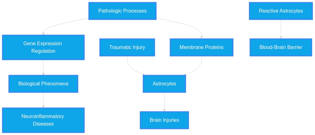
Neuroinflammatory astrocyte subtypes in the mouse brain
Plausibility Verdicts
Astrocyte subtypes in the mouse brain are best described as dynamic functional states along a continuum, rather than fixed, rigid categories.
Mouse brain astrocytes display transcriptional diversity, with specific reactive subtypes orchestrating neuroinflammation and BBB integrity.
Astrocyte subtypes are not binary; they are highly dynamic, state-dependent functional cells influenced by specific signaling hubs like STING, FGF13, and Tweak.
Dataset Summary
Novel & Overlooked Insights
- Astrocyte activation is not exclusively a proliferative process; in models such as peripheral nerve injury, spinal astrocytes respond primarily through remodeling rather than cell division.
- The astrocyte-microglia network, rather than individual cell activation, serves as the critical functional unit for containing lesions and restoring homeostasis.
- Perisynaptic astrocyte processes represent unique "hotspots" for local protein synthesis that may bypass global cellular transcriptional states.
- The expression of specific proteins, such as MINK1 and PLEKHB1, provides a spatial coordinate system for astrocyte functional identity across different brain regions.
- Lipid metabolism (e.g., long-chain fatty acids) and mitochondrial function are primary drivers of the neurotoxic astrocyte phenotype in ischemic injury.
- The "neurotoxic" vs. "neuroprotective" paradigm for astrocyte activation is being replaced by the understanding that states are highly state-dependent and cannot be explained by simplified paradigms.
- Mechanical signaling via Piezo1, regulated by microglia-derived cytokines, links physical tissue alterations to the inflammatory profile of astrocytes.
- Transcriptional Heterogeneity:** Astrocytes exist in at least five distinct subpopulations following traumatic injury, with Osmr+ variants exhibiting specific neurotoxic and protective metabolic signatures.
- Mechanical Sensing:** Endothelial Piezo1 sensors translate mechanical stress into astrocytic apoptosis via cAMP-Epac1 microvesicular signaling.
- Gut-Brain Signaling:** Chronic enteric gliosis in Parkinson's disease-model mice (A53T) precedes CNS inflammation, driven by LRRK2 up-regulation.
- Barrier Regulation:** Astrocytes serve as primary regulators of the blood-brain barrier, often utilizing the cGAS-STING pathway to govern tight junction stability.
- Regenerative Potential:** "Direct in situ astrocyte-to-neuron reprogramming offers a compelling regenerative alternative by leveraging the abundant endogenous glial reservoir," though this is hindered by existing epigenetic memory.
- Stress Resilience:** Structural depolymerization of AQP4 orthogonal array particles in A25Q mutant mice confers resilience to chronic stress by dampening glial-mediated neuroinflammation.
- Developmental Plasticity:** Adolescent intermittent ethanol exposure disrupts the physical and functional coupling of astrocytes to synapses, a deficit that persists into adulthood.
- Metabolic Rewiring:** Astrocytes undergo significant metabolic transitions during reprogramming, shifting from glycolysis to oxidative phosphorylation to support nascent neuronal survival.
- Astrocyte reactivity is not merely a binary 'A1/A2' state; modern transcriptomic analysis reveals finer gradations of cellular activation.
- The TRPC6-STING pathway represents a specific, druggable hub for stabilizing the blood-brain barrier via astrocytes during ischemia.
- Peripheral inflammation, as seen in atopic dermatitis or respiratory infection, directly reshapes cortical astrocytic transcriptional landscapes.
- Senescence markers in astrocytes and neurons represent a distinct, aging-associated inflammatory pathway mediated by cGAS-STING.
- Dietary interventions, such as a nut-enriched diet, can actively suppress pro-inflammatory astrocyte markers in AD mouse models.
- Clusterin (CLU) secretion from astrocytes, triggered by STING activation, is a primary driver of oligodendrocyte apoptosis in MS.
- FGF13 acts as a critical molecular switch that prevents astrocytic apoptosis and associated depression-like behavioral deficits.
- The Tweak/Fn14 and Stat1 signaling loop constitutes a positive feedback mechanism specifically fueling astrocytic activation in TLE.
Extracted Discoveries
- Perform spatial transcriptomics on astrocyte perisynaptic processes in multi-hit models of neurodegeneration to map the influence of local vs. global signals.
- Test the therapeutic efficacy of temporal-specific inhibition of LMP2 in late-stage chronic neuroinflammatory models.
- Spatial transcriptomic profiling of Osmr+ astrocytes in chronic versus acute neurodegeneration models.
- Chemogenetic activation/silencing of identified reactive astrocyte subpopulations to measure synaptic recovery.
- Perform single-cell spatial transcriptomics on astrocyte populations in the Tweak/Snhg3-knockout mouse TLE model.
- Assess the effect of astrocyte-specific FGF13 supplementation on hippocampal synaptic density in aging models.
- Use patch-clamp electrophysiology on astrocytes sorted by specific disease-associated markers to determine functional shifts in glutamate homeostasis.
- Longitudinal analysis of astrocyte proteomic signatures in aging populations vs. disease-associated models using spatial proteomics.
- Comparative analysis of human vs. mouse astrocyte reactivity markers to bridge translational gaps in current CNS research.
- Cross-species transcriptomic meta-analysis to determine if mouse astrocyte states correlate with human pathological lesions.
- Longitudinal imaging of astrocyte-neuron crosstalk using sensors for calcium and neurotransmitter uptake post-injury.
- A comparative longitudinal transcriptomic study of astrocytic subtypes across various stages of Alzheimer's disease progression.
- Meta-analysis of astrocyte-specific transcriptomic datasets to reconcile nomenclature differences between injury-reactive models.
- S100B inhibition in astrocyte perisynaptic processes may mitigate pre-symptomatic synaptic loss in non-KLEFS1 neurodegenerative conditions.
- EHMT1 deficiency in astrocytes increases S100B levels leading to network hyperactivity (Source: 42378039).
- Early translational dysregulation in PAPs in AD precedes plaque deposition (Source: 42425228).
- S100B regulation within astrocyte sub-compartments via JAK-STAT3 signaling.
- Since S100B is a marker for inflammatory reactive states and JAK-STAT3 is a known driver of Serpina3n expression in PAPs, it is plausible that S100B accumulation is a downstream target of this early translational pathway in broader neurodegenerative models.
- Discovered Hypothesis (A to C): Inhibition of AQP4 orthogonal array particle stabilization via site-specific mutations could mitigate astrocyte-driven neurotoxicity in traumatic brain injury.
Literature A (Origin): AQP4-A25Q mutations prevent OAP assembly and improve stress resilience (42595228).
Literature C (Target): Osmr+ reactive astrocytes and neurotoxicity in TBI (42603599).
The Intersecting Bridge B: Reactive gliosis and neuroinflammatory pathway suppression (GFAP/cGAS-STING).
Biological Rationale: Reducing AQP4-dependent OAP formation likely limits the astrocytic stress response that precipitates the pathological reactive states observed in TBI, thereby preventing the transition to the Osmr+ neurotoxic phenotype. - Snhg3-mediated astrocytic activation is a key metabolic driver of synaptic vulnerability in aging-related neurodegenerative niches.
- Tweak/Snhg3 positive feedback loop in astrocytes drives TLE (ID 42456384).
- Astrocytic energy metabolism genes are critical for neuron protection in AD pathology (ID 42403013).
- Snhg3/Tweak-driven metabolic reprogramming.
- The Tweak/Snhg3 loop alters gene transcription; if this loop is active in AD, it likely impairs the metabolic homeostasis required for healthy OL-astrocyte-neuron communication.
- Conflicting roles of zafirlukast: ID 42557520 reports zafirlukast exacerbates seizure activity despite reducing neurodegeneration markers, while other studies (e.g., 42458512, 42398271) suggest inflammatory modulation is consistently protective, indicating target-specific complexities in epileptogenesis.
- Evidence regarding astrocyte roles varies from 'protective/homeostatic' to 'deleterious/pro-inflammatory' based on the insult type and temporal window (42589548), indicating that astrocyte phenotype is highly conditional.
- There is a tension in the literature between viewing astrocytes as a binary A1 (toxic)/A2 (protective) paradigm versus the emerging evidence from single-cell transcriptomics which suggests a vast, fluid landscape of reactive states depending on the specific inflammatory trigger and disease stage.
- The use of HFn-ApoE130-149 nanocarriers to target the LRP1-NF-κB signaling axis represents a repurposed solution for modulating astrocyte reactivity in various neuroinflammatory disorders, not just NMOSD, by crossing the BBB to restore astrocytic homeostasis.
- Intranasal NPY and intranasal insulin are identified as potent therapeutic strategies for modulating astrocyte reactive states and preserving the neurovascular unit after injury (42575454, 42600992).
- Repurpose Tweak/Snhg3 inhibitors developed for epilepsy (42456384) to mitigate glial reactivity and cognitive decline in AD models, as common pathways involving inflammatory signaling are implicated in both.
PathMap is funded by sales of datasets and coversheets to researchers of any kind who wish to discover the most viable routes and paths to accelerate cures. We do not make theoretical molecules, we expose the truth in current PubMed literature. Commission a trace today.
PathMap Scores
How are these metrics evaluated?
Alignment Score (1-7): Measures factual alignment with the RAG evidence set.
[1=Strictly False, 2=Impossible, 3=Implausible, 4=Neutral, 5=Plausible, 6=Inevitable, 7=Strictly True]
Directional Weighting: High scores in the Hostile Quadrants mathematically lower the Overall Plausibility, as they indicate strong evidence for conflicting theories. Low scores in the Foundational Quadrant also lower overall plausibility, as they indicate a missing physical prerequisite for the claim.
Veridicality Audit Report
All Extracted Datapoints
Evaluated Perspectives & Quadrants
CLAIM EVALUATED AND ANSWER TO USER
The claim evaluated is that neuroinflammatory astrocyte subtypes in the mouse brain are well-defined entities that mediate pathology in neurodegenerative disorders. The evidence set indicates that while astrocyte reactivity is a hallmark of neuroinflammatory responses in various mouse models, the categorization into distinct, stable subtypes remains an area of active investigation. The provided literature suggests that astrocytic responses exist along a functional continuum rather than being confined to simple binary "neurotoxic" or "neuroprotective" states.ABSTRACT & REWRITTEN CLAIM
Neuroinflammatory astrocyte activation is a complex, state-dependent phenomenon. Rather than functioning as static subtypes, astrocytes exhibit dynamic transcriptomic and proteomic remodeling in response to diverse pathological stimuli (e.g., ischemia, amyloid-beta, pro-inflammatory cytokines). Evidence supports a "continuum model" of astrocyte states, where functional programming is influenced by cell-type-specific regulators, local metabolic demands, and bidirectional crosstalk with microglia.INTRODUCTION & JUSTIFICATION
Astrocyte activation, often termed astrogliosis, is an essential mechanism in the central nervous system (CNS) response to injury and neurodegeneration. In mouse models, this reactivity is characterized by the upregulation of intermediate filament proteins such as GFAP. Recent high-resolution molecular profiling has challenged the existence of binary astrocyte phenotypes. Instead, research indicates that reactive astrocytes undergo heterogeneous remodeling, influenced by specific upstream signaling pathways and metabolic shifts. For instance, the immunoproteasome subunit LMP2 has been identified as a critical regulator that modulates the balance between inflammatory and reparative gene programs in astrocytes following ischemic injury. Similarly, local translation of specific mRNAs in perisynaptic astrocyte processes provides an early, compartment-specific layer of control that contributes to synaptic dysfunction in Alzheimer’s disease. Crucially, the functional outcome of astrocytic activation is highly context-dependent, often necessitating coordinated interactions with other glial populations, such as microglia, through signaling axes like the CX3CR1 or the LRP1-NF-κB pathway. Consequently, current research is moving toward a framework where reactive states are viewed as dynamic "motifs" shaped by spatial and temporal microenvironmental factors.Novel & Overlooked
EVIDENCE, METHODOLOGY & CITATIONS
1. ID: 42504987 - Application: LMP2 regulates the transition of astrocyte states. - "LMP2 coordinately modulates inflammatory and reparative signaling networks by promoting nuclear factor kappa-B (NF-κB)-dependent inflammatory activation while constraining transforming growth factor-β1(TGF-β1)/SMAD family member 3 (Smad3)-associated reparative responses" 2. ID: 42547642 - Application: Notes the limitations of binary classification. - "exosome function is highly state-dependent and cannot be fully explained by simplified pro-inflammatory microglia anti-inflammatory microglia (M1/M2) or A1/A2 paradigms" 3. ID: 42467524 - Application: Spatial heterogeneity of astrocytes. - "MINK1 and PLEKHB1 showed preferential expression in hippocampal and cortical astrocytes, respectively, highlighting their potential as region-specific astrocyte markers." 4. ID: 42362040 - Application: LPI effects on astrocytic activation in AD. - "Exogenous LPI treatment reduced cerebral Aβ deposition, improved performance in learning and memory behavioral tasks, reduced pathological microglial aggregation, inhibited astrocyte proliferation, and ameliorated hippocampal oxidative stress." 5. ID: 42539240 - Application: Metabolic profiling of astrocytes. - "Integrated epigenomic, proteomic, and metabolomic profiling of cortical astrocytes isolated 28 days after injury revealed a pronounced metabolic restriction in wild type astrocytes" 6. ID: 42552048 - Application: Metabolic support in AD. - "While the brain relies on lactate as a prominent energy substrate, astrocytic metabolic defects lead to impaired neuronal energy homeostasis, thereby promoting neurodegeneration." 7. ID: 42511849 - Application: Tay-Sachs astrocyte model features. - "The resulting cell line recapitulates key pathological features, including lysosomal accumulation, increased neutral lipid content, reduced mitochondrial mass, and elevated reactive oxygen species production." 8. ID: 42462474 - Application: circSLC8A1 localization. - "circSLC8A1 was significantly upregulated in the hippocampus of TLE mice and predominantly localized in astrocytes." 9. ID: 42560948 - Application: P2X7 mechanism in heat stress. - "Mild hyperthermia upregulated P2X7 expression in astrocytes and induced a substantial calcium influx, leading to activation of the Calcineurin-NFAT pathway." 10. ID: 42523300 - Application: AQP4 in vascular disease. - "Aquaporin 4 (AQP4) water channels are polarized to astrocytic endfeet at blood vessel interfaces, and lose polarity in vascular diseases" 11. ID: 42449389 - Application: Nanotherapeutic impact on astrocyte phenotype. - "The treatment induced a spatial and phenotypic restructuring of the astrocytic response, notably reducing excessive astrocyte accumulation around lesions while encouraging a proliferative and reparative phenotype." 12. ID: 42378039 - Application: S100B in EHMT1 models. - "EHMT1+/- astrocytes displayed elevated levels of S100B, a neuroinflammatory marker contributing to neuronal network hyperactivity." 13. ID: 42369041 - Application: Cx50 mediated crosstalk. - "Cx50 knockdown differentially modulates pro-inflammatory cytokines in BDSO-treated conditions, indicating strain-dependent effects and Cx50-mediated neuron-astrocyte crosstalk" 14. ID: 42439282 - Application: LPS effects on astrocyte cell lines. - "LPS reduced cell viability in a concentration-dependent manner in both AWT and MG6 cells, inducing apoptotic cell death in AWT cells" 15. ID: 42599550 - Application: Astrocyte reactive state in PD. - "Activated astrocytes accelerate neuronal degeneration by releasing inflammatory mediators, disrupting glutamate homeostasis, and triggering oxidative stress." 16. ID: 42456384 - Application: Tweak/Snhg3 feedback loop. - "Tweak and Snhg3 mutually upregulated each other and synergistically activated the Stat1 pathway, forming a positive feedback loop in MAs that collectively drived astrocyte activation." 17. ID: 42425228 - Application: Local translation in PAPs. - "Mechanistically, early Serpina3n upregulation was partially driven by JAK-STAT3 signaling, with preferential effects in astrocyte processes." 18. ID: 42365203 - Application: Distinct glial phenotypes. - "However, there is growing evidence that human and mouse glia adopt distinct phenotypes in response to neurodegeneration." 19. ID: 42502884 - Application: Piezo1 regulation. - "Microglial depletion in 5xFAD mice consistently and substantially reduced astrocytic Piezo1 expression in vivo, supporting the physiological relevance of this microglia-astrocyte signaling axis during pathology." 20. ID: 42557483 - Application: circRNAs in PD. - "Recent research has indicated that circRNAs participate in PD pathophysiology by modulating neuroinflammation, immune response, mitochondrial dysfunction, and reactive oxygen species accumulation."CLAIM EVALUATED AND ANSWER TO USER
"Neuroinflammatory astrocyte subtypes in the mouse brain"ABSTRACT & REWRITTEN CLAIM
Neuroinflammatory astrocyte subtypes are defined by heterogeneous transcriptional states that modulate central nervous system (CNS) homeostasis, blood-brain barrier (BBB) integrity, and neuronal repair. Research identifies specific reactive astrocyte subpopulations (e.g., Osmr+ astrocytes) that emerge following traumatic injury, exhibiting distinct metabolic and inflammatory gene signatures. These astrocytes function as critical nodes in orchestrating neuroimmune responses through the release of cytokines, interactions with microglia, and regulation of the glymphatic system.INTRODUCTION & JUSTIFICATION
Astrocytes are no longer viewed merely as structural support cells; they are highly diversified, homeostatic regulators of the CNS. Recent transcriptomic evidence, particularly in mouse models of injury and neurodegeneration, has mapped distinct reactive profiles. A landmark reanalysis of single-cell RNA-sequencing data identified that "We identified five astrocyte subpopulations." Among these, "Descriptive analysis of the pooled cells showed a greater relative proportion of C3 Osmr+ astrocytes in the TBI group than in the Sham group." This specific subtype exhibits a "highly reactive transcriptional state with concurrent protection-associated and neurotoxicity-associated features." The reactivity of astrocytes is context-dependent. While they can be neuroprotective, "Persistent biomechanical and ischemic insults shift this profile into a typically deleterious one." In the context of inflammatory responses, "These findings emphasize the ability of astrocytes to act as immunocompetent cells that coordinate inflammatory responses through mechanisms such as the NOD-like receptor and NF-κB pathways." This capacity extends to the regulation of innate immunity, as "Astrocytic TRPC6 maintains BBB integrity by negatively regulating the cGAS-STING innate immune pathway in the early phase of CIRI." The functional consequences of these inflammatory states are profound: "Mechanistically, DNA synergized with pro-inflammatory cytokines to trigger astrocytic STING signaling, driving Clusterin (CLU) secretion that directly induced oligodendrocyte apoptosis and demyelination."Novel & Overlooked
EVIDENCE, METHODOLOGY & CITATIONS
1. ID: 42603599 - "We identified five astrocyte subpopulations. Descriptive analysis of the pooled cells showed a greater relative proportion of C3 Osmr+ astrocytes in the TBI group than in the Sham group." 2. ID: 42603599 - "This subpopulation exhibited a highly reactive transcriptional state with concurrent protection-associated and neurotoxicity-associated features" 3. ID: 42604981 - "Astrocytes are fundamental elements of the pathophysiology of neurological, neuropsychiatric, and neurodegenerative diseases" 4. ID: 42589548 - "Initially, the macroglial response is adaptive and neuroprotective. Persistent biomechanical and ischemic insults shift this profile into a typically deleterious one" 5. ID: 42586471 - "Astrocytic TRPC6 maintains BBB integrity by negatively regulating the cGAS-STING innate immune pathway in the early phase of CIRI." 6. ID: 42601829 - "Mechanistically, DNA synergized with pro-inflammatory cytokines to trigger astrocytic STING signaling, driving Clusterin (CLU) secretion that directly induced oligodendrocyte apoptosis and demyelination." 7. ID: 42576543 - "The vesicular cAMP payload subsequently activated the downstream effector Epac1, dictating a spatially restricted wave of astrocyte apoptosis." 8. ID: 42593416 - "A53T mice, without central neuroinflammation, showed intestinal disturbances, colonic α-synuclein accumulation and an increase in colonic GFAP+/LRRK2+ glial cells before brain pathology." 9. ID: 42574907 - "In ICH, cGAS-STING activation in microglia and astrocytes drives neuroinflammation, promotes pyroptosis via inflammasome assembly, disrupts blood-brain barrier (BBB) integrity, and exacerbates secondary brain injury." 10. ID: 42604624 - "genetic knockdown as well as inhibition of NQO2 in astrocytes promote neurite regrowth of injured cortical neurons." 11. ID: 42576592 - "The role of the Purinergic receptor (P2X7 receptor), a ligand-gated ion channel activated by extracellular ATP, was examined across existing cellular mechanisms and possible pathways involved in AD" 12. ID: 42579790 - "Direct intrathecal IL-6 blockade was associated with qualitative cellular-level neuroprotection after permanent focal cerebral ischemia." 13. ID: 42573852 - "Direct in situ astrocyte-to-neuron reprogramming offers a compelling regenerative alternative by leveraging the abundant endogenous glial reservoir." 14. ID: 42595228 - "CUMS induced marked astrocytic (GFAP, S100β) and microglial (Iba-1, CD68) activation in WT hippocampus, but these responses were largely absent in mutants." 15. ID: 42603821 - "AIE induces structural and functional decoupling of astrocytes from synapses and astrocyte dysregulation that persists into adulthood." 16. ID: 42568651 - "After SCI, autonomic dysfunction, impaired gut motility, and neurogenic bowel dysfunction may disrupt the homeostasis of gut microbiota and barrier" 17. ID: 42591297 - "These findings emphasize the ability of astrocytes to act as immunocompetent cells that coordinate inflammatory responses through mechanisms such as the NOD-like receptor and NF-κB pathways." 18. ID: 42600992 - "Intranasal rhInsulin treatment significantly attenuated HI-induced behavioral deficits by 100% and increased myelination (MBP+) by 64% in cingulate white matter, restored dendritic expression (MAP2+) by 56%, and reduced astrocytes (GFAP+) by 70% in hippocampal regions" 19. ID: 42574907 - "In SAH, the pathway is engaged through cytosolic mtDNA release and transcellular cGAMP transfer from neurons to microglia, amplifying neuroinflammation, ferroptosis, NLRP3 inflammasome activation, and autophagic dysfunction." 20. ID: 42576543 - "Although Piezo1 was broadly detected in glial cells, genetic ablation of Piezo1 exclusively in endothelial cells, but not in astrocytes or microglia, significantly reduced infarct volume and improved neurological deficits."CLAIM EVALUATED AND ANSWER TO USER
Neuroinflammatory astrocyte subtypes in the mouse brainABSTRACT & REWRITTEN CLAIM
This assessment evaluates the evidence regarding distinct neuroinflammatory astrocyte subtypes (often termed A1/A2-like or disease-associated) within the murine central nervous system. The literature identifies that astrocytes undergo profound transcriptional and morphological reprogramming under pathological conditions, such as traumatic brain injury (TBI), ischemia, and neurodegeneration. These subtypes are characterized by specific molecular markers and signaling axes that either promote tissue damage or facilitate repair, though recent data emphasize that these classical binary labels (A1/A2) are simplified representations of a complex, heterogeneous cellular state.INTRODUCTION & JUSTIFICATION
Astrocytes are no longer viewed merely as passive support cells; they are dynamic participants in CNS pathology, capable of adopting distinct transcriptional states in response to injury. "These findings emphasize the ability of astrocytes to act as immunocompetent cells that coordinate inflammatory responses through mechanisms such as the NOD-like receptor and NF-κB pathways." This immune competence is further refined through specific genetic pathways. "The study identified the "Astrocyte TRPC6-STING-Tight Junction" axis, offering a precise and promising novel therapeutic target for CIRI." Under aging or injury, these cells demonstrate marked morphological remodeling. "Data show that brain aging alters the tridimensional structure of microglia and astrocytes in the hippocampus and prefrontal cortex." Furthermore, the "A1/A2" paradigm, while historically used to categorize reactive states, is now recognized as insufficient to capture the full diversity of these responses, particularly when interventions like electroacupuncture modulate them. "EA at GV20 and GV24 improves cognitive impairment and attenuate neuroinflammation in VD rats possibly by inhibiting TLR4/MyD88/NF-κB signaling and regulating astrocytic A1/A2-like phenotypic imbalance."Novel & Overlooked
EVIDENCE, METHODOLOGY & CITATIONS
1. ID: 42591297 - These findings emphasize the ability of astrocytes to act as immunocompetent cells that coordinate inflammatory responses through mechanisms such as the NOD-like receptor and NF-κB pathways. 2. ID: 42586471 - The study identified the "Astrocyte TRPC6-STING-Tight Junction" axis, offering a precise and promising novel therapeutic target for CIRI. 3. ID: 42582005 - In contrast, PE more consistently reverses age-related microglial gene expression changes and induces region-specific remodeling of astrocytic morphology. 4. ID: 42576490 - EA at GV20 and GV24 improves cognitive impairment and attenuate neuroinflammation in VD rats possibly by inhibiting TLR4/MyD88/NF-κB signaling and regulating astrocytic A1/A2-like phenotypic imbalance. 5. ID: 42547491 - Studies suggest that in schizophrenia, inflammatory changes may influence brain cells, chemical signals, stress pathways, and the systems involved in sensing and responding to pain. 6. ID: 42462474 - Our findings identify a novel circSLC8A1/PTBP1/CEBPB signaling axis that mediates astrocytic inflammation and pyroptosis in TLE. 7. ID: 42444329 - These findings support a role for C1q in astrocytic C3 induction and the engulfment of both synapses and amyloid. 8. ID: 42438359 - Data show that brain aging alters the tridimensional structure of microglia and astrocytes in the hippocampus and prefrontal cortex. 9. ID: 42421017 - Astrocyte-specific knockout of FGF13 induces astrocytic apoptosis, exacerbates inflammatory levels, and aggravates depression-like behaviors in mice. 10. ID: 42418159 - In APP mice, NED enhanced hippocampal-dependent memory, reduced microglia and astrocyte reactivity, decreased cortical and hippocampal Aβ plaque burden, and preserved dendritic spine density. 11. ID: 42401926 - In addition, elevated expression of β-galactosidase, a senescence marker, was predominantly observed in neurons compared to microglia and astrocytes, indicating a primary role for neurons in infection-associated senescence. 12. ID: 42446255 - In the control, microglial cells possessed a large number of processes typical of nonactivated cells. 13. ID: 42599550 - Recent studies indicate that mutations in PD-associated genes may induce functional alterations in astrocytes. 14. ID: 42575454 - The ipsilateral hippocampus exhibited early blood-brain barrier (BBB) disruption and astrocytic alterations, whereas the contralateral hippocampus developed a more pronounced and sustained inflammatory response characterized by microglial activation and increased expression of inflammatory and endothelial activation markers. 15. ID: 42567990 - TBI significantly and temporarily increased the expression of PDCD1 in vivo, with PDCD1 mostly expressed in microglia and neurons, but not in astrocytes. 16. ID: 42552556 - However, a significant increase in Galectin-3/GFAP colocalization compared to control at the same timepoint in the rat additionally associates Galectin-3 production with astrocytes and Müller glia. 17. ID: 42557563 - Spatial transcriptomics revealed that CXCL10 is mainly expressed by disease-associated astrocytes, defining an astrocytic CXCL10-rich inflammatory niche within the tauopathy brain. 18. ID: 42456384 - Conversely, up-regulation of Tweak or Snhg3 promoted proliferation, migration, and inflammatory factor secretion in mouse astrocytes (MAs), indicating that TWEAK and Snhg3 each induce glial activation in vitro. 19. ID: 42484902 - We demonstrated morphological alterations of BAM populations in the meninges and parallel disintegration of the astrocyte barrier due to haemorrhage. 20. ID: 42427668 - E4FAD mice demonstrated plaque reductions with accompanying increases in microhemorrhages (measured on both MRI and histology), and increases in microglial and astrocyte reactivity - especially in the perivascular compartment.Verbatim Quote Audit Console
Mapped Reference Directory (APA)
- [1] ID: 42504987 - Mao Y, Ma R, Lin Z, Zhao H, Liu Y et al. (2026). Astrocytic LMP2 Coordinates NF-κB and TGF-β1/Smad3 Signaling to Drive Neuroinflammation after Cerebral Ischemia/Reperfusion.. Advanced science (Weinheim, Baden-Wurttemberg, Germany). ID: 42504987.
- [2] ID: 42547642 - Yang Z, Chen H, Zhang Z, Wei X, Han W et al. (2026). The Dual Roles of Microglia- and Astrocyte-Derived Exosomes in Cerebral Ischemia-Reperfusion Injury: from Intercellular Communication to Therapeutic Prospects.. Molecular neurobiology. ID: 42547642.
- [3] ID: 42467524 - Huang CC, Chang CY, Chan PC, Chong WM, Chang HJ et al. (2026). Single Cell-Type Spatial Proteomics Uncovers Regional Heterogeneity of Astrocytes.. Journal of proteome research. ID: 42467524.
- [4] ID: 42362040 - Xu W, Cao J, Liu Y, Wei Z, Zha X et al. (2026). LPI alleviates Alzheimer's disease pathology via the GPR55 receptor.. Neuroscience. ID: 42362040.
- [5] ID: 42539240 - Delgado T, Arefin TM, Pagan I, Weekley BH, Rodwell-Bullock J et al. (2026). Transglutaminase 2 Deletion Enhances Astrocyte-to-Neuron Metabolic Support and Attenuates Subacute Pathology Following Repetitive Mild Traumatic Brain Injury.. bioRxiv : the preprint server for biology. ID: 42539240.
- [6] ID: 42552048 - Baskar G, Kandasamy M (2026). Energetic crisis, mitochondrial vulnerability and disruption of lactate shuttle in Alzheimer's disease.. International review of neurobiology. ID: 42552048.
- [7] ID: 42511849 - Suárez-García DA, Espejo-Mojica AJ, Alméciga-Díaz CJ (2026). Modeling Tay-Sachs Disease in Astrocyte-like Cells Reveals Significant Changes in the Transcriptomic Profile.. International journal of molecular sciences. ID: 42511849.
- [8] ID: 42462474 - Chai W, Wan Y, Nie Y, Kang Q (2026). Astrocytic circular RNA SLC8A1 boosted CEBPB/NLRP3-triggered pyroptosis by stabilizing PTBP1 to drive neuroinflammation in temporal lobe epilepsy.. International immunopharmacology. ID: 42462474.
- [9] ID: 42560948 - Wang Q, Chen X, Zhang J, Zhang Y, Yang H (2026). Heat stress-activated P2X7 receptor induces astrocyte activation and regulates glioma tumor microenvironment via calcium signaling pathway.. Neuroimmunomodulation. ID: 42560948.
- [10] ID: 42523300 - Flores S, Wilpitz A, Ojeda-Juarez D, Wang J, Danque G et al. (2026). Aquaporin-4 mislocalization from astrocyte endfeet prolongs survival in a prion-cerebral amyloid angiopathy model.. bioRxiv : the preprint server for biology. ID: 42523300.
- [11] ID: 42449389 - Zhao X, Liang Q, Lin K, Jiang S, Yang T et al. (2026). Ferritin-ApoE nanocarrier for targeted therapy of neuromyelitis optica spectrum disorder in mice.. Journal of neuroinflammation. ID: 42449389.
- [12] ID: 42378039 - Vermeulen-Kalk K, Wang S, Kummeling J, Mossink B, Wijnant K et al. (2026). Astrocytes contribute to olanzapine-mediated reversal of kleefstra syndrome-associated neurodevelopmental regression.. The Journal of clinical investigation. ID: 42378039.
- [13] ID: 42369041 - Puangmalai N, Sengupta U, Bhatt N, Suthprasertporn N, Al-Shaebi F et al. (2026). Connexin 50 mediates disease-relevant alpha-synuclein oligomer propagation and neuroinflammation in neurodegenerative disease.. iScience. ID: 42369041.
- [14] ID: 42439282 - Eguchi R, Ishida A, Suzuki-Yamamoto T, Maru I (2026). S-allyl cysteine suppresses lipopolysaccharide-induced microglial inflammation accompanied by attenuation of JNK1/2 and STAT3 signaling.. Nutritional neuroscience. ID: 42439282.
- [15] ID: 42599550 - Li Y, Li Q, Wang Y, Wang X, Di G et al. (2026). Astrocytes orchestrate neuroprotection and pathogenesis via bidirectional neuronal interactions in Parkinson's disease.. Journal of physiology and biochemistry. ID: 42599550.
- [16] ID: 42456384 - Li P, Cao B (2026). Tweak regulates glial cell activation in temporal lobe epilepsy through a positive feedback circuit.. Tissue & cell. ID: 42456384.
- [17] ID: 42425228 - Avila-Gutierrez K, de Sauvage MÁC, Depret N, Oudart M, Thompson R et al. (2026). Local translation controls early reactive changes in perisynaptic astrocyte processes at pre-symptomatic stages of Alzheimer's disease.. Neurobiology of disease. ID: 42425228.
- [18] ID: 42365203 - MacLean M, Lydon SD, Gomes C, Pizzi EM, Diemler CA et al. (2026). Neuroinflammation in glaucoma: a myriad of cellular pathways and players.. Mammalian genome : official journal of the International Mammalian Genome Society. ID: 42365203.
- [19] ID: 42502884 - Bai Y, Choi HB, Wendt S, Towriss M, Lin AJ et al. (2026). A Microglia-Astrocyte Signaling Axis Regulates Astrocyte Piezo1 Expression and Inflammatory Responses.. Glia. ID: 42502884.
- [20] ID: 42557483 - El-Ashmawy NE, Khedr EG, Darwish RT, Ibrahim AO (2026). Cross-link Between CircRNAs and Neuroinflammation in Parkinson's Disease.. Molecular neurobiology. ID: 42557483.
- [21] ID: 42603599 - Zhao F, Cao L, Chen J, Li G, Guo Y et al. (2026). Single-cell reanalysis characterizes an Osmr+ astrocyte state and predicts midkine signaling to Cox6b1+ glutamatergic neurons at 24 h after traumatic brain injury.. Brain research. ID: 42603599.
- [22] ID: 42604981 - Falcone C, Arckens L, Baiula M, Bedini A, Bocchi R et al. (2026). Written in the Stars: Astrocyte Biology From Evolution to Disease.. Acta physiologica (Oxford, England). ID: 42604981.
- [23] ID: 42589548 - Teixeira GR, Costa AGA, Mattos ACL, Souza Monteiro de Araújo D, Brito R et al. (2026). The Dual Role of Macroglia in Glaucoma: Deciphering the Contributions of Astrocytes and Müller Cells to Retinal Neurodegeneration and Neuroprotection.. International journal of molecular sciences. ID: 42589548.
- [24] ID: 42586471 - Li Y, Li Y, Qiu S, Gu L, Zhang Y et al. (2026). Astrocytic TRPC6 protects against cerebral ischemia-reperfusion injury by inhibiting cGAS-STING pathway.. Experimental neurology. ID: 42586471.
- [25] ID: 42601829 - Hu S, Xiao X, Cheng X, Huang Y, Cui T et al. (2026). The cGAS-STING Pathway Drives Astrocyte-Mediated Demyelination in Multiple Sclerosis Through Clusterin Secretion.. CNS neuroscience & therapeutics. ID: 42601829.
- [26] ID: 42576543 - Liu Y, Sun M, Shen M, Yang X, Sheng Z et al. (2026). Mechanochemical endothelial-astrocyte signalling via Piezo1-Epac1 drives neurovascular injury after stroke.. Brain : a journal of neurology. ID: 42576543.
- [27] ID: 42593416 - D'Antongiovanni V, Pierucci C, Segnani C, Ippolito C, Di Salvo C et al. (2026). Up-regulation of the kinase LRRK2, in enteric glia contributes to mucosal barrier impairment in Parkinson's disease via secretory autophagy.. British journal of pharmacology. ID: 42593416.
- [28] ID: 42574907 - Wang Y, Dou L, Gao Y, Zhang M, Wang Z et al. (2026). cGAS-STING targeting offers a novel therapeutic paradigm in hemorrhagic stroke.. Tissue & cell. ID: 42574907.
- [29] ID: 42604624 - Wang Y, Liao WL, Wang C, Li YC, Lu TH et al. (2026). Isorhoifolin regulates S1PR3-CK2-GSK3β axis and promotes neurite regrowth and functional recovery after traumatic brain injury.. Phytomedicine : international journal of phytotherapy and phytopharmacology. ID: 42604624.
- [30] ID: 42576592 - Jeeru TR, Palathoti N, Swaminathan G (2026). The Multifaceted Role of the P2X7 Receptor in Alzheimer's Disease: A Unifying Pathological Link.. CNS & neurological disorders drug targets. ID: 42576592.
- [31] ID: 42579790 - Zeren M, İldan F (2026). Ultrastructural neuroprotection by intrathecal interleukin-6 antagonism in a rat model of permanent focal cerebral ischemia.. Ultrastructural pathology. ID: 42579790.
- [32] ID: 42573852 - Nguyen HM, Nguyen LDT (2026). Towards Structural Restoration: Epigenetic Reprogramming and Direct Astrocyte-to-Neuron Lineage Conversion as Next-Generation Regenerative Neurotherapeutics.. Molecular neurobiology. ID: 42573852.
- [33] ID: 42595228 - Kundu S, Ai Y, Huang YL, Lu JC, Wu T et al. (2026). Depolymerization of aquaporin-4 orthogonal array particles via the A25Q mutation does not cause behavioral deficits but confers resilience to chronic unpredictable mild stress.. Journal of affective disorders. ID: 42595228.
- [34] ID: 42603821 - Coulter O, Walker CD, Carter T, Sexton HG, Denvir J et al. (2026). Adolescent alcohol exposure disrupts astrocyte-synaptic structural and functional coupling in the male dorsal hippocampus.. Molecular psychiatry. ID: 42603821.
- [35] ID: 42568651 - Yi B, Chen W, Chi Z, Mao Q, Li X et al. (2026). Gut-derived signals regulating glial activation and secondary neuroinflammation after spinal cord injury: an evidence mapping and mechanistic framework.. Frontiers in cellular neuroscience. ID: 42568651.
- [36] ID: 42591297 - Luque-Bolivar A, Ruiz-Araujo K, Aristizábal-Pachón AF, González J (2026). Integrated meta-analysis of human astrocytes transcriptomes reveals a candidate recurrent inflammatory signature in response to inflammatory and immune stimuli.. Frontiers in cellular neuroscience. ID: 42591297.
- [37] ID: 42600992 - Dankhara N, Lee JW, Ojeda NB, Tucci MA, Lu S et al. (2026). Intranasal insulin reduces ADHD-like behaviors and neurodevelopmental deficits following neonatal hypoxia-ischemia in juvenile rats.. Neurochemistry international. ID: 42600992.
- [38] ID: 42582005 - Singhal G, Baune BT (2026). Differential effects of environmental enrichment and physical exercise on glial biology in aging and aging-related conditions: a systematic review.. Frontiers in cellular neuroscience. ID: 42582005.
- [39] ID: 42576490 - Gao J, Shi C, Li W, Shang X, Wang F et al. (2026). [Electroacupuncture ameliorates cognitive impairment and suppresses TLR4/MyD88/NF-κB pathway-mediated astrocyte activation in rats with vascular dementia].. Nan fang yi ke da xue xue bao = Journal of Southern Medical University. ID: 42576490.
- [40] ID: 42547491 - Sedghi Esfahani S, Mahdinia E, Dehkhodaei S, Abedi Oumali N, Taherkhani S et al. (2026). Neuroinflammatory pathways linking pain and rehabilitation outcomes in schizophrenia: a narrative review.. Pain management. ID: 42547491.
- [41] ID: 42444329 - Petrisko TJ, Chu SH, Gomez-Arboledas A, Zhang B, Tenner AJ (2026). Young Adult Microglial Deletion of C1q Reduces Engulfment of Synapses and Partially Mitigates Cognitive Impairment in an Aggressive Alzheimer's Disease Mouse Model.. Glia. ID: 42444329.
- [42] ID: 42438359 - Lopes CR, Ferreira SG, Cunha RA, Agostinho P (2026). Genetic Deletion of Adenosine A2A Receptors Attenuates Aged-Related Alterations of Glial Cells Morphology and of Inflammasome in the Hippocampus and Prefrontal Cortex of Mice.. Glia. ID: 42438359.
- [43] ID: 42421017 - Zheng R, Zhang Y, Tu Z, Luo Y, Lin H et al. (2026). FGF13 alleviates astrocytic apoptosis via JIP2 inhibition in the hippocampus and mitigates depression-like behavior.. Journal of neuroinflammation. ID: 42421017.
- [44] ID: 42418159 - Panisello L, Millet-Sigalat M, Novau-Ferré N, Mateu-Fabregat J, Carrasco M et al. (2026). Nut consumption as a therapeutic strategy to preserve brain function, attenuate neuropathology, and modulate cross-tissue microRNAs in a mouse model of Alzheimer's disease.. Food & function. ID: 42418159.
- [45] ID: 42401926 - Xing Y, Lv H, He P, Xu Y, Shen W et al. (2026). Targeting the cGAS-STING pathway alleviates neuroinflammation and cognitive impairment induced by chronic infection of Toxoplasma gondii.. Journal of neuroinflammation. ID: 42401926.
- [46] ID: 42446255 - Stelmashook EV, Genrikhs EE, Kapkaeva MR, Alexandrova OP, Isaev NK (2026). Methylene blue reduces the severity of lipopolysaccharide-induced morphological changes in microglia in rat cerebral cortex glial cell cultures.. Biomeditsinskaia khimiia. ID: 42446255.
- [47] ID: 42575454 - Leitão RA, Alves JL, Bernardo AL, Mota-Pinto A, Silva AP (2026). Differential consequences of traumatic brain injury in the hippocampal hemispheres of male rats and the beneficial effect of neuropeptide Y.. Brain, behavior, and immunity. ID: 42575454.
- [48] ID: 42567990 - Tian J, Wang Y, Zhao J, Guo Z, Jiang L et al. (2026). PDCD1 Signaling in Microglia Can Reduce Neuroinflammation and Apoptosis Induced by Traumatic Brain Injury by Regulating PI3K/Akt Signaling Pathway, Thereby Alleviating Neurological Dysfunction.. Molecular neurobiology. ID: 42567990.
- [49] ID: 42552556 - Rombaut A, Wang L, Lardner E, Wong RC, Taul C et al. (2026). Galectin-3 is elevated in Müller glia in human glaucomatous eyes and ocular hypertensive rat eyes and associated with phagocytosing states.. Acta neuropathologica communications. ID: 42552556.
- [50] ID: 42557563 - Uenishi R, Kawata R, Manabe T, Matsuba Y, Mihira N et al. (2026). CXCL10 contributes to female-specific pathological progression in tauopathy model mice.. Journal of neuroinflammation. ID: 42557563.
- [51] ID: 42484902 - Masood T, Lakatos S, Ignácz M, Rosta J (2026). Simultaneous activation of border-associated immune cells and glial cells at the CNS-meningeal interface after subarachnoid haemorrhage in rats.. Brain structure & function. ID: 42484902.
- [52] ID: 42427668 - Pallerla AV, Lucido CC, Saito K, Nolt GL, Arbones-Mainar J et al. (2026). Anti-amyloid immunotherapy drives APOE4 specific increases in glial reactivity, perivascular immune activation, and ARIA-like events.. bioRxiv : the preprint server for biology. ID: 42427668.
Abstract Repository (Raw Full-Texts) Show Database Collapse Database
ID: 42362040
Title: LPI alleviates Alzheimer's disease pathology via the GPR55 receptor.
Abstract: Lysophosphatidylinositol (LPI) is an endogenous GPR55 agonist, yet its role in Alzheimer's disease (AD) remains unclear. Here, we performed serum metabolomic profiling in 5xFAD mice and observed a reduction in multiple LPI species prior to the onset of overt Aβ pathology, and this decrease was further corroborated in human cohort samples. Exogenous LPI treatment reduced cerebral Aβ deposition, improved performance in learning and memory behavioral tasks, reduced pathological microglial aggregation, inhibited astrocyte proliferation, and ameliorated hippocampal oxidative stress. Mechanistically, administration of the GPR55 antagonist ML191 blocked the protective effects of LPI, while the GPR55 agonist O-1602 recapitulated these benefits, indicating that LPI acts through GPR55. Collectively, our findings suggest that reduced LPI represents an early metabolic vulnerability in the 5xFAD model and establish the LPI-GPR55 axis as a potential therapeutic target for early intervention in AD.
View on PubMed
ID: 42365203
Title: Neuroinflammation in glaucoma: a myriad of cellular pathways and players.
Abstract: Glaucoma is a complex neurodegenerative disease with multiple subtypes, yet all are characterized by the progressive dysfunction and loss of retinal ganglion cells (RGCs), which ultimately results in vision impairment and blindness. Elevated intraocular pressure (IOP) is a major risk factor for glaucoma; however, it is neither necessary nor sufficient for glaucomatous neurodegeneration, as patients can exhibit high IOP without developing glaucoma and patients can develop glaucoma with normal IOP. Yet FDA-approved treatment options are largely limited to approaches to minimize risk and reduce IOP. Thus, there is a critical need to target other aspects of glaucoma pathophysiology. Neuroinflammation is broadly defined here as immune-relevant responses, often involving microglia and astrocytes, within the central nervous system which may include peripheral immune cell infiltration. Burgeoning evidence has implicated glia in the development and progression of glaucoma in human tissues and mouse models. Most mouse models of glaucoma to date have shown that microglia and astrocytes are reactive in early stages of glaucomatous neurodegeneration prior to overt RGC loss. However, there is growing evidence that human and mouse glia adopt distinct phenotypes in response to neurodegeneration. Thus, there is critical need to expand our studies to include the new generations of human cell culture models. In this review, we discuss: 1) the evidence of neuroinflammatory processes in human glaucoma; 2) models of glaucoma relevant neuroinflammation; and the evidence specifically for 3) innate immune cell-driven and 4) macroglia-driven processes.
View on PubMed
ID: 42369041
Title: Connexin 50 mediates disease-relevant alpha-synuclein oligomer propagation and neuroinflammation in neurodegenerative disease.
Abstract: Connexins, fundamental components of gap junctions and hemichannels, regulate intercellular communication and are emerging neurodegeneration regulators. Primary synucleinopathies and co-morbid synuclein pathologies feature pathological α-synuclein (α-Syn) aggregation, yet mechanisms driving pathogenic α-Syn propagation remain unclear. We identify that connexin 50 (Cx50) interacts with α-Syn aggregates in synucleinopathy-affected human brain tissue. Ex vivo dye uptake assays show markedly elevated hemichannel activity in synucleinopathy mouse brain tissue versus wild-type controls, suppressed by selective Cx50 inhibition. Cx50-expressing cell models exhibit strain-dependent brain-derived α-Syn oligomers (BDSOs) uptake, confirmed pharmacologically. In primary neuron-astrocyte co-cultures from mice expressing human wild-type α-Syn, Cx50 knockdown markedly reduced BDSO uptake and α-Syn aggregation. Cx50 knockdown differentially modulates pro-inflammatory cytokines in BDSO-treated conditions, indicating strain-dependent effects and Cx50-mediated neuron-astrocyte crosstalk in regulating neuroinflammation. This identifies Cx50 as a plausible target for modulating initiation and early spread of α-Syn pathology, supporting Cx50-directed interventions for early-stage disease modification.
View on PubMed
ID: 42378039
Title: Astrocytes contribute to olanzapine-mediated reversal of kleefstra syndrome-associated neurodevelopmental regression.
Abstract: Kleefstra syndrome (KLEFS1) results from EHMT1 haploinsufficiency and is characterized by variable neurodevelopmental delays and psychopathology. Developmental regression, marked by the sudden loss of previously acquired daily life skills during late puberty or early adulthood, has emerged as a severe complication in individuals with KLEFS1. To investigate the clinical and molecular mechanisms underlying developmental regression and assess the therapeutic potential of olanzapine, we conducted a sequential study in an international cohort of 54 individuals with KLEFS1. Among 16 individuals treated with olanzapine, 10 exhibited a beneficial response based upon improvement of their adaptive functioning, and 4 showed temporary improvement. These clinical findings informed preclinical studies using human induced pluripotent stem cell-derived and ex-vivo cortical slices from a mouse model of KLEFS1. We identified hyperactivity in EHMT1+/- neuronal networks cocultured with EHMT1+/- astrocytes, a dysfunction reversible by olanzapine. Mechanistically, EHMT1+/- astrocytes displayed elevated levels of S100B, a neuroinflammatory marker contributing to neuronal network hyperactivity. Notably, olanzapine treatment reduced S100B levels, and pharmacological inhibition or genetic knockdown of S100B in EHMT1+/- astrocytes was sufficient to rescue the neuronal hyperactivity phenotype. These findings underscore a critical role for astrocytes in KLEFS1 pathophysiology and identify a potential cellular target for olanzapine in mitigating developmental regression.
View on PubMed
ID: 42401926
Title: Targeting the cGAS-STING pathway alleviates neuroinflammation and cognitive impairment induced by chronic infection of Toxoplasma gondii.
Abstract: Chronic infection of Toxoplasma gondii has been established as a contributor to cognitive impairment via inducing sustained neuroinflammation and synaptic damage. However, the underlying mechanisms remain poorly understood. As a key regulator of both neuroinflammation and cellular senescence, Cyclic GMP-AMP synthase-stimulator of interferon genes (cGAS-STING) pathway is implicated in pathogenesis induced by T. gondii infection. Here, we found that cGAS-STING pathway was activated in the cerebral cortex of mouse chronically infected with T. gondii, as indicated by the elevated protein levels of cGAS and STING, and increased phosphorylation of TBK1 and IRF3. Pharmacological inhibition of this pathway with RU.521 and H151, specific inhibitors of cGAS and STING, significantly alleviated T. gondii-induced cognitive impairment and neuronal damage. Moreover, chronic T. gondii infection was shown to trigger senescence characterized by increased expression of senescence markers P16, P21 and P53, and senescence-associated secretory phenotypes (SASPs), including Il-1β, Il-6, Tnf-α, Cxcl1, Cxcl10 and Mmp9. In addition, elevated expression of β-galactosidase, a senescence marker, was predominantly observed in neurons compared to microglia and astrocytes, indicating a primary role for neurons in infection-associated senescence. Notably, these phenotypes of senescence were rescued by inhibition of the cGAS-STING pathway. Collectively, our findings demonstrate that chronic infection of T. gondii activates the cGAS-STING pathway, which in turn drives neuroinflammation and cognitive dysfunction in which neuronal senescence plays a contributory role. Targeting this pathway alleviates T. gondii-induced cognitive decline, highlighting its therapeutic potential against infection-triggered neurodegenerative diseases.
View on PubMed
ID: 42418159
Title: Nut consumption as a therapeutic strategy to preserve brain function, attenuate neuropathology, and modulate cross-tissue microRNAs in a mouse model of Alzheimer's disease.
Abstract: Nutritional modulation of brain metabolism is emerging as a key strategy for preventing Alzheimer's Disease (AD), with potential to influence key pathologies such as amyloid beta/β (Aβ) accumulation, tau phosphorylation, and neuroinflammation. However, the biological mechanisms linking diet, metabolism, and AD remain poorly understood. The aim of this study is to investigate the neuroprotective effects of a nut-enriched diet (NED) on AD-like pathology using APPswe/PS1dE9 (APP) transgenic mice, focusing on cognition, neuroinflammation, Aβ burden, and the potential regulatory role of circulating and brain-tissue specific microRNA (miRNA). APP and wild-type (WT) male mice were fed either a control diet (CD) or NED providing 10% of total energy from mixed nuts. Behavioral performance, Aβ deposition, glial activation, and synaptic integrity were assessed, alongside miRNA profiling in serum, cortex, and hippocampus. In APP mice, NED enhanced hippocampal-dependent memory, reduced microglia and astrocyte reactivity, decreased cortical and hippocampal Aβ plaque burden, and preserved dendritic spine density. Multi-compartment miRNA analyses revealed that NED modulated several AD-relevant miRNAs involved in insulin signaling, neuroinflammation, and synaptic function. These miRNA alterations correlated with improved cognitive outcomes and attenuated neuropathology, suggesting coordinated metabolic and molecular reprogramming in response to dietary intervention. A nut-enriched diet exerted significant neuroprotective effects in an AD mouse model, potentially mediated through coordinated miRNA regulation and related metabolic pathways. These findings support nut consumption as a feasible nutrition-based strategy for AD prevention and identify candidate miRNAs that may serve as biomarkers or mechanistic mediators at the intersection of diet, metabolism, and neurodegeneration.
View on PubMed
ID: 42421017
Title: FGF13 alleviates astrocytic apoptosis via JIP2 inhibition in the hippocampus and mitigates depression-like behavior.
Abstract: Major depressive disorder (MDD) is one of the leading causes of disability worldwide and significantly increases the risk of premature death and other diseases. Astrocyte loss is a key pathological hallmark of MDD, yet the underlying mechanisms remain unclear. Here, we identify fibroblast growth factor 13 (FGF13) as a critical regulator of astrocyte apoptosis in depression, which is closely associated with depression-like behaviors in mice. In depressive models, FGF13 expression is markedly reduced, particularly in astrocytes, accompanied by astrocyte apoptosis in the hippocampal region and decreased synaptic protein levels in the nervous system. Astrocyte-specific knockout of FGF13 induces astrocytic apoptosis, exacerbates inflammatory levels, and aggravates depression-like behaviors in mice. In contrast, astrocyte-specific overexpression of FGF13 significantly attenuates both astrocyte apoptosis and inflammation, and effectively ameliorates depression-like behaviors. Mechanistically, FGF13 directly binds to JIP2 protein, inhibits its activity, and subsequently blocks the downstream JIP2-JNK signaling pathway, thereby suppressing Bax/Bcl-2-mediated astrocyte apoptosis. These findings reveal a key mechanism by which FGF13 regulates astrocyte death in depression and highlight its potential as a therapeutic target for MDD, offering new insights for the development of antidepressant drugs targeting astrocytes.
View on PubMed
ID: 42425228
Title: Local translation controls early reactive changes in perisynaptic astrocyte processes at pre-symptomatic stages of Alzheimer's disease.
Abstract: Early synaptic dysfunction is a hallmark of Alzheimer's disease (AD), yet the astrocytic mechanisms underlying these alterations remain poorly defined. Here, we identify astrocyte perisynaptic processes (PAPs) as subcellular hotspots of early translational dysregulation in AD. Soluble Aβ₁-₄₂ rapidly enhanced global and local protein synthesis in primary astrocytes. In 5.5-month-old APP/PS1-dE9 (APP) mice, translating ribosome affinity purification (TRAP) revealed widespread remodeling of the PAP translatome, while whole-astrocyte translation remained largely unchanged. Dysregulated mRNAs were linked to neuroinflammation, synaptic remodeling, and endoplasmic reticulum stress, and alterations emerged prior to amyloid plaque deposition. Among them, Serpina3n encoding α1-antichymotrypsin exhibited increased mRNA abundance in PAPs, uncovering spatially restricted translational control. Mechanistically, early Serpina3n upregulation was partially driven by JAK-STAT3 signaling, with preferential effects in astrocyte processes. These findings provide a conceptual advance by demonstrating that local translation in astrocyte PAPs is an early and compartment-specific mechanism that may contribute to synaptic dysfunction and disease initiation in AD.
View on PubMed
ID: 42427668
Title: Anti-amyloid immunotherapy drives APOE4 specific increases in glial reactivity, perivascular immune activation, and ARIA-like events.
Abstract: Anti-amyloid antibodies represent the first disease modifying therapeutics for Alzheimer's disease (AD). Adoption of these novel treatments has been slowed by the occurrence of amyloid related imaging abnormalities (ARIA) - treatment-associated edema (ARIA-E) or microhemorrhages (ARIA-H) that disproportionately affect carriers of the E4 allele of apolipoprotein E (APOE). With E4 carriers comprising nearly 70% of the AD population, there is a critical need to understand the unique vulnerability of E4 carriers to these events. To address this gap, we utilized the EFAD mouse model - which expresses human APOE isoforms on the 5xFAD background of amyloidosis - to directly compare the effects of anti-amyloid therapy across APOE genotypes. 9-month-old E2, E3, and E4FAD mice received weekly injections of chimeric Aducanumab (chAdu) or IgG control for 12 weeks, to assess APOE isoform-specific effects on amyloid dynamics, ARIA-H-like microhemorrhages, and underlying cellular and transcriptomic responses. E4FAD mice demonstrated plaque reductions with accompanying increases in microhemorrhages (measured on both MRI and histology), and increases in microglial and astrocyte reactivity - especially in the perivascular compartment. Additionally, vascular branching analysis and parallel single cell and spatial transcriptomics revealed a loss of vascular plasticity and increased inflammatory and immune signaling in the neurovascular units of E4FAD mice. Together, these findings suggest the cerebrovasculature of E4s is uniquely susceptible to antibody mediated vascular damage and provide immunological targets for the assessment or mitigation of ARIA risk in this highest need population.
View on PubMed
ID: 42438359
Title: Genetic Deletion of Adenosine A2A Receptors Attenuates Aged-Related Alterations of Glial Cells Morphology and of Inflammasome in the Hippocampus and Prefrontal Cortex of Mice.
Abstract: Although brain disorders are the major burden of disease in Western countries and their incidence increases sharply with aging, the biological basis of brain aging is still poorly explored. Glial cells, namely microglia and astrocytes, maintain brain homeostasis and mount neuroinflammation that can contribute to age-related deterioration of brain functions. The purinergic system, particularly adenosine A2A (A2AR) and P2X7 (P2X7R) receptors, modulates glial function and neuroinflammation. The present study aims to investigate how aging affects microglia and astrocytes morphology and the NRLP3 inflammasome complex, a key driver of the inflammatory process, and if the genetic deletion of A2AR has a protective role in inflammaging. We resorted to wild-type and A2AR knockout mice with 3- and 24- month-old to investigate alterations in microglia and astrocytes morphology, in P2X7R, and in related NRLP3 inflammasome components in the hippocampus and prefrontal cortex. Data show that brain aging alters the tridimensional structure of microglia and astrocytes in the hippocampus and prefrontal cortex. Aging decreased the levels of P2X7R and of inflammasome components, NLRP3 and caspase 1, in the hippocampus. Remarkably, A2AR knockout abrogated age-related morphological changes of glial cells in both brain structures. Also, the decreased hippocampal P2X7R levels and the alterations in NLRP3 levels in both hippocampus and cortex, were no longer present in aged A2AR knockout mice. These findings indicate that A2AR might bolster NRLP3 inflammasome activation associated with an age-related neuroinflammation, and A2AR blockade might promote healthy brain aging.
View on PubMed
ID: 42439282
Title: S-allyl cysteine suppresses lipopolysaccharide-induced microglial inflammation accompanied by attenuation of JNK1/2 and STAT3 signaling.
Abstract: S-allyl-L-cysteine (SAC) is a garlic-derived organosulfur compound with reported anti-inflammatory properties. SAC has been detected in the brain after oral administration in animal studies, suggesting relevance to neuroinflammatory processes; however, its direct effects on nutrient-responsive glial cells remain unclear. Previous human studies suggest that SAC-enriched garlic extracts alleviate subjective mental fatigue by modulating glial inflammation. The present study aimed to examine whether SAC directly modulates lipopolysaccharide (LPS; 1-100 ng/ml)-induced inflammatory responses in astrocyte (AWT) and microglial (MG6) cell lines. LPS reduced cell viability in a concentration-dependent manner in both AWT and MG6 cells, inducing apoptotic cell death in AWT cells but not in MG6 cells. SAC at physiologically relevant concentrations did not prevent LPS-induced reduction in AWT cell viability, whereas it significantly attenuated the reduction in MG6 cell viability induced by LPS at 10 ng/ml. Using the Olink Target 48 Mouse Cytokine Panel, LPS markedly increased the secretion of eight inflammatory cytokines and chemokines, including CCL5, CXCL1, CXCL2, G-CSF, IL-1α, IL-1β, IL-6, and TNFα, in MG6 cells. Additionally, SAC significantly suppressed LPS-induced mRNA expression of these inflammatory mediators. SAC also attenuated LPS-induced phosphorylation of JNK1/2 and STAT3, while NF-κB phosphorylation was unaffected. Furthermore, JNK-IN-8, a selective JNK inhibitor, but not STAT3 knockdown by RNA interference, significantly suppressed the LPS-induced IL-1β protein expression. These findings provide insight into the cellular mechanisms by which a dietary garlic-derived compound modulates microglial inflammatory responses and support a nutritional basis for the potential neuroprotective effects of SAC.
View on PubMed
ID: 42444329
Title: Young Adult Microglial Deletion of C1q Reduces Engulfment of Synapses and Partially Mitigates Cognitive Impairment in an Aggressive Alzheimer's Disease Mouse Model.
Abstract: C1q is a multifunctional protein, including its role as the initiating protein of the classical complement cascade. While classical pathway activation is involved in synaptic pruning during nervous system development, it also contributes to inflammation and cognitive decline in Alzheimer's disease (AD). Constitutive genetic C1q deficiency has been shown to reduce glial activation and attenuate neuronal loss in AD mouse models, but the specific contributions of microglial C1q to AD pathology while avoiding deficits during post-natal development remain unaddressed. To dissect specific role(s) of microglial C1q in AD progression, we crossed the Cx3cr1CreERT2 mouse model that deletes C1q from microglia in young adulthood (8 weeks of age) to the aggressive Arctic48 (Arc) amyloidosis mouse model. At 10 months, young adult microglial C1q deletion (Arc C1qΔMG) was associated with improved spatial memory performance, despite unchanged amyloid plaque burden. Furthermore, Arc C1qΔMG mice exhibited reduced hippocampal C3 protein levels without altering C3 mRNA. No changes were observed in C5aR1, astrocyte GFAP, or microglial Iba1 protein expression. However, Arc C1qΔMG mice demonstrated region specific reductions in microglial synaptic engulfment, alongside decreased phagolysosome-associated amyloid in both microglia and astrocytes, and reduced hippocampal amyloid compaction. These findings support a role for C1q in astrocytic C3 induction and the engulfment of both synapses and amyloid. Importantly, young adult microglial C1q inhibition confers cognitive benefits without exacerbating amyloid pathology, suggesting a therapeutic window in which targeting microglial C1q may help preserve synaptic integrity and modulate the neuroinflammatory processes during the later stages of AD.
View on PubMed
ID: 42446255
Title: Methylene blue reduces the severity of lipopolysaccharide-induced morphological changes in microglia in rat cerebral cortex glial cell cultures.
Abstract: Neuroinflammation is a process implicated in the development of many neurodegenerative diseases. It involves microglia, astrocytes, and cytokines. The aim of this study was to investigate the effects of neuroprotectors on morphology of microglial cell during lipopolysaccharide (LPS)-induced neuroinflammation. Immunocytochemical detection of microglia using the IBA1 marker in glial cell cultures obtained from rat cerebral cortex revealed the presence of a significant number of microglial cells in the studied culture. In the control, microglial cells possessed a large number of processes typical of nonactivated cells. In cultures treated with LPS (10 μg/ml, 24 h), microglia had a flattened amoeboid morphology, characteristic of activated cells. Furthermore, LPS treatment also resulted in an increase in the profile field area of the cell body, while the perimeter did not increase significantly, indicating a more rounded cell body shape compared to the control. In cultures treated with methylene blue (1 μM, 24 h) in the presence of LPS, microglial cells had a larger number of processes and a smaller body profile area than microglia treated with LPS alone, and their perimeter did not differ significantly from that of control cells. In the case of menadione (1 μM, 24 h) in the presence of LPS, the cells retained an amoeboid shape, and their size did not change significantly compared to the LPS group. Microglia treated with methylene blue alone did not differ from control microglia in morphology, body profile area, or perimeter, whereas menadione caused a significant increase in the cell's body profile area and a shift in their morphology toward an activated phenotype. Methylene blue, a substance whose anti-inflammatory action is associated with Nrf2 activation, is capable of not only reducing the production of proinflammatory cytokines but also preventing the transition of microglia to the activated phenotype.
View on PubMed
ID: 42449389
Title: Ferritin-ApoE nanocarrier for targeted therapy of neuromyelitis optica spectrum disorder in mice.
Abstract: Neuromyelitis optica spectrum disorder (NMOSD) is a chronic inflammatory autoimmune disease affecting the central nervous system (CNS), characterized by anti-aquaporin 4 (AQP4) antibody-mediated damage to astrocytes, resulting in subsequent demyelination. Our prior work identified the protective effects of the apolipoprotein E130-149 (ApoE130-149) peptide in NMOSD mice by promoting astrocyte-microglia intercellular communication. However, its therapeutic potential is restricted due to the limited penetration of the blood-brain barrier (BBB) with systemic administration. Here, we designed a heavy-chain ferritin (HFn)-based nanocarrier containing the ApoE130-149 peptide (HFn-ApoE130-149), specifically engineered for CNS delivery. HFn-ApoE130-149 was constructed through genetic engineering by fusing the coding sequence of HFn with that of the ApoE130-149 peptide in a recombinant plasmid. An acute NMOSD mouse model was induced by transcranial co-injection of AQP4-IgG and human complement (hC) into the brain. The distribution of Cy5.5-labeled HFn-ApoE130-149 post intravenous injection was tracked using in vivo fluorescence imaging to confirm its presence in the brain and peripheral organs. Lesions in the brain were quantified using T2-weighted 7 Tesla magnetic resonance imaging (7T-MRI). Neuropathological features of NMOSD were evaluated by immunostaining of brain sections. Neuroinflammation and immune cell infiltration were analyzed via flow cytometry. The key signaling pathways regulated by HFn-ApoE130-149 were investigated through Western blot (WB) analysis. The interaction between HFn-ApoE130-149 and its receptors was validated through co-immunoprecipitation and visualized on microglia using proximity ligation assay (PLA). Finally, the therapeutic effect on spatial learning and memory was evaluated using the Morris water maze (MWM) test. The HFn-ApoE130-149 effectively crossed the BBB, attenuated lesion progression and demyelination, as well as preserved AQP4 expression and astrocytic integrity in NMOSD mice. The treatment induced a spatial and phenotypic restructuring of the astrocytic response, notably reducing excessive astrocyte accumulation around lesions while encouraging a proliferative and reparative phenotype. Furthermore, HFn-ApoE130-149 influenced microglial polarization towards an anti-inflammatory state, reducing infiltration of peripheral immune cells. Mechanistically, HFn-ApoE130-149 exerted its anti-inflammatory effects through the low-density lipoprotein receptor-related protein 1 (LRP1) -nuclear factor kappa B (NF-κB) signaling axis in microglia. Functional binding of HFn-ApoE130-149 to LRP1 suppressed inhibitor of NF-κB (IκBα) phosphorylation, thereby inhibiting NF-κB nuclear translocation and the subsequent release of pro-inflammatory cytokines, including interleukin-1 beta (IL-1β), interleukin-6 (IL-6), and tumor necrosis factor-alpha (TNF-α). Knocking down LRP1 reversed these effects, highlighting the importance of the LRP1-NF-κB signaling axis in the nanotherapeutic's efficacy. Treatment with HFn-ApoE130-149 improved spatial learning and rescued memory deficits in NMOSD mice. This study demonstrates that the engineered nanodrug HFn-ApoE130-149 is a promising targeted therapy for alleviating NMOSD pathology by enhancing BBB penetration and suppressing neuroinflammation through the LRP1-NF-κB signaling axis.
View on PubMed
ID: 42456384
Title: Tweak regulates glial cell activation in temporal lobe epilepsy through a positive feedback circuit.
Abstract: Gliosis is a hallmark of temporal lobe epilepsy (TLE) and contributes to disease progression and cognitive deficits, yet its regulatory mechanisms remain poorly understood. Tweak (tumor necrosis factor-related weak inducer of apoptosis) has been implicated in glial activation and inflammation, but its role in TLE remains unclear. In this study, a TLE mouse model was established by intraperitoneal injection of pilocarpine. Knockdown of either Tweak or long non-coding RNA Snhg3 (small nucleolar RNA host gene 3), a lncRNA co-expressed with Tweak, alleviated glial activation, neuroinflammatory, and cognitive behavioral deficits in TLE mice. Conversely, up-regulation of Tweak or Snhg3 promoted proliferation, migration, and inflammatory factor secretion in mouse astrocytes (MAs), indicating that TWEAK and Snhg3 each induce glial activation in vitro. Mechanistically, Tweak/Fn14 and Stat1 signaling reciprocally promoted each other, with Stat1 directly binding to the Snhg3 promoter to enhance its transcription, while Tweak and Snhg3 mutually upregulated each other and synergistically activated the Stat1 pathway, forming a positive feedback loop in MAs that collectively drived astrocyte activation. In conclusion, this study identifies a positive feedback regulation loop involving Tweak/Stat1/Snhg3 that contributes to glial cell activation in TLE mice. These findings highlight Tweak and Snhg3 as potential therapeutic targets for gliosis-related cognitive impairment in epilepsy.
View on PubMed
ID: 42462474
Title: Astrocytic circular RNA SLC8A1 boosted CEBPB/NLRP3-triggered pyroptosis by stabilizing PTBP1 to drive neuroinflammation in temporal lobe epilepsy.
Abstract: Temporal lobe epilepsy (TLE) is the most common form of chronic focal epilepsy in adults and is often associated with pharmacoresistance and cognitive impairment. Accumulating evidence suggests that neuroinflammation and glial cell dysfunction play pivotal roles in TLE pathogenesis. However, the molecular mechanisms underlying astrocyte-mediated inflammation remain poorly defined. A mouse model of TLE was established using kainic acid-induced seizures. circSLC8A1 expression and cell distribution were assessed in the hippocampus by RT-qPCR, in situ hybridization, and immunostaining. Primary astrocytes were manipulated to overexpress or knock down circSLC8A1, and inflammatory and pyroptotic responses were evaluated. RNA pull-down and RNA immunoprecipitation (RIP) assays were performed to identify RNA-binding partners. mRNA stability assays and dual-luciferase reporter experiments were used to validate the circSLC8A1/PTBP1/CEBPB regulatory axis. circSLC8A1 was significantly upregulated in the hippocampus of TLE mice and predominantly localized in astrocytes. Gain- and loss-of-function studies demonstrated a promotive role of circSLC8A1 in astrocytic inflammation and pyroptosis. Mechanistically, circSLC8A1 directly interacted with the RNA-binding protein PTBP1, protecting it from ubiquitin/proteasome-dependent degradation. The circSLC8A1/PTBP1 complex enhanced the stability of CEBPB mRNA. CEBPB subsequently promoted NLRP3 inflammasome activation, contributing to pyroptosis in astrocytes. Our findings identify a novel circSLC8A1/PTBP1/CEBPB signaling axis that mediates astrocytic inflammation and pyroptosis in TLE. Targeting circSLC8A1 may represent a promising therapeutic strategy for epilepsy.
View on PubMed
ID: 42467524
Title: Single Cell-Type Spatial Proteomics Uncovers Regional Heterogeneity of Astrocytes.
Abstract: Astrocytes are a subset of glial cells in the central nervous system (CNS) that support numerous processes essential for brain function. Their functional diversity is thought to arise from specialized subpopulations with distinct molecular profiles. Although single-cell and single-nucleus RNA sequencing (scRNA-seq and snRNA-seq) have greatly advanced our understanding of astrocyte transcriptomic heterogeneity, mRNA abundance does not always correlate with protein levels because of post-transcriptional and translational regulation. Therefore, studying protein profiles remains essential to accurately capture astrocyte functional states and heterogeneity. Here, we used Microscoop Mint, a microscopy-guided spatial proteomics platform that integrates subcellular, region-specific sample preparation with LC-MS/MS-based mass spectrometry, enabling direct protein profiling of astrocytes in paraformaldehyde-fixed, optimal cutting temperature (OCT)-embedded mouse brain tissue. By applying this approach, we uncovered distinct region-associated astrocyte proteomic signatures in the cerebral cortex and hippocampus and selected novel candidate protein markers for subsequent validation by immunofluorescence. Notably, MINK1 and PLEKHB1 showed preferential expression in hippocampal and cortical astrocytes, respectively, highlighting their potential as region-specific astrocyte markers. Overall, this strategy enables high-precision, unbiased spatial proteomic discovery at subcellular resolution, providing a powerful framework for linking molecular diversity to functional specialization in astrocyte biology.
View on PubMed
ID: 42484902
Title: Simultaneous activation of border-associated immune cells and glial cells at the CNS-meningeal interface after subarachnoid haemorrhage in rats.
Abstract: Border-associated macrophages (BAM) and mast cells are resident immune cells at the peripheral CNS borders, strategically located close to the brain surface, potentially influencing the homeostasis of the underlying parenchyma. Subarachnoid haemorrhage (SAH), when blood enters between the meningeal layers that cover the brain, is associated with neuroinflammation, which has been shown to play a critical role in subsequent brain damage; however, the impact of the activation of border-associated immune cells on the pathomechanism of the disease has not been investigated. Our aim was to examine inflammatory reactions that occur simultaneously at the cellular level in various compartments of the CNS: meningeal, subdural space, and parenchyma after experimental SAH in rats. Using immunohistochemistry, we performed the morphological characterisation of the BAM subpopulations in meningeal preparations. Additionally, confocal microscopy and image analysis were used to evaluate the reactive state of microglia cells and the integrity of the glial boundary in the upper fronto-parietal cortex of the rat 72 h after SAH. We demonstrated morphological alterations of BAM populations in the meninges and parallel disintegration of the astrocyte barrier due to haemorrhage. Furthermore, we confirmed the crucial role of mast cells in subsequent glial reactions. Our results suggest that activation of border-associated immune cells, contemporaneously with the early neuroinflammatory reactions that take place in the brain parenchyma, proposes a feasible signalling between these compartments following haemorrhage. Further studies are to be performed to reveal the importance of CNS meningeal border as a communication interface in the pathomechanism of SAH.
View on PubMed
ID: 42502884
Title: A Microglia-Astrocyte Signaling Axis Regulates Astrocyte Piezo1 Expression and Inflammatory Responses.
Abstract: Structural tissue alterations in numerous brain disorders can initiate mechanosensory signaling pathways and influence neuropathology. Astrocytes are highly mechanosensitive cells that play essential roles in maintaining brain homeostasis; however, the molecular mechanisms underlying astrocyte mechanosensation during pathological conditions remain largely unexplored. In this study, we investigated how the expression of the mechanosensitive ion channel Piezo1 in astrocytes is modulated by inflammatory triggers. We found that direct exposure of primary astrocyte cultures to inflammatory stimuli, including lipopolysaccharide (LPS) or oligomeric amyloid-β (oAβ), had minimal impact on astrocytic Piezo1 expression. In contrast, when LPS or oAβ were applied to primary microglia cultures, Piezo1 expression was increased in microglia, and conditioned media from these microglia cultures significantly upregulated Piezo1 expression in astrocytes. We further identified that microglia released pro-inflammatory cytokines (IL-1α, IL-1β, and TNF-α) that can directly enhance Piezo1 expression and Piezo1-mediated Ca2+ signaling in both rodent and human astrocytes. Microglial depletion in 5xFAD mice consistently and substantially reduced astrocytic Piezo1 expression in vivo, supporting the physiological relevance of this microglia-astrocyte signaling axis during pathology. Activation of Piezo1 with Yoda2 did not alter astrocytic inflammatory gene expression under basal conditions but reduced TNF-α, CCL2, and C3 expression following cytokine pretreatment. Conversely, Piezo1 knockdown increased GFAP expression at baseline and enhanced pro-inflammatory gene expression under cytokine stimulation, indirectly promoting microglial activation. These findings demonstrate that astrocytic Piezo1 expression is regulated by microglia-derived inflammatory signals and plays a context-dependent role in modulating astrocyte reactivity and neuroinflammatory responses.
View on PubMed
ID: 42504987
Title: Astrocytic LMP2 Coordinates NF-κB and TGF-β1/Smad3 Signaling to Drive Neuroinflammation after Cerebral Ischemia/Reperfusion.
Abstract: Astrocyte reactivity critically shapes neuroinflammatory outcomes after ischemic stroke, yet the upstream regulators governing astrocyte state transitions remain incompletely defined. Here, we identify the immunoproteasome subunit low molecular weight protein 2 (LMP2) as an important modulator of astrocyte functional remodeling following cerebral ischemia/reperfusion (I/R). Using global and astrocyte-specific knockout models, we demonstrate that LMP2 deficiency markedly reduces infarct volume, attenuates neuroinflammation, and improves neurological and cognitive outcomes. Mechanistically, LMP2 coordinately modulates inflammatory and reparative signaling networks by promoting nuclear factor kappa-B (NF-κB)-dependent inflammatory activation while constraining transforming growth factor-β1(TGF-β1)/SMAD family member 3 (Smad3)-associated reparative responses, thereby biasing astrocyte reactive states toward more inflammatory and maladaptive programs along the inflammatory-reparative continuum. Conversely, LMP2 inhibition promoted more adaptive and neuroprotective astrocyte-associated programs, enhanced neurotrophic support, and limited apoptosis under ischemic stress. Integrative transcriptomic and single-cell analyses further revealed that astrocyte responses exist along a continuum of functional states, with LMP2 influencing the distribution of astrocyte states rather than acting as a binary switch. Collectively, these findings uncover a previously unrecognized immunoproteasome-astrocyte regulatory axis involved in neuroinflammatory remodeling and highlight LMP2 as a promising target for precision modulation of post-ischemic brain injury.
View on PubMed
ID: 42511849
Title: Modeling Tay-Sachs Disease in Astrocyte-like Cells Reveals Significant Changes in the Transcriptomic Profile.
Abstract: Tay-Sachs disease is a rare genetic disorder characterized by the accumulation of GM2 ganglioside in neuronal lysosomes due to deficient β-hexosaminidase A (HexA) activity. Progressive GM2 storage leads to severe neurodegeneration, including developmental delay, motor weakness, seizures, ataxia, and early death, typically by five years of age. Previous studies have elucidated several neuronal mechanisms, including apoptosis, endoplasmic reticulum stress, neuroinflammation, and demyelination, these investigations have focused almost exclusively on neurons. However, other components of the central nervous system, particularly astroglia, may play a critical role in disease pathophysiology as suggested by studies in related lysosomal storage disorders. To address this gap, we generated an astrocyte-like model deficient in HexA by targeted knockdown of the HEXA gene in U87MG astrocytoma cells. The resulting cell line recapitulates key pathological features, including lysosomal accumulation, increased neutral lipid content, reduced mitochondrial mass, and elevated reactive oxygen species production. Transcriptomic analysis revealed significant alterations in pathways associated with neuronal degeneration, synaptic organization, mitochondrial dysfunction, and ganglioside metabolism. In summary, this model reproduces some classical cellular alterations reported in Tay-Sachs disease and could potentially provide novel insight into astrocyte involvement in its pathophysiology. These findings support the relevance of non-neuronal cells in disease pathophysiology and establish this system as a valuable platform for screening potential novel mechanisms and therapeutic approaches. Furthermore, this approach highlights the importance of integrating cell type specific models to better understand disease heterogeneity and providing insights into the progressive neurodegeneration of Tay-Sachs disease, positioning this model as a valuable tool for studying its underlying pathophysiology.
View on PubMed
ID: 42523300
Title: Aquaporin-4 mislocalization from astrocyte endfeet prolongs survival in a prion-cerebral amyloid angiopathy model.
Abstract: Aquaporin 4 (AQP4) water channels are polarized to astrocytic endfeet at blood vessel interfaces, and lose polarity in vascular diseases, including stroke, chronic traumatic encephalopathy, and Alzheimer's disease. AQP4 modulates water influx and efflux in the interstitial fluid, yet how AQP4 localization impacts cerebral amyloid angiopathy (CAA) remains poorly understood. Here we show that astrocytic end feet and AQP4 are displaced from amyloid-bearing vessels in a prion-CAA mouse model that expresses GPI-anchorless PrPC. Displacing AQP4 genetically through deleting alpha-syntrophin (Snta1 -/-) led to a marked prolongation in survival, together with reduced microglial inflammation and C1q, in prion-CAA-affected mice. Additionally, synaptic structural proteins were better maintained. Finally, the level and distribution of prion aggregates were similar among the mice, indicating that prion conversion and spread was not affected. These results suggest that reducing AQP4 water channel function slows the decline in a vascular amyloid disease by reducing neuroinflammation.
View on PubMed
ID: 42539240
Title: Transglutaminase 2 Deletion Enhances Astrocyte-to-Neuron Metabolic Support and Attenuates Subacute Pathology Following Repetitive Mild Traumatic Brain Injury.
Abstract: Mild traumatic brain injury (mTBI) is the most common form of central nervous system (CNS) injury and is often characterized by persistent neuroinflammation, metabolic dysregulation, and oxidative stress. Repetitive injuries compound these pathologies and lead to multifocal axonal injuries and long-term functional deficits. Despite the prevalence of mTBIs, the cellular mechanisms that facilitate or prevent recovery following injury remain poorly defined. Here, we extend our previous work on the role of the protein transglutaminase 2 (TG2) in CNS injury and we hypothesize that transcriptional regulation by TG2 restricts metabolic versatility in astrocytes following TBI, thereby impairing neuronal energetic support and worsening pathological outcomes. We utilized an established weight-drop model of repetitive mTBI followed by multi-parametric analysis of TBI pathology in complete TG2 knockout (TG2-/-) and wild type mice. At 28 days post-injury, TG2-/- mice showed marked attenuation of TBI pathology, compared to wild type mice, in vulnerable white matter and default mode network (DMN) regions, as assessed by diffusion magnetic resonance imaging (MRI), resting-state functional MRI, and immunohistochemistry. Integrated epigenomic, proteomic, and metabolomic profiling of cortical astrocytes isolated 28 days after injury revealed a pronounced metabolic restriction in wild type astrocytes which was remarkably attenuated in the TG2-/- mice. This rescue was associated with a de-repression of gene networks involved in glutamate recycling, lipid metabolism, and metabolic homeostasis. Together, these studies provide novel mechanistic insights into the metabolic dysregulation that characterizes persistent TBI pathology, and establish a foundation for evaluating TG2 as a therapeutic target for TBI.
View on PubMed
ID: 42547491
Title: Neuroinflammatory pathways linking pain and rehabilitation outcomes in schizophrenia: a narrative review.
Abstract: Schizophrenia is a chronic and disabling neuropsychiatric disorder traditionally defined by psychotic and cognitive symptoms. Increasing evidence suggests that neuroinflammatory mechanisms contribute to its pathophysiology and may also underlie common but underrecognized somatic manifestations. These include altered pain perception, characterized by both diminished sensitivity and chronic pain, with important implications for functional outcomes and rehabilitation. This narrative review examines clinical, preclinical, and translational studies addressing the role of neuroinflammation in schizophrenia, with a specific focus on microglial and astrocytic activation, cytokine signaling, oxidative stress pathways, and their interactions with central pain processing circuits. The review was informed by targeted searches of PubMed, Scopus, Web of Science, and Google Scholar, covering articles published from database inception to January 2026, with emphasis on studies relevant to pain modulation, symptom expression, and neurobiological heterogeneity in schizophrenia. Neuroinflammation represents a biologically plausible link between core schizophrenia pathology and altered pain perception. Recognition of pain as an integrated component of disease biology, rather than a secondary complaint, may improve clinical assessment and treatment planning. Investigating and targeting neuroinflammatory pathways holds promise for personalized interventions that address neuropsychiatric symptoms and pain, potentially enhancing rehabilitation outcomes and quality of life. Schizophrenia is a long-term mental health condition that is usually known for symptoms such as changes in thinking, perception, emotions, and memory. However, people with schizophrenia may also experience physical problems that receive less attention, including unusual pain responses. Some may seem less sensitive to pain, while others may live with ongoing pain that affects daily functioning and recovery.This review explores whether inflammation in the brain and body may help explain this pattern. Inflammation is part of the body’s defense system, but when it becomes persistent or unbalanced, it may affect how the brain works. Studies suggest that in schizophrenia, inflammatory changes may influence brain cells, chemical signals, stress pathways, and the systems involved in sensing and responding to pain. We reviewed findings from human and animal research on schizophrenia, inflammation, and pain-related processes. The evidence suggests that inflammation may be one of the biological mechanisms linking schizophrenia with altered pain experience. Understanding pain as part of the illness, rather than as a separate or secondary problem, may help clinicians provide better care. It may also support more personalized treatment and rehabilitation strategies. In the future, treatments that target inflammatory pathways may improve both mental health symptoms and pain-related outcomes, leading to better quality of life for people living with schizophrenia.
View on PubMed
ID: 42547642
Title: The Dual Roles of Microglia- and Astrocyte-Derived Exosomes in Cerebral Ischemia-Reperfusion Injury: from Intercellular Communication to Therapeutic Prospects.
Abstract: Cerebral ischemia-reperfusion injury (CIRI) is a complex pathological process characterized by metabolic dysfunction, oxidative stress, neuroinflammation, and structural and functional alterations of the neurovascular unit (NVU). Across different studies, CIRI has been reported to be associated, to varying degrees, with neuronal injury and neurological dysfunction. Increasing evidence suggests that exosomes (EXOs) derived from glial cells, particularly microglia and astrocytes, play critical roles in mediating intercellular communication and regulating injury progression in CIRI. This review systematically summarizes the context-dependent and heterogeneous functions of glia-derived EXOs in CIRI. Microglia-derived EXOs exhibit diverse and context-dependent functions depending on the activation state of donor cells and the surrounding microenvironmental conditions. Under pro-inflammatory conditions, EXOs released from microglia may exacerbate inflammation by carrying cargo components such as circular RNAs (circRNAs) and pro-inflammatory proteins, whereas EXOs associated with reparative states may support tissue recovery through the delivery of functional non-coding RNAs. These cargo components may participate in pathological regulation through multiple signaling pathways. Among them, the nuclear receptor coactivator 4 (NCOA4) axis is associated with ferroptosis, ubiquitin-specific protease 14 (USP14) with proteostasis/apoptosis, and thioredoxin-interacting protein (TXNIP) with inflammasome activity, all of which have been linked to reduced neuronal injury and functional recovery. In addition, M2-type-derived EXOs may participate in the regulation of synaptic plasticity and axonal regeneration by modulating the plexin A2 (PLXNA2)/RhoA/ROCK2 signaling pathway. Astrocyte-derived EXOs (ATC-EXOs) further contribute to NVU regulation. A2-type-derived EXOs have been reported in multiple experimental models to be associated with reduced NLR family pyrin domain containing 3 (NLRP3) inflammasome activity and alterations in the PI3K/Akt and MAPK signaling pathways, accompanied by attenuated inflammatory responses and improved blood-brain barrier (BBB) integrity in these models. Some studies suggest that these effects may be related to the transition of microglial phenotypes toward reparative states; however, sufficient in vivo mechanistic evidence supporting their direct regulatory effects remains lacking. In contrast, neurotoxic astrocytes (A1)-derived EXOs exhibit limited or context-dependent effects. Importantly, exosome function is highly state-dependent and cannot be fully explained by simplified pro-inflammatory microglia anti-inflammatory microglia (M1/M2) or A1/A2 paradigms. Moreover, extracellular vesicle heterogeneity and methodological limitations remain major challenges. Despite promising therapeutic potential, including the ability to cross the BBB and enable multi-target regulation, significant barriers to clinical translation persist, such as delivery efficiency, biodistribution, and standardization. Overall, glia-derived EXOs represent a dynamic and multi-level regulatory system in CIRI and a promising platform for precision therapeutic strategies.
View on PubMed
ID: 42552048
Title: Energetic crisis, mitochondrial vulnerability and disruption of lactate shuttle in Alzheimer's disease.
Abstract: Alzheimer's disease (AD) has traditionally been characterized by amyloid-beta (Aβ) plaques and neurofibrillary tangles. Emerging evidence reveals that metabolic dysfunction represents a key pathological feature central to disease progression. Mitochondrial dysfunction in AD leads to impaired electron transport chain activity and reduced level of adenosine triphosphate (ATP) synthesis, preceding neurodegeneration and structural abnormalities in cognitive centres of the brain. Early glucose hypometabolism and lactate deprivation or interference with their utilization represent a primary bioenergetic failure driving mitochondrial dysfunction and neuroinflammation prior to the clinical manifestation of AD. While the brain relies on lactate as a prominent energy substrate, astrocytic metabolic defects lead to impaired neuronal energy homeostasis, thereby promoting neurodegeneration. In turn, this metabolic uncoupling could also be associated with defects in regenerative mechanisms by impairing adult neurogenesis in the hippocampus due to energy deprivation, accounting for memory deficits. This chapter discusses the evidence for the energetic crisis in AD, focusing on the disruption of the astrocyte-neuron lactate shuttle (ANLS), hypometabolism of glucose, and mitochondrial vulnerability, as interconnected pathogenic mechanisms. We emphasise cerebral bioenergetic metabolic failure as a central driver of cognitive decline, arising from irreversible reactive gliosis and neuroblastosis mechanisms and highlight various therapeutic options, including restoration of ANLS to mitigate the pathogenesis and memory loss in AD.
View on PubMed
ID: 42552556
Title: Galectin-3 is elevated in Müller glia in human glaucomatous eyes and ocular hypertensive rat eyes and associated with phagocytosing states.
Abstract: Glaucoma is a leading cause of irreversible blindness worldwide, yet available treatments fail to prevent disease progression for all patients. It is characterized by a progressive dysfunction and loss of retinal ganglion cells. Neuroinflammation has been recognized as an underlying neurodegenerative mechanism of glaucoma in animal models and human post-mortem samples, and targeting neuroinflammation may provide additional means to neuroprotection. Galectin-3, a pro-inflammatory mediator encoded by the LGALS3 gene in humans, holds promise as a treatable target as its pharmacological and genetic inhibition is neuroprotective in multiple models of experimental glaucoma. However, the role of Galectin-3 in glaucoma remains unclear, particularly whether its emergence is a consequence of degeneration, or occurs at earlier time points. To address these knowledge gaps, we labeled IBA1, GFAP, and Galectin-3 in retina sections at early glaucoma stages in the rat bead glaucoma model, and in human retina from glaucoma donors. In the rat, IBA1 volume, but not GFAP, increased at an early, pre-degenerative timepoint. Accompanying this, we identified a significant increase of Galectin-3/IBA1 colocalization compared to control at the same timepoint, supporting the upregulation of Galectin-3 in early inflammation, preceding retinal ganglion cell degeneration in experimental glaucoma. However, a significant increase in Galectin-3/GFAP colocalization compared to control at the same timepoint in the rat additionally associates Galectin-3 production with astrocytes and Müller glia. This Galectin-3 to Müller glia relationship was significantly pronounced in human glaucomatous retina, predominating over microglia co-labelling. We further demonstarted that human MIO-M1 Müller glia in vitro express Galectin-3, but this is not altered in response to glaucoma relevant stimuli (TNF-α or mild-metabolic stress from rotenone). Instead, Galectin-3 expression was altered in phagocytosing states from exposure to E. coli particles, brain synaptosomes, or apoptotic neuronal debris. These findings provide further insight into Galectin-3 and gross inflammatory responses in glaucoma pathology.
View on PubMed
ID: 42557483
Title: Cross-link Between CircRNAs and Neuroinflammation in Parkinson's Disease.
Abstract: Parkinson's disease (PD) is a major neurodegenerative disorder affecting a large number of people worldwide. PD has been characterized by motor abnormalities, as well as non-motor abnormalities that lower patients' quality of life. The pathological features of PD include the substantia nigra's dopaminergic neurons degradation, leading to a progressive clinical course, Lewy bodies and Lewy neurites, which are primarily composed of α-synuclein, and chronic neuroinflammatory changes that contribute to disease progression. Circular RNAs (circRNAs) are a type of circular single-stranded RNAs possessing high stability. Their expression varies depending on tissue type, cell type, and developmental stage, suggesting their roles in regulating biological processes. Recent research has indicated that circRNAs participate in PD pathophysiology by modulating neuroinflammation, immune response, mitochondrial dysfunction, and reactive oxygen species accumulation. Mechanistically, many circRNAs appear to act as molecular sponges for microRNAs, thereby influencing the expression of key genes involved in inflammatory signaling, synaptic regulation, and neuronal survival. This review summarizes the impact of circRNAs on neuroinflammation, astrocyte/microglia dysfunction, mitochondrial damage, and oxidative stress in PD. It also summarizes experimental evidence from cellular and animal models showing that multiple circRNAs can modulate inflammatory pathways in PD and related neurological disorders. However, only a limited number of studies have evaluated circRNAs as biomarkers or therapeutic targets in patient samples, and comprehensive in vivo validation of circRNA-miRNA-target network remains insufficient. A better understanding of these regulatory pathways may help identify clinically relevant biomarkers and support the development of circRNA-based therapeutic strategies for PD.
View on PubMed
ID: 42557563
Title: CXCL10 contributes to female-specific pathological progression in tauopathy model mice.
Abstract: Neuroinflammation plays a central role in the progression of tauopathy via the glial activation and T cell accumulation in the brain parenchyma. However, the key molecular mediators that link these processes to tau pathology remain poorly understood.Here, we identify C-X-C motif chemokine ligand 10 (CXCL10) as a critical inflammatory mediator that is markedly upregulated in the brains of P301S-mutant tau transgenic mice and associated with regions of severe tau pathology. Spatial transcriptomics revealed that CXCL10 is mainly expressed by disease-associated astrocytes, defining an astrocytic CXCL10-rich inflammatory niche within the tauopathy brain.Genetic ablation of Cxcl10 significantly attenuated soluble and insoluble tau accumulation selectively in 9-month-old female mice, whereas no attenuation of tau accumulation was observed at 11-12 months of age. In addition, Cxcl10 deficiency significantly prolonged survival specifically in female tauopathy mice. Although Cxcl10 deficiency reduced the number of parenchymal T cells in both sexes, this reduction did not explain the female-specific effects. Furthermore, Cxcl10 deficiency did not alter neurodegeneration and motor dysfunction, suggesting that downstream sex-dependent regulatory mechanisms govern tauopathy progression. Moreover, CXCL10-dependent inflammatory activation within the local microenvironments was observed in both sexes. Although the molecular mechanisms underlying the sex-dependent effects of CXCL10 remain unclear, these findings suggest that CXCL10 contributes to tau pathology through multiple inflammatory pathways.In summary, our findings identify CXCL10 as a key inflammatory mediator of sex specific tau-associated pathology.
View on PubMed
ID: 42560948
Title: Heat stress-activated P2X7 receptor induces astrocyte activation and regulates glioma tumor microenvironment via calcium signaling pathway.
Abstract: The effects of adjuvant hyperthermia on glioblastoma-associated astrocytes remain poorly characterized. This study aimed to investigate the role of the purinergic P2X7 receptor, an ATP-gated ion channel, in mediating heat-induced astrocyte activation and its impact on tumor progression. Primary mouse astrocytes were subjected to heat stress (mild hyperthermia at 42°C). P2X7 signaling was examined using a specific antagonist (A-740003), siRNA-mediated knockdown, and live-cell calcium imaging. Astrocyte activation was evaluated by assessing Glial Fibrillary Acidic Protein (GFAP) expression and pro-inflammatory markers. The pro-tumorigenic potential of astrocyte-conditioned medium was tested on U87 glioblastoma cells. An orthotopic mouse model was used to validate the effects of local hyperthermia, with or without P2X7 inhibition. Mild hyperthermia upregulated P2X7 expression in astrocytes and induced a substantial calcium influx, leading to activation of the Calcineurin-NFAT pathway. This process promoted a reactive astrocyte phenotype and a pro-tumorigenic secretory profile, enhancing U87 cell proliferation, migration, and invasion. In vivo, mild hyperthermia was associated with increased tumor progression, which was attenuated by pharmacological inhibition of P2X7. Heat stress facilitates glioblastoma progression by activating astrocytes through the P2X7-mediated calcium-calcineurin-NFAT signaling pathway. These findings highlight P2X7 as a potential therapeutic target for optimizing hyperthermia-based strategies in glioblastoma treatment.
View on PubMed
ID: 42567990
Title: PDCD1 Signaling in Microglia Can Reduce Neuroinflammation and Apoptosis Induced by Traumatic Brain Injury by Regulating PI3K/Akt Signaling Pathway, Thereby Alleviating Neurological Dysfunction.
Abstract: Following traumatic brain injury (TBI), inflammation of the nerve and death of nerve cells are intimately associated with the unfavorable prognosis of TBI patients. This study aims to examine the function of programmed cell death protein-1 (PDCD1) signaling in neuroinflammation and nerve cell death following TBI in mice, as well as its impact on the recuperation of cognitive, memory, and motor capabilities, and to initially analyze its underlying mechanism. In vivo investigations employed a controlled cortical impact (CCI) murine model. BV-2 cells were activated with lipopolysaccharide (LPS) to create an in vitro model of microglial inflammation. The outcome indicates that TBI significantly and temporarily increased the expression of PDCD1 in vivo, with PDCD1 mostly expressed in microglia and neurons, but not in astrocytes. Knockdown of PDCD1 led to an increase in the protein expression levels of IL-1β, iNOS, and Bax, whereas the levels of Bcl-2, p-PI3K, and p-Akt dropped. Nonetheless, the overexpression of PDCD1 yielded contrary outcomes; furthermore, LY294002 may partially counteract the effects of PDCD1 overexpression and diminish its expression levels. And the results of further cell experiments in vitro were consistent with those in vivo. PDCD1 expression is elevated in both in vivo TBI models and in vitro microglial inflammation models. Moreover, PDCD1 mitigates neuroinflammation and nerve cell death, at least partially, via the PI3K/Akt pathway.
View on PubMed
ID: 42568651
Title: Gut-derived signals regulating glial activation and secondary neuroinflammation after spinal cord injury: an evidence mapping and mechanistic framework.
Abstract: Secondary neuroinflammation after spinal cord injury (SCI) is a key pathological process that affects neuronal survival, axonal regeneration, and functional recovery. Increasing evidence suggests that dysbiosis of the gut microbiota, disruption of the intestinal barrier, and abnormal microbial inflammatory and metabolic signals may promote the progression of secondary injury after SCI. However, direct, continuous, and cell-type-specific evidence explaining how gut-derived signals influence glial and neurovascular unit responses within the injured spinal cord through peripheral immune imbalance, blood-spinal cord barrier (BSCB) disruption, and local molecular pathways remains limited. In this narrative review, we organize the existing literature into an evidence map and propose a mechanistic hypothesis: After SCI, autonomic dysfunction, impaired gut motility, and neurogenic bowel dysfunction may disrupt the homeostasis of gut microbiota and barrier, leading to lipopolysaccharide (LPS) overflow, reduced short-chain fatty acids (SCFAs), altered tryptophan metabolism, and increased trimethylamine N-oxide (TMAO). These signals may modulate the responses of microglia/infiltrating macrophages, astrocytes, and the neurovascular unit via peripheral immunity, BSCB, and pathways, including TLR4/NF-κB, NLRP3, and AhR. We also distinguish direct SCI evidence, single-study support, and extrapolated evidence, and specifically avoid presenting the tryptophan metabolite-AhR axis or TMAO-NLRP3 axis as established SCI pathways. Overall, the gut-spinal cord axis may provide a useful framework for understanding and targeting secondary neuroinflammation after SCI. Still, its causal chain, temporal characteristics, and cell-specific effects require further validation.
View on PubMed
ID: 42573852
Title: Towards Structural Restoration: Epigenetic Reprogramming and Direct Astrocyte-to-Neuron Lineage Conversion as Next-Generation Regenerative Neurotherapeutics.
Abstract: While the recent clinical approval of amyloid-targeting monoclonal antibodies represents a landmark in Alzheimer's disease (AD) management, these immunotherapies fundamentally function as agents of mitigation rather than restoration, failing to reconstitute decimated neural circuitry. Direct in situ astrocyte-to-neuron reprogramming offers a compelling regenerative alternative by leveraging the abundant endogenous glial reservoir. However, translating this cellular plasticity in vivo is severely bottlenecked by the hostile pathological microenvironment and the deeply entrenched epigenetic memory of reactive astrocytes. In this review, we delineate a tripartite neuroregenerative framework. First, we evaluate the prerequisite use of senotherapeutics to engineer a permissive parenchymal niche for nascent neuronal survival. Second, we explore epigenomic editing strategies-including CRISPR-dCas9 platforms and targeted pharmacological modulators-required to dismantle repressive heterochromatin and unlock sequestered neurogenic loci. Third, we dissect the molecular execution of reprogramming via pioneer transcription factors (TFs), emphasizing the obligatory metabolic rewiring from astrocytic glycolysis to neuronal oxidative phosphorylation (OXPHOS). Finally, to overcome formidable translational hurdles, we highlight the convergence of AI-optimized lipid nanoparticles (LNPs) for non-viral blood-brain barrier (BBB) transcytosis alongside Neurological Digital Twins (NDTs) to computationally predict the optimal presymptomatic intervention window. By harmonizing microenvironmental conditioning, epigenetic rejuvenation, and precision delivery, this systems-level blueprint provides a promising rationale for transitioning AD therapeutics from passive deceleration to active structural restoration.
View on PubMed
ID: 42574907
Title: cGAS-STING targeting offers a novel therapeutic paradigm in hemorrhagic stroke.
Abstract: As a pivotal module of the innate immune system, the cGAS-STING signaling pathway is responsible for sensing cytosolic DNA and triggering inflammatory reactions, and it exerts a vital function in the pathological progression of hemorrhagic stroke.This review synthesizes current evidence on the involvement of cGAS-STING in both intracerebral hemorrhage (ICH) and subarachnoid hemorrhage (SAH), highlighting its activation by damage-associated molecular patterns (DAMPs) such as neutrophil extracellular traps (NETs) and mitochondrial DNA (mtDNA). In ICH, cGAS-STING activation in microglia and astrocytes drives neuroinflammation, promotes pyroptosis via inflammasome assembly, disrupts blood-brain barrier (BBB) integrity, and exacerbates secondary brain injury. In SAH, the pathway is engaged through cytosolic mtDNA release and transcellular cGAMP transfer from neurons to microglia, amplifying neuroinflammation, ferroptosis, NLRP3 inflammasome activation, and autophagic dysfunction. Therapeutic targeting of cGAS-STING with pharmacological inhibitors (e.g., RU.521, H-151), genetic interventions, and cell-based strategies demonstrates significant neuroprotection in preclinical models, attenuating inflammation, preserving BBB function, and improving neurological outcomes. Collectively, the cGAS-STING axis emerges as a pivotal integrative mechanism and promising therapeutic target for mitigating brain injury following hemorrhagic stroke.
View on PubMed
ID: 42575454
Title: Differential consequences of traumatic brain injury in the hippocampal hemispheres of male rats and the beneficial effect of neuropeptide Y.
Abstract: Traumatic brain injury (TBI) initiates a complex cascade of secondary injury mechanisms, including neurovascular dysfunction, neuroinflammation, and glial activation, which progressively contribute to long-term neurological deficits. Although the primary mechanical insult is typically unilateral, secondary pathological processes can extend beyond the impact site. However, the spatiotemporal evolution of these bilateral alterations remains poorly understood. Neuropeptide Y (NPY) is an endogenous neuromodulator with anti-inflammatory and neuroprotective properties, making it a promising candidate for limiting secondary brain injury. Here, we characterized the bilateral hippocampal response to experimental TBI and evaluated whether early intranasal NPY administration post-TBI attenuates neurovascular and neuroinflammatory alterations while improving behavioral outcomes. Male Sprague-Dawley rats were subjected to a closed-head weight-drop model of TBI and treated intranasally with NPY (100 μg/animal) or vehicle 30 min after injury. Molecular, histological, and behavioral analyses were performed 48 h and 7 days post-injury. We concluded that TBI induced distinct spatiotemporal pathological responses in the hippocampi. The ipsilateral hippocampus exhibited early blood-brain barrier (BBB) disruption and astrocytic alterations, whereas the contralateral hippocampus developed a more pronounced and sustained inflammatory response characterized by microglial activation and increased expression of inflammatory and endothelial activation markers. Early intranasal NPY administration attenuated these bilateral pathological alterations by preserving BBB integrity, reducing neuroinflammatory responses, and normalizing glial morphology. These neurobiological effects were accompanied by improvements in spatial working memory and anxiety-related behaviors. Collectively, our findings demonstrate that unilateral TBI induces distinct bilateral secondary injury responses within the hippocampus and identify early intranasal NPY administration as a promising strategy. Further investigation is warranted to clarify the underlying mechanisms and establish the long-term therapeutic potential of NPY.
View on PubMed
ID: 42576490
Title: [Electroacupuncture ameliorates cognitive impairment and suppresses TLR4/MyD88/NF-κB pathway-mediated astrocyte activation in rats with vascular dementia].
Abstract: To investigate the effects of electroacupuncture (EA) on cognitive function and neuroinflammation in a rat model of vascular dementia (VD) and the underlying mechanism. Sixty male SD rats were randomly assigned to sham-operated group (n=10) and VD model group (n=50) receiving bilateral common carotid artery occlusion. Thirty rats with successful VD modeling were randomized into model group, EA group, and donepezil treatment group (n=10). EA treatment was administered at the acupoints Baihui (GV20) and Shenting (GV24) with a disperse-dense wave (2/15 Hz, 1 mA, 30 min/day), and donepezil was given by gavage at 0.45 mg/kg. Both interventions lasted 28 days. Cognitive function of the rats was assessed using Morris water maze test, and neuronal pathologies were observed using HE and Nissl staining. GFAP-labeled astrocyte activation was assessed by immunohistochemistry, and astrocytic ultrastructure was examined with transmission electron microscopy. GFAP/p-NF-κB colocalization was detected by immunofluorescence staining. Hippocampal IL-1β, IL-6, and TNF-α levels were measured by ELISA, and the protein expression levels of C3, S100A10, TLR4, and MyD88 and the p-NF-κB/NF‑κB ratio were detected by Western blotting. Compared with the sham-operated rats, VD rats showed significant cognitive impairment, obvious neuronal disorganization and pyknosis in the hippocampus, excessive astrocyte activation, increased GFAP/p-NF‑κB colocalization, inflammatory cytokine levels and expressions of C3 and TLR4/MyD88/NF-κB pathway proteins, and decreased expression of S100A10. Treatment with EA and donepezil significantly improved the performance of the rats in Morris water maze test, alleviated neuronal injury, inhibited astrocyte overactivation and ultrastructural damage, reduced inflammatory cytokine levels, expressions of C3, TLR4, and MyD88 proteins and the p-NF-κB/NF-κB ratio, and increased the expression of S100A10 in the hippocampus. EA at GV20 and GV24 improves cognitive impairment and attenuate neuroinflammation in VD rats possibly by inhibiting TLR4/MyD88/NF-κB signaling and regulating astrocytic A1/A2-like phenotypic imbalance. 目的: 探讨电针对血管性痴呆(VD)大鼠认知功能及神经炎症反应的影响,并观察其对Toll样受体4/髓样分化初级反应蛋白88/核因子κB(TLR4/MyD88/NF-κB)通路介导星形胶质细胞异常活化的调控作用。方法: 60只SPF级雄性SD大鼠随机分为假手术组(n=10)和造模组(n=50)。采用双侧颈总动脉结扎术(2-VO)制备VD模型,筛选造模成功大鼠30只,随机分为模型组、电针组及西药组(每组10只)。电针组选取“百会”、“神庭”穴,采用疏密波(2 Hz/15 Hz,1 mA,30 min/d)干预;西药组灌胃盐酸多奈哌齐(0.45 mg/kg),连续治疗28 d。通过Morris水迷宫评估认知功能;苏木精-伊红和尼氏染色观察神经元病理损伤;免疫组织化学法及透射电子显微镜检测胶质纤维酸性蛋白(GFAP)标记的星形胶质细胞活化状态及超微结构;免疫荧光检测GFAP与磷酸化NF-κB(p-NF-κB)共定位;ELISA测定海马炎症因子白细胞介素1β(IL-1β)、白细胞介素6(IL-6)和肿瘤坏死因子α(TNF-α)水平;Western blotting检测补体成分3(C3)、S100钙结合蛋白A10(S100A10)、TLR4、MyD88蛋白表达及p-NF-κB/NF-κB比值。结果: 与假手术组相比,模型组大鼠逃避潜伏期延长、平台穿越次数减少、目标象限停留时间缩短(P<0.01);海马神经元排列紊乱、核固缩;星形胶质细胞呈异常激活状态,超微结构受损,GFAP与p-NF-κB共定位表达增强;炎症因子水平、C3及TLR4/MyD88/NF-κB通路蛋白表达均显著升高(P<0.01),S100A10的表达量显著降低(P<0.01)。与模型组相比,电针与西药干预均能显著缩短逃避潜伏期,增加平台穿越次数(P<0.01);减轻神经元病理损伤,抑制星形胶质细胞过度活化及超微结构破坏;降低促炎因子含量,下调C3、TLR4、MyD88蛋白表达及p-NF-κB/NF-κB比值(P<0.05,P<0.01),上调S100A10的表达(P<0.05,P<0.01)。结论: 电针“神庭”、“百会”可改善VD大鼠认知障碍,减轻神经炎症反应,其作用机制可能与下调TLR4/MyD88/NF-κB通路相关蛋白表达、调节星形胶质细胞A1/A2样表型失衡有关。.
View on PubMed
ID: 42576543
Title: Mechanochemical endothelial-astrocyte signalling via Piezo1-Epac1 drives neurovascular injury after stroke.
Abstract: Limited therapies exist to preserve tissue function in ischemia-reperfusion injury, particularly for ischemic stroke, where intravenous thrombolysis remains a primary but risky treatment option. During stroke reperfusion, mechanical forces including hemodynamic shear stress and tissue stiffness change rapidly. However, how the neurovascular endothelium senses and responds to these physical cues to drive pathological injury remains unclear. Using a transient middle cerebral artery occlusion and reperfusion mouse model, we mapped acute shear stress and stiffness remodeling via near-infrared II imaging and atomic force microscopy. In vivo fiber photometry, single-cell transcriptomics, electron microscopy, biochemical assays and cell-type-specific conditional knockout mice were utilized to decode the Piezo1-dependent mechanochemical signaling. Reperfusion-induced disturbed blood flow and aberrant tissue stiffening robustly over-activated the mechanosensitive channel Piezo1 specifically in vascular endothelial cells. Although Piezo1 was broadly detected in glial cells, genetic ablation of Piezo1 exclusively in endothelial cells, but not in astrocytes or microglia, significantly reduced infarct volume and improved neurological deficits. Mechanistically, endothelial Piezo1 hyperactivation induced adenylyl cyclase 1, driving a surge in intracellular cyclic AMP (cAMP). This triggered the assembly and release of cAMP-enriched extracellular microvesicles, which preferentially accumulated within adjacent perivascular astrocytes. The vesicular cAMP payload subsequently activated the downstream effector Epac1, dictating a spatially restricted wave of astrocyte apoptosis. Concordantly, astrocyte-specific genetic ablation of Epac1 replicated the neuroprotective phenotype, significantly alleviating ischemic brain injury. These findings delineate a pathogenic mechanochemical cascade at the neurovascular interface, establishing that endothelial Piezo1 translates post-ischemic mechanical stress into an apoptotic chemical signal via microvesicular cAMP-Epac1 communication. Targeting the upstream endothelial Piezo1 mechanosensor or the downstream astrocytic Epac1 effector offers a promising therapeutic strategy to preserve neurovascular unit integrity following stroke reperfusion.
View on PubMed
ID: 42576592
Title: The Multifaceted Role of the P2X7 Receptor in Alzheimer's Disease: A Unifying Pathological Link.
Abstract: Alzheimer's Disease (AD) is a neurodegenerative disorder that characterizes depletion of memory, cognition, and a change in behavioural patterns. There is no standard treatment that completely cures this prevalent disease. This review delves into the existing pathologies of AD, which include the Aβ plaques accumulation, neurofibrillary tangles and Lewy bodies formation, and the influence of the P2X7 receptor on cellular mechanisms of neuronal cells like microglial cells, astrocytes and oligodendrocytes and also its influence on pathways such as JAK2/STAT3, NGF signalling, (Transactive response DNA binding protein) TDP-43 Proteinopathy, Wnt/β-Catenin signalling, and FGF7/FGFR2/PI3K/Akt causing AD. It discusses the unifying role of the P2X7 receptor mediating these pathways that link to the occurrence and progression of AD. The role of the Purinergic receptor (P2X7 receptor), a ligand-gated ion channel activated by extracellular ATP, was examined across existing cellular mechanisms and possible pathways involved in AD, as well as the co-pathologies encompassed and their hypothetical relationship with the P2X7 receptor. Additionally, the current P2X7 receptor antagonists treating neurotoxicity are discussed along with existing pre-clinical and clinical data. This may further advance drug development by targeting the P2X7 receptor to mitigate AD across multiple mechanisms.
View on PubMed
ID: 42579790
Title: Ultrastructural neuroprotection by intrathecal interleukin-6 antagonism in a rat model of permanent focal cerebral ischemia.
Abstract: This study aimed to determine whether intrathecal administration of an interleukin-6 (IL-6) neutralizing antibody could reduce ultrastructural neuronal and vascular damage in a rat model of permanent middle cerebral artery occlusion (MCAO). Forty male Wistar rats were randomly assigned to four groups: Control, Sham-operated, Occlusion (MCAO + saline), and Treatment (MCAO + anti-rat IL-6 antibody). One week later, ischemic core brain tissue was processed for transmission electron microscopy to evaluate neuronal, axonal, and microvascular integrity. The Occlusion group showed severe ischemic injury, including mitochondrial swelling with cristolysis, cytoplasmic vacuolization, axonal edema, endothelial swelling, and perivascular astrocyte edema. By contrast, the Treatment group demonstrated marked ultrastructural preservation. Endothelial swelling and perivascular edema were reduced, neuronal nuclei were more preserved, and myelin sheath separation in white matter fibers was less pronounced than in the Occlusion group. Direct intrathecal IL-6 blockade was associated with qualitative cellular-level neuroprotection after permanent focal cerebral ischemia. The treatment attenuated inflammatory vascular injury and white matter damage, supporting IL-6 as a potential therapeutic target for limiting secondary stroke injury.
View on PubMed
ID: 42582005
Title: Differential effects of environmental enrichment and physical exercise on glial biology in aging and aging-related conditions: a systematic review.
Abstract: Aging is associated with progressive changes in glial cell dynamics, including altered morphology, activation states, and neuroimmune interactions of microglia, astrocytes, and other glial populations. These changes contribute to chronic neuroinflammation, impaired brain homeostasis, and increased vulnerability to cognitive decline and neurodegenerative disorders. Non-pharmacological lifestyle interventions such as environmental enrichment (EE) and physical exercise (PE) have shown promise in modulating brain aging, but their comparative and combined effects on glial cells remain incompletely understood. This systematic review aimed to synthesize and compare the effects of EE, PE, and their combination on glial cell dynamics during aging. Specific aims included evaluating their individual and combined impacts on microglial and astrocytic morphology and function, identifying molecular mechanisms and neuroimmune crosstalk, benchmarking experimental paradigms, and examining regional, temporal, and lifespan variations in outcomes. A systematic search was conducted in PubMed, Scopus, and Google Scholar up to November 2025, following PRISMA 2020 guidelines. Preclinical (primarily rodent) studies were included if they examined well defined EE (cognitive, sensory, and social stimulation), isolated PE, or combined interventions in physiological aging models or in disease, injury, or stress paradigms considered relevant to aging because they shared glial mechanisms such as chronic neuroinflammation or impaired cellular homeostasis. These model classes were interpreted separately during synthesis, and studies were required to report glial relevant outcomes. A structured risk-of-bias assessment using the SYRCLE tool was conducted. Data were narratively synthesized due to anticipated heterogeneity. Included studies showed that EE is consistently associated with increase in microglial number and morphological complexity and modulates peripheral T cell subsets, with stronger effects observed after long-term exposure. In contrast, PE more consistently reverses age-related microglial gene expression changes and induces region-specific remodeling of astrocytic morphology. Combined EE+PE interventions produced additive benefits on neurogenesis but yielded variable and non-superior effects on glial modulation. Molecular pathways such as BDNF-TrkB signaling and inflammatory cascades mediated these effects, with neuroimmune crosstalk (particularly involving peripheral T cells) influencing central glial states. Methodological heterogeneity and limited sex-specific analyses constrained generalizability. Environmental enrichment and PE exert distinct yet partially overlapping effects on glial plasticity and neuroinflammation across physiological aging and aging relevant pathological contexts, with EE showing greater strength in modulating glial-immune interfaces and PE in metabolic/anti-inflammatory glial remodeling. Combined interventions do not consistently outperform single modalities for glial outcomes.
View on PubMed
ID: 42586471
Title: Astrocytic TRPC6 protects against cerebral ischemia-reperfusion injury by inhibiting cGAS-STING pathway.
Abstract: Cerebral ischemia-reperfusion injury (CIRI) is complicated by BBB breakdown and neuroinflammation, processes partially regulated by astrocytes. This study aimed to investigate the neuroprotective mechanism of astrocyte-specific TRPC6, focusing on elucidating its molecular link to the cGAS-STING pathway and BBB integrity. MCAO mouse models were established, with astrocyte-specific TRPC6 overexpression achieved via stereotactic injection of AAV-GFAP-Trpc6. Neurological function, infarct volume, apoptosis, and BBB integrity (including tight junction proteins and AQP4) were systematically assessed. In vitro, OGD/R conditioned medium culture and co-culture were used for mechanistic validation, with the STING agonist ADU-S100 employed for intervention and causality confirmation. Astrocyte TRPC6 overexpression significantly improved neurological function and behavioral outcomes, reduced infarct volume, and inhibited neuronal apoptosis. TRPC6 overexpression also stabilized the BBB, shown by reduced cerebral edema, reversed tight junction protein (ZO-1/Occludin) loss, and decreased AQP4 expression. Mechanistic analysis confirmed that TRPC6 overexpression significantly suppressed CIRI-induced activation of the astrocytic cGAS-STING pathway. The STING agonist ADU-S100 partially reversed the neuroprotective and BBB-stabilizing effects of TRPC6. Astrocytic TRPC6 maintains BBB integrity by negatively regulating the cGAS-STING innate immune pathway in the early phase of CIRI. The study identified the "Astrocyte TRPC6-STING-Tight Junction" axis, offering a precise and promising novel therapeutic target for CIRI.
View on PubMed
ID: 42589548
Title: The Dual Role of Macroglia in Glaucoma: Deciphering the Contributions of Astrocytes and Müller Cells to Retinal Neurodegeneration and Neuroprotection.
Abstract: Glaucoma is a leading cause of irreversible vision loss characterized by the progressive degeneration of retinal ganglion cells (RGCs) and structural and biochemical remodeling of the optic nerve head. Although lowering intraocular pressure remains the primary clinical intervention, neurodegeneration often persists, highlighting the complexity and multiple mechanisms involved in the disease's pathophysiology. In the healthy retina, astrocytes and Müller cells maintain structural integrity, homeostatic balance, and metabolic support. However, sustained pathological stress triggers reactive gliosis, a phenomenon with a dichotomous phenotype. Initially, the macroglial response is adaptive and neuroprotective. Persistent biomechanical and ischemic insults shift this profile into a typically deleterious one, characterized by extracellular matrix remodeling, complement system activation, and heightened neuroinflammation, factors that intensify RGC death. Mechanosensitive pathways, notably Piezo1 and various transient receptor potential (TRP) channels, emerge as critical sensors translating physical stress into these reactive cascades within interconnected multicellular networks. This review examines the crucial role of astrocytes and Müller cells in the dynamic modulation of the retinal microenvironment during glaucomatous progression. Finally, it discusses the therapeutic potential of macroglia-directed pharmacological or gene therapies to reprogram the retinal environment.
View on PubMed
ID: 42591297
Title: Integrated meta-analysis of human astrocytes transcriptomes reveals a candidate recurrent inflammatory signature in response to inflammatory and immune stimuli.
Abstract: Astrocytes are key regulators of inflammatory and immune responses in the central nervous system, particularly under pathological conditions. We conducted a systematic search of the NCBI GEO and ENA databases to identify transcriptomic studies of stimulated astrocytes. This meta-analysis integrates 11 RNA-Seq datasets, encompassing a total of 153 samples (91 stimulated, and 62 controls) exposed to pro-inflammatory stimuli such as cytokines (TNF-α, IL-6, and IL-1β), palmitic acid, and pathogens like SARS-CoV-2 and Borrelia burgdorferi. Through robust rank aggregation (RRA), we identified 130 differentially expressed genes (DEGs), including 125 upregulated and 5 downregulated. Functional enrichment analyses revealed that these DEGs are primarily involved in immune and inflammatory pathways, such as cytokine signaling, interferon responses, and NF-κB activation. Network analysis revealed five hub nodes, CXCL10, DDX58, IFIH1, IL-1β, and TLR3, underscoring their importance in astrocytic inflammatory signaling. These findings emphasize the ability of astrocytes to act as immunocompetent cells that coordinate inflammatory responses through mechanisms such as the NOD-like receptor and NF-κB pathways. Although chronic activation of NF-κB has been linked to inflammation, this pathway also plays essential roles in synaptic plasticity. Moreover, the consistent upregulation of DDX58 and IFIH1 across varied inflammatory stimuli suggests that astrocytes transition into a common 'reactive' state that may contribute to chronic neuroinflammation. This study identifies a candidate gene signature and underscores the dual protective and pathological roles of astrocytes in inflammatory processes.
View on PubMed
ID: 42593416
Title: Up-regulation of the kinase LRRK2, in enteric glia contributes to mucosal barrier impairment in Parkinson's disease via secretory autophagy.
Abstract: Patients with Parkinson's disease (PD) show intestinal epithelial barrier (IEB) alterations, enteric gliosis and inflammation that could contribute to gastrointestinal symptoms. Moreover, changes in leucine rich-repeat kinase 2 (LRRK2) expression/activity have been associated with PD development and related intestinal inflammation. However, the molecular determinants linking LRRK2, enteric gliosis and IEB impairment remain unclear. Therefore, we investigated the role of LRRK2 in IEB changes associated with PD, focusing on its role in the interplay between enteric glial cells (EGCs) and intestinal epithelial cells (IECs). Human A53T α-synuclein transgenic (Tg) mice (9 months old) were provided a model of early PD. Central neuroinflammation was studied by IBA-1 staining. Intestinal motility, colonic α-synuclein and LRRK2 expression were assessed. Enteric gliosis was evaluated by detection of GFAP+ cells co-expressing LRRK2; IEB was tested by mucins detection and quantification of Muc-2, tight junction proteins and secretory autophagy. In vitro co-cultures between EGCs and IECs were performed to investigate glial LRRK2-mediated gut barrier alterations. A53T mice, without central neuroinflammation, showed intestinal disturbances, colonic α-synuclein accumulation and an increase in colonic GFAP+/LRRK2+ glial cells before brain pathology. Moreover, PD animals displayed IEB alterations and increased colonic autophagosomes, suggesting a shift towards secretory autophagy. In co-culture experiments, α-synuclein and lipopolysaccharide promoted enteric gliosis and LRRK2 up-regulation in glial cells, contributing to IEB impairment via secretory autophagy. These changes could influence bowel symptoms and central pathology associated with PD, via the gut-brain axis.
View on PubMed
ID: 42595228
Title: Depolymerization of aquaporin-4 orthogonal array particles via the A25Q mutation does not cause behavioral deficits but confers resilience to chronic unpredictable mild stress.
Abstract: Aquaporin-4 (AQP4) formed orthogonal array particles (OAPs) is critical for brain water homeostasis and astrocytic function, but whether OAP structural integrity influences behavior or stress susceptibility is unknown. Using knock-in mice carrying the AQP4-A25Q mutation, which depolymerizes OAPs without altering AQP4 expression, we investigate baseline behavior and responses to chronic unpredictable mild stress (CUMS). Naïve AQP4-A25Q mice showed no anxiety- or depression-like behavior differences from wild-type (WT) mice, indicating OAP disassembly alone does not cause behavior deficit disorders. However, after CUMS, AQP4-A25Q mice exhibited significant resilience: reduced immobility in the tail suspension and forced swimming tests, preserved locomotor activity and central-zone exploration in the open field, and decreased anxiety-like responses in elevated plus maze compared to post stress WT mice. CUMS induced marked astrocytic (GFAP, S100β) and microglial (Iba-1, CD68) activation in WT hippocampus, but these responses were largely absent in mutants. Consistently, CUMS elevated pro-inflammatory cytokine (IL-1β, IL-6, TNF-α) in WT but not mutant mice. Although CUMS reduced the pAkt/Akt ratio in both genotypes, AQP4-A25Q mice maintained significantly higher pAkt levels after stress. Moreover, CUMS caused neuronal damage in WT hippocampus and cortex, whereas AQP4-A25Q mice were protected and even showed increased hippocampal neuronal density after stress. Collectively, OAP depolymerization does not intrinsically disrupt behavior but confers resilience to chronic stress by attenuating glial activation, neuroinflammation, and pAkt decline, preserving neuronal integrity. This identifies AQP4 OAP structure as a novel molecular determinant of stress susceptibility and highlights therapeutic potential for targeting OAP assembly in stress-related neuropsychiatric disorders.
View on PubMed
ID: 42599550
Title: Astrocytes orchestrate neuroprotection and pathogenesis via bidirectional neuronal interactions in Parkinson's disease.
Abstract: Parkinson's disease (PD), a prevalent neurodegenerative disorder, is characterized by the degeneration of dopaminergic neurons in the substantia nigra and striatum of the midbrain, manifesting as distinct motor impairments. While conventional theories attribute PD's development to neuronal damage, astrocytes have garnered significant attention for their potential protective role. As the most abundant glial cells in the central nervous system (CNS), astrocytes may exert neuroprotective effects through mechanisms including secretion of neurotrophic factors, regulation of the blood-brain barrier (BBB), and maintenance of water-electrolyte balance. Recent studies indicate that mutations in PD-associated genes may induce functional alterations in astrocytes. Activated astrocytes accelerate neuronal degeneration by releasing inflammatory mediators, disrupting glutamate homeostasis, and triggering oxidative stress. Furthermore, bidirectional regulatory interactions exist between astrocytes and both dopaminergic neurons and other glial cells in PD. This review systematically summarizes current research on astrocyte involvement in PD pathology and their neuronal interaction mechanisms, further exploring their interconnections to elucidate disease pathogenesis. The findings provide novel theoretical frameworks for developing astrocyte-targeted therapies and preventive strategies against PD.
View on PubMed
ID: 42600992
Title: Intranasal insulin reduces ADHD-like behaviors and neurodevelopmental deficits following neonatal hypoxia-ischemia in juvenile rats.
Abstract: Neonatal hypoxia-ischemia (HI) is a leading cause of long-term neurodevelopmental impairment and is increasingly associated with a heightened risk of attention-deficit/hyperactivity disorder (ADHD) and related behavioral abnormalities. Beyond its metabolic role, insulin functions as a neurotrophic and immunomodulatory factor in the developing brain. However, whether early enhancement of central insulin signaling can mitigate the neuroinflammatory and behavioral sequelae of HI remains unclear. Male and female Sprague-Dawley rats were subjected to HI (right common carotid artery ligation followed by 90 minutes of 8% oxygen) at P10 and randomized to Sham+Vehicle, Sham+Insulin, HI+Vehicle, or HI+Insulin groups (n = 12 males and 12 females/group). Recombinant human insulin (rhInsulin) (50 μg/day) was administered intranasally once daily from P10 to P12, and behavioral and histological outcomes were assessed at P21-P25. Neonatal HI produced persistent ADHD-like behavioral abnormalities and deficits in neurobiological outcomes. Notably, sex-specific responses were observed: males exhibited greater deficits in inattention, spatial working memory, impulsivity, adaptive social development, myelination and vascularization, whereas females showed more pronounced increases in repetitive and compulsive-like behaviors. Intranasal rhInsulin treatment significantly attenuated HI-induced behavioral deficits by 100% and increased myelination (MBP+) by 64% in cingulate white matter, restored dendritic expression (MAP2+) by 56%, and reduced astrocytes (GFAP+) by 70% in hippocampal regions, indicating suppression of chronic astrogliosis neuroinflammation. Furthermore, intranasal rhInsulin increased cerebral vascular volume by 49% and normalized vessel diameters as assessed by micro-computed tomography (microCT) imaging, suggesting enhanced neurovascular integrity. While our previous study demonstrated that intranasal rhInsulin attenuated acute brain injury, neuronal apoptosis, and short-term sensorimotor deficits following neonatal hypoxia-ischemia (HI), its effects on long-term neurodevelopmental outcomes remained unclear. The present study addresses this important knowledge gap by evaluating juvenile behavioral and neurobiological outcomes through P25, including ADHD-like behaviors, social deficits, repetitive behaviors, white matter integrity, astrogliosis, cerebrovascular development, and sex-specific treatment responses. Collectively, these findings identify central insulin signaling as a key regulator of post-HI neuroimmune and neurodevelopmental trajectories and support intranasal insulin as a promising, minimally invasive therapeutic approach to reduce the long-term neurobehavioral sequelae of neonatal brain injury.
View on PubMed
ID: 42601829
Title: The cGAS-STING Pathway Drives Astrocyte-Mediated Demyelination in Multiple Sclerosis Through Clusterin Secretion.
Abstract: Multiple sclerosis (MS) is a chronic neuroinflammatory disorder characterized by oligodendrocyte injury and demyelination. The disease progresses from peripheral immune attacks to compartmentalized central nervous system (CNS) inflammation, culminating in irreversible neurodegeneration. Although current immunotherapies suppress peripheral relapses, they inadequately address compartmentalized CNS inflammation and progressive neurodegeneration. We reanalyzed published single-nucleus RNA-seq datasets from human MS lesions. Primary astrocytes, oligodendrocytes, and organotypic cultures were used for in vitro studies. Outcomes were assessed by immunofluorescence, Western blot, qRT-PCR, RNA-seq, cell viability assay, and behavioral scoring. The STING inhibitor H-151 was administered in preventive and therapeutic paradigms. Single-nucleus RNA-seq showed inflammatory astrocytes accumulate preferentially at chronic active lesion edges in MS. These astrocytes exhibited STING pathway activation, coinciding with elevated DNA concentrations in cerebrospinal fluid. Mechanistically, DNA synergized with pro-inflammatory cytokines to trigger astrocytic STING signaling, driving Clusterin (CLU) secretion that directly induced oligodendrocyte apoptosis and demyelination. Pharmacological inhibition of STING with H-151 prevented and ameliorated established clinical deficits in experimental autoimmune encephalomyelitis mice. DNA elevation in inflammatory microenvironments activates the astrocytic STING-CLU axis to promote disease pathogenesis, validating STING targeting as a treatment strategy for MS.
View on PubMed
ID: 42603599
Title: Single-cell reanalysis characterizes an Osmr+ astrocyte state and predicts midkine signaling to Cox6b1+ glutamatergic neurons at 24 h after traumatic brain injury.
Abstract: Traumatic brain injury (TBI) remains a leading cause of mortality and long-term neurological disability worldwide. The cellular heterogeneity and intercellular communication in the injured brain remain incompletely defined, particularly the astrocyte-neuron crosstalk that could drive potential interventions. We reanalyzed the publicly available single-cell RNA-sequencing dataset GSE290150, comprising 60,962 high-quality cells from the ipsilateral cortex of mice at 24 h after TBI or sham surgery. Integrated bioinformatic analyses, including unsupervised clustering, gene-set activity scoring, pseudotime inference, transcriptional regulatory network analysis using SCENIC, and cell-cell communication inference using CellChat, were performed to characterize the early post-TBI cellular landscape. We identified five astrocyte subpopulations. Descriptive analysis of the pooled cells showed a greater relative proportion of C3 Osmr+ astrocytes in the TBI group than in the Sham group. This subpopulation exhibited a highly reactive transcriptional state with concurrent protection-associated and neurotoxicity-associated features, together with relatively high oxidative-phosphorylation- and glutamate-metabolism-related activity scores and elevated inferred Tfe3 regulon activity. Among ten neuronal subpopulations, C0 Cox6b1+ glutamatergic neurons displayed oxidative-phosphorylation- and aerobic-respiration-related features. CellChat analysis prioritized Mdk-Ncl as a candidate ligand-receptor interaction contributing to inferred communication from C3 Osmr+ astrocytes to C0 Cox6b1+ neurons, suggesting a potential astrocyte-to-neuron communication pattern after TBI. This study identifies a TBI-associated C3 Osmr+ astrocyte subpopulation characterized by the highest pan-reactive signature together with protection-associated, neurotoxicity-associated, and metabolic gene expression features, and identifies C0 Cox6b1+ glutamatergic neurons as a candidate recipient population of astrocyte-derived MK signaling. Tfe3 was further prioritized as a candidate transcriptional regulator associated with the C3 Osmr+ astrocyte state. These findings provide a valuable framework for advancing experimental studies of astrocyte-neuron communication after TBI.
View on PubMed
ID: 42603821
Title: Adolescent alcohol exposure disrupts astrocyte-synaptic structural and functional coupling in the male dorsal hippocampus.
Abstract: Adolescence is a window of heightened vulnerability to the neurotoxic effects of binge ethanol exposure. Adolescent intermittent ethanol (AIE) exposure has been shown to induce long-lasting cognitive and behavioral impairments in patients and rodent models that increase the risk of developing alcohol use disorder (AUD). Our previous work shows that these behavioral deficits coincide with persistent astrocyte dysfunction. Here, we aim to understand how astrocyte-synaptic structural and functional crosstalk are disrupted following AIE to provide better mechanistic understanding of why behavioral impairments persist into adulthood. Male Sprague-Dawley rats received AIE, a variety of adeno-associated viruses encoding astrocyte-specific sensors, and fiber implantation in the dorsal hippocampal (dHipp) for in vivo photometry. A subset of rats received hM3D(Gq) to chemogenetically activate astrocytes. Following AIE and a forced abstinence period that allowed growth into adulthood, rats underwent assessment in the contextual fear conditioning (CFC) task with simultaneous fiber photometry recordings. By combining immunohistochemistry (IHC), Stimulated Emission Depletion (STED) microscopy, fiber photometry, chemogenetics, and slice physiology, we show that AIE induces structural and functional decoupling of astrocytes from synapses and astrocyte dysregulation that persists into adulthood. Remarkably, stimulating astrocytic calcium signaling via chemogenetic activation partially attenuates heightened fear responding and increases gliotransmitter availability. These findings highlight a critical role for astrocyte-synaptic crosstalk in regulating fear learning and underscore the untapped therapeutic potential of targeting astrocytes to improve behavioral outcomes following substance use.
View on PubMed
ID: 42604624
Title: Isorhoifolin regulates S1PR3-CK2-GSK3β axis and promotes neurite regrowth and functional recovery after traumatic brain injury.
Abstract: Traumatic brain injury (TBI) disrupts anatomical structure and cellular signaling, yet the molecular mechanisms governing endogenous repair remain incompletely defined. Accumulating evidence implicate an increased risk of developing to neurodegenerative diseases for TBI patients, in part through chronic neuroinflammation, protein aggregation, and progressive synaptic dysfunction. However, a critical unmet need is that no approved medicine directly promotes neurite regrowth and functional recovery after TBI. To identify candidate compounds that can promote neurite regrowth of injured brain neurons and improve functional outcome of TBI mice. The mechanism of action of the lead compound will be determined. Through an extensive screening of plant extracts, we have identified a nature compound, isorhoifolin, that promotes neurite regrowth of injured cortical and hippocampal neurons. Functional assays were conducted to assess behavioral efficacy and the direct protein targets of isorhoifolin were identified. Using complementary in vitro, ex vivo, and in vivo models of TBI, we demonstrated that isorhoifolin attenuated both cytosolic and mitochondrial reactive oxygen species, highlighting its role in redox homeostasis. Comparative structure-activity analyses revealed that the closely related flavonoids exhibited divergent biological efficacy, indicating that specific chemical features determine functional outcomes. In vivo, isorhoifolin crossed the blood-brain barrier and significantly improved motor coordination following experimental TBI. Transcriptomic profiling and cellular thermal shift assay (CETSA) further revealed that isorhoifolin bound directly to sphingosine-1-phosphate receptor-3 (S1PR3) and exerted temporally structured effects on injury-responsive networks. In human transcriptomic data, we found activation of S1P receptor-related pathways in TBI patients and the expression of S1PR3 was increased approximately 40%. Importantly, the current work delineates a neuron-centric role for S1PR3 in regulating structural repair that is mechanistically distinct from the known functions of S1PRs in immune cells. Biochemical assays supported a model in which isorhoifolin facilitates neurite repair through inhibiting neuronal S1PR3-CK2-GSK3β signaling axis. In parallel, isorhoifolin interacted directly with N-ribosyldihydronicotinamide:quinone reductase 2 (NQO2) based on proteomic CESTA, and genetic knockdown as well as inhibition of NQO2 in astrocytes promote neurite regrowth of injured cortical neurons. Together, these findings define mechanistically distinct yet coordinated neuronal and astrocytic pathways that are responsible for isorhoifolin-enhanced structural and functional recovery after TBI, and identify S1PR3 and NQO2 as direct and druggable targets.
View on PubMed
ID: 42604981
Title: Written in the Stars: Astrocyte Biology From Evolution to Disease.
Abstract: In the 21st century, neuroglial research has entered a period of Renaissance, extending the views of prominent neuroanatomists and neurologists of the 19th and early 20th centuries, who assigned to glial cells numerous physiological functions and highlighted their fundamental role in the pathophysiology of nervous system diseases. Astrocytes are highly diversified in structure and function; they control brain homeostasis, support synaptic connectivity, and enable information processing in neural networks. Evolutionary diversification of astrocytes, initially emerging as supportive cells of primitive sensory organs, drove a continuous expansion of astroglial complexity and functional versatility, ultimately making them indispensable neuroprotectors and homeostatic regulators. The large, morphologically elaborate astrocytes of the human brain arguably reflect an evolutionary response to increased neuronal homeostatic demands. Astrocytes are indispensable for synaptic function, serving as the principal regulators of neurotransmitter turnover and neuronal excitability. Astrocytes also govern brain energy metabolism, mitochondrial dynamics, and calcium signaling, thereby actively shaping cortical plasticity and circuits. Astrocytes are fundamental elements of the pathophysiology of neurological, neuropsychiatric, and neurodegenerative diseases, including Alzheimer's and Parkinson's disease, amyotrophic lateral sclerosis, Rett syndrome, genetic astrocytopathies, and neurotrauma, where they demonstrate complex reactive changes directed at tissue preservation and regeneration, but which can also contribute to disease progression. Advances in single-cell transcriptomics, calcium imaging, chemogenetics, and iPSC-based models have transformed our understanding of astrocyte diversity and disease-specific dysfunction, opening new avenues of investigation. Given that no CNS disorder is known to occur without astrocyte involvement, multiple astrocyte-specific molecules represent compelling targets for cell-directed therapeutic strategies.
View on PubMed
Investigator Profile