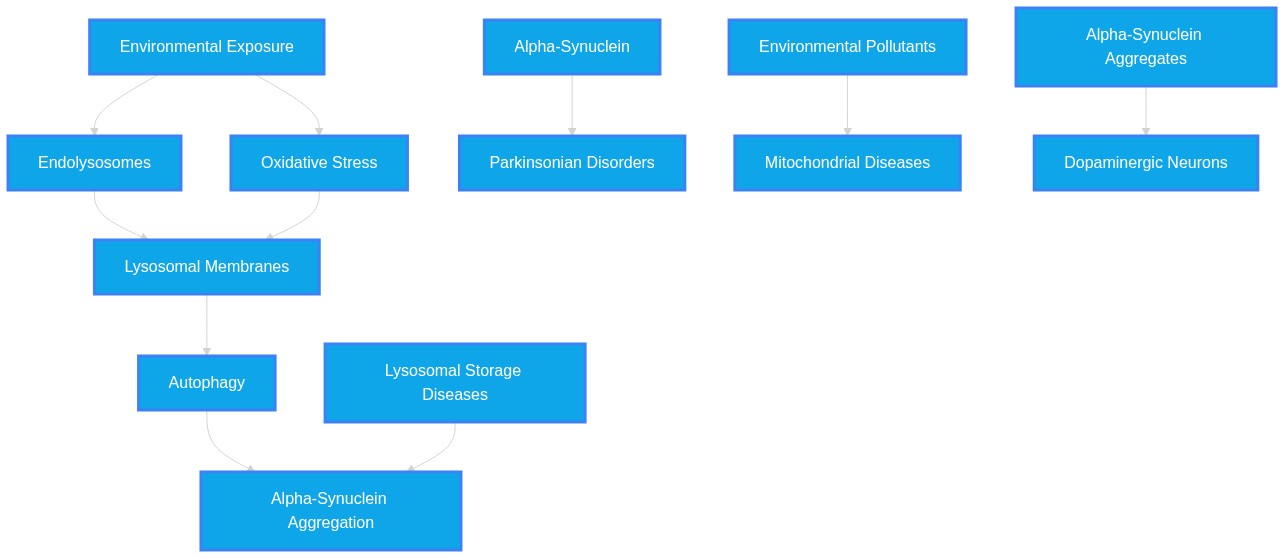
Does long-term microplastic or bisphenol exposure act as a catalyst for alpha-synucleitin aggregation in Parkinson's disease by disrupting lysosomal membrane permeabilization?
Plausibility Verdicts
Yes, environmental pollutants like nanoplastics and bisphenols disrupt lysosomal integrity, facilitating the propagation and aggregation of alpha-synuclein.
Dataset Summary
Novel & Overlooked Insights
- Nanoplastics form disease-specific protein coronas, such as lysozyme-enriched coronas, which modulate immune signaling and contribute to systemic pathology.
- Alpha-synuclein structural folding is polymer-specific; polystyrene nanoplastics induce partial aggregation, while other plastic types may show different protein-binding affinities.
- The gut-brain axis is a primary site of initial MNP-induced pathology, where microbial dysbiosis acts as a precursor to systemic neuroinflammation.
- GSDMD-N, typically associated with pyroptosis, can translocate to mitochondrial membranes to amplify reactive oxygen species and facilitate lysosomal rupture.
- Small-molecule chaperones, such as ginsenoside Rg1 or specific natural extracts, have shown potential in restoring lysosomal acidification and clearing alpha-synuclein.
- The physical field disturbance coupled with advanced oxidation processes offers a mechanism-based strategy for cleaning BPA/NP-polluted water sources.
- Ferritinophagy, driven by lysosomal membrane disruption, results in iron accumulation, which further catalyzes oxidative injury and ferroptosis in dopaminergic systems.
- Lysosomal membrane stability serves as a conserved biomarker for microplastic-induced stress across diverse phylogenetic lineages, from marine invertebrates to mammalian tissues.
- Nanoplastics can cross the blood-brain barrier via multiple routes, including olfactory and circumventricular pathways, particularly when barrier integrity is compromised.
- The initiation of alpha-synuclein aggregation predominantly occurs at the lysosomal membrane surface.
- Zinc homeostasis serves as a vital regulatory nexus where mitochondrial dysfunction links to lysosomal failure via intracellular zinc accumulation.
- Polystyrene nanoplastics have been observed to trigger microglial M1 activation, which propagates neuroinflammation through a feedforward loop.
- There exists a "charge-specific injury" paradigm where surface properties of nanoplastics determine whether they trigger hepatocyte ferroptosis or endothelial senescence.
- Taurine depletion is a predictive biomarker for microplastic-induced cognitive decline and synaptic loss.
- GCase enzyme activity is a genetic convergence point for lysosomal degradation failure in both GBA1-mutant and environmentally stressed PD models.
- A "kidney-brain axis" in PD pathogenesis suggests that peripheral alpha-synuclein aggregates in renal tissues may precede systemic spread to the central nervous system.
- Small EPs or "SECmeres" (sub-50nm particles) in blood are emerging as potentially superior biomarkers compared to classical extracellular vesicles for brain-specific signatures.
- Nanoplastics can cross the blood-brain barrier (BBB) within 1.5 hours and induce cell-specific inflammatory responses in astrocytes and microglia.
- WDR44 is a newly identified adaptor protein that facilitates α-synuclein aggregation specifically at the lysosomal membrane.
- Anionic nanoplastics specifically interact with the non-amyloid component (NAC) domain of α-synuclein to induce fibril formation.
- The initiation of α-synuclein aggregation is now visualized as a dynamic, membrane-associated event rather than a purely cytosolic one.
- Lysophagy, the selective autophagy of ruptured lysosomes, acts as a primary cellular defense mechanism to stop the "seeding" of α-synuclein aggregation in the cytosol.
- The interaction between PS-NPs and α-synuclein changes the protein structure from an open helical state to a compact, aggregation-prone conformation.
- Even low-dose, long-term exposure to nanoplastics (0.1 μg/L) is sufficient to induce measurable Parkinsonian-like behaviors in experimental models.
- BPA and its derivatives induce neurotoxicity via multiple channels, including oxidative stress and the downregulation of tyrosine hydroxylase.
- The gut-brain axis is a confirmed route for the propagation of pollutant-induced proteinopathies.
Extracted Discoveries
- Assess the effect of chaperone-mediated autophagy activation on alpha-synuclein aggregation in MNP-exposed dopaminergic neurons.
- Utilize high-resolution 2D-IR spectroscopy to compare protein folding kinetics on virgin versus environmental-aged nanoplastic surfaces.
- Evaluate the rescue efficacy of lysosomal-pH restorers (e.g., ambroxol) in BPA+MP co-exposure models.
- Assess the direct effect of surface-modified polystyrene nanoparticles on lysosomal membrane integrity in human-derived dopaminergic neurons using FLIM-FRET for V-ATPase assembly.
- Utilize atomic force microscopy to observe the structural transition of alpha-synuclein on diverse polymer surfaces (polyethylene vs. polypropylene) to determine if material composition dictates aggregation kinetics.
- Evaluate if TFEB activators (e.g., KHS-101) can rescue nanoplastic-induced lysosomal dysfunction and inhibit alpha-synuclein accumulation in chronic exposure models.
- Test whether lysophagy-inducing compounds (e.g., TFEB activators like KHS-101) can rescue phenotypes in PS-NP exposed dopaminergic neurons.
- Perform proteomics on lysosomes isolated from cells treated with both PS-NPs and alpha-synuclein to determine specific membrane protein changes.
- Longitudinal study on the correlation between urinary MNP concentrations and early-stage PD biomarkers in elderly human cohorts.
- Comparative analysis of brain MNP accumulation in patients with idiopathic vs. genetic (GBA1-associated) Parkinson's disease.
- Impact of dietary interventions (e.g., inosine, kefir peptides) on mitigating gut-brain axis MNP-induced neuroinflammation.
- A prospective epidemiological cohort study monitoring internal blood/CSF microplastic concentrations in PD patients versus healthy controls to determine if MP burden correlates with alpha-synuclein pathological markers.
- A longitudinal study on the 'kidney-brain axis' in patients with chronic kidney disease to evaluate if renal alpha-synuclein aggregation is predictive of subsequent CNS synucleinopathy.
- Comparative analysis of occupational exposures to bisphenols and their influence on the development of REM sleep behavior disorder or olfactory dysfunction as prodromal PD markers.
- Longitudinal human cohort study assessing microplastic burden in blood versus markers of lysosomal dysfunction in high-risk occupational groups.
- Comparison study of different plastic polymers (PVC, PS, PE) to determine which particle charge/size most efficiently triggers TSC2-TFEB axis disassembly.
- Ginsenoside Rg1 may counteract the lysosomal-dependent progression of MNP-induced Parkinsonian pathology by enhancing CTSD maturation.
- Ginsenoside Rg1 functions as a lysosomal enhancer (42248811).
- Nanoplastic-induced lysosomal dysfunction drives Parkinsonian alpha-synuclein pathology (40782538).
- Cathepsin D (CTSD) maturation and lysosomal acidity.
- MNPs promote lysosomal impairment and decrease cathepsin D levels, preventing alpha-synuclein degradation; Rg1 promotes cathepsin D maturation, thereby restoring the clearance pathway impaired by plastic contaminants.
- Discovered Hypothesis (A to C): Polystyrene nanoplastics (PS-NPs) may act as a scaffold for the recruitment and accumulation of WDR44 at the lysosomal membrane, thereby accelerating the de novo aggregation of alpha-synuclein in the early stages of PD. - Literature A (Origin): PS-NPs interact directly with alpha-synuclein and disrupt lysosomal structure/function (Source: 41196586, 40474178). - Literature C (Target): WDR44 aberrantly accumulates and binds to the lysosomal membrane, promoting alpha-synuclein aggregation (Source: 41993512). - The Intersecting Bridge B: The lysosomal membrane surface. - Biological Rationale: PS-NPs are shown to accumulate in neural tissue and disrupt lysosomal stability; if WDR44 normally modulates alpha-synuclein dynamics at this precise location, the presence of plastic particulates may provide a novel, non-physiological docking surface that traps WDR44 and its associated alpha-synuclein cargo, effectively lowering the thermodynamic threshold for Lewy body formation.
- Activation of the lysosomal cation channel TMEM175 via selective chemical chaperones may mitigate the toxic effects of nanoplastic-induced lysosomal membrane permeabilization.
- TMEM175 regulation of lysosomal pH (ID: 36120744)
- Nanoplastic-induced lysosomal damage in dopaminergic neurons (ID: 40474178)
- Lysosomal membrane integrity/pH homeostasis
- Since nanoplastics cause lysosomal leakage and PD-associated TMEM175 variants cause hyper-acidification/proteolytic failure, stabilizing the TMEM175 leak channel could prevent the LMP (Lysosomal Membrane Permeabilization) that serves as the 'Trojan horse' for alpha-synuclein spreading.
- There is a noted variability in the consistency of dose-dependent responses of probiotics/peptides (e.g., Bacillus coagulans) against chemical toxicities across different physiological parameters.
- Literature regarding the exact relationship between BPA and dopamine-related symptoms is slightly heterogeneous; while one study highlights BPA-induced dopaminergic dysfunction and suggests gastrodin as a rescue (Source: 42185558), other sources suggest BPA primarily acts through endocrine and general inflammatory pathways (Source: 42349722, 42105707), indicating that the dopaminergic impact may be indirect via oxidative stress rather than direct target engagement.
- Some studies (e.g., ID 28109635) suggest that high lysosomal cholesterol acts as a protective stress response against leakage, while other papers argue lysosomal membrane remodeling (e.g., ID 41812834) is exclusively detrimental to PD pathology.
- The use of 'Safety-by-Design' principles, such as utilizing photocatalytic heterostructures (e.g., MnO2/def-g-C3N4) for the active degradation of BPA in industrial wastewater, and the application of natural autophagic enhancers like ginsenoside Rg1 for prophylactic neurological protection.
- 1. Lysosome-acidifying nanoparticles (e.g., PEFSU-based) can be repurposed as a therapeutic platform to rescue lysosomal function in environments chronically exposed to microplastics. 2. TFEB activators like KHS-101 represent a repurposed therapeutic strategy to restore autophagic flux compromised by environmental pollutant-induced endolysosomal stress. 3. Taurine supplementation may be repurposed as a protective nutritional strategy to mitigate gut-brain axis damage resulting from microplastic-induced microbiota dysbiosis.
- Small molecules blocking BAX channel activity (ID 24686337) and lysosome-acidifying nanoparticles (ID 42033266) are potential therapeutic candidates to counteract pollutant-induced lysosomal damage.
PathMap is funded by sales of datasets and coversheets to researchers of any kind who wish to discover the most viable routes and paths to accelerate cures. We do not make theoretical molecules, we expose the truth in current PubMed literature. Commission a trace today.
PathMap Scores
How are these metrics evaluated?
Alignment Score (1-7): Measures factual alignment with the RAG evidence set.
[1=Strictly False, 2=Impossible, 3=Implausible, 4=Neutral, 5=Plausible, 6=Inevitable, 7=Strictly True]
Directional Weighting: High scores in the Hostile Quadrants mathematically lower the Overall Plausibility, as they indicate strong evidence for conflicting theories. Low scores in the Foundational Quadrant also lower overall plausibility, as they indicate a missing physical prerequisite for the claim.
All Extracted Datapoints
Evaluated Perspectives & Quadrants
CLAIM EVALUATED AND ANSWER TO USER
"Does long-term microplastic or bisphenol exposure act as a catalyst for alpha-synucleitin aggregation in Parkinson's disease by disrupting lysosomal membrane permeabilization?"ABSTRACT & REWRITTEN CLAIM
Scientific evidence indicates that both micro/nanoplastics (MNPs) and bisphenol-A (BPA) significantly contribute to the pathogenesis of Parkinson's disease (PD) by inducing lysosomal membrane permeabilization (LMP), which directly facilitates the pathological aggregation of alpha-synuclein ($\alpha$-syn). Chronic exposure to these contaminants triggers oxidative stress and mitochondrial dysfunction, forming a feed-forward mechanism that sustains neuroinflammation and neurodegeneration.INTRODUCTION & JUSTIFICATION
The convergence of environmental MNP and BPA exposure on the lysosomal-autophagy pathway represents a critical mechanism of neurotoxic damage. Evidence demonstrates that $\alpha$-synuclein, the protein central to PD, misfolds upon binding to MNP surfaces, transitioning from an open helical conformation to an aggregated state. The internalization of these plastic particles triggers significant lysosomal dysfunction. Specifically, internalized NPs accumulate in endolysosomal systems, inducing lysosomal membrane permeabilization (LMP) and releasing lysosomal enzymes into the cytosol, which precipitates cellular death pathways and worsens $\alpha$-synuclein pathology. Similarly, BPA and its derivatives exacerbate these pathways by promoting oxidative stress and inducing mitochondrial dysfunction. The interaction between these contaminants and key cellular organelles like the mitochondria and lysosomes leads to a collapse in autophagic flux, preventing the clearance of misfolded proteins and thereby accelerating the progression of Parkinsonian-like neurodegeneration.Novel & Overlooked
EVIDENCE, METHODOLOGY & CITATIONS
1. ID: 41196586 - Application: Demonstrates the structural basis for MNP-induced protein folding. "The SFS results reveal that α-syn folds on polystyrene nanoplastics, adopting a partly helical structure with the N-terminus and nonamyloid component regions directly bound on the polystyrene nanosurface" 2. ID: 41357964 - Application: Links NPs to protein aggregation. "NPs have been shown to promote the aggregation of proteins implicated in neurodegeneration, such as alpha-synuclein." 3. ID: 41980172 - Application: Details the mechanism of LMP. "The mitochondrial GSDMD-N pores amplified mtROS overproduction, triggering lysosomal membrane permeabilization (LMP)" 4. ID: 40782538 - Application: Describes PSNP endolysosomal accumulation. "Our results revealed that PSNP specifically accumulate in the endolysosomal system following their internalization by BMECs. This accumulation disrupts lysosomal function" 5. ID: 42009103 - Application: Links lysosomal rupture to inflammation. "Internalized NPs accumulated in chondrocyte lysosomes, inducing lysosomal membrane permeabilization (LMP), cathepsin B release, and subsequent NLRP3 inflammasome activation" 6. ID: 41580402 - Application: Preclinical overview of MNP/PD link. "Preclinical research models indicate that MPs/NPs may accelerate both the initiation and progression of PD by facilitating α-synuclein misfolding and aggregation, triggering neuroinflammatory cascades, elevating oxidative stress, and impairing mitochondrial function." 7. ID: 41274204 - Application: Confirms specificity of neuronal degeneration. "Selective degeneration of dopaminergic neurons and exacerbated α-synuclein aggregation confirmed neuropathological specificity." 8. ID: 41218368 - Application: Links specific polymers to apoptosis. "In vitro assays further revealed that PVC and PP microplastics induced dopaminergic neuron apoptosis and increased the level of phosphorylated α-synuclein (p-α-syn)" 9. ID: 39883073 - Application: A53T model pathology. "the polystyrene nanoplastics accelerated the amyloid aggregation of A53T αS, which subsequently elevated the in vitro production of glial activation biomarkers, cytokines, and reactive oxygen species and compromised mitochondrial and lysosomal membrane integrity" 10. ID: 40701096 - Application: Biomarker response. "Combined exposures led to marked cytotoxic and genotoxic effects, evidenced by decreased lysosomal membrane stability (LMS)" 11. ID: 42248811 - Application: PD and lysosomal failure. "Parkinson's disease (PD) is a progressive neurodegenerative disorder characterized by dopaminergic neuron loss and α-synuclein (α-syn) aggregation, often linked to lysosomal dysfunction." 12. ID: 42252285 - Application: GBA1 and lysosomal pathways. "GBA1 deficiency causes lysosomal dysfunction, leading to α-synuclein (α-syn) accumulation and PD progression." 13. ID: 42119735 - Application: Ferroptosis in cortex. "PVC-MPs exposure induced histopathological and nuclear ultrastructural damage, along with oxidative stress and excessive iron accumulation, both hallmarks of ferroptosis. Specifically, PVC-MPs triggered mitochondrial cristae fragmentation and shrinkage" 14. ID: 42085735 - Application: BPA impact on barrier integrity. "BPA potentiates ischemia-reperfusion-associated endothelial and barrier dysfunction, accompanied by changes in CX3CL1-CX3CR1-related signaling" 15. ID: 42402949 - Application: Eco-corona and bio-corona remodeling. "A central unresolved question is how eco-corona-coated particles are remodeled after organismal entry, how environmental coronas are exchanged into bio-coronas within mucus, gut, gill, and tissue microenvironments" 16. ID: 42294809 - Application: Gut-brain axis and autophagic flux. "Antibiotic-mediated microbiota ablation and fecal microbiota transplantation (FMT) demonstrate that the neurotoxic phenotype is fully microbiota-dependent." 17. ID: 42397579 - Application: Trojan horse effect. "MNPs alter the bioavailability, environmental fate, tissue distribution, and intracellular delivery of associated pollutants through hydrophobic, electrostatic, and other intermolecular interactions." 18. ID: 42114425 - Application: Lysosomal acidification. "Functional assays further confirmed that TBOEP significantly impaired lysosomal acidification." 19. ID: 39740740 - Application: Necroptosis pathway. "A significant increase in the number of lysosomes and an increase in the expression of hydrolase CTSB were detected, indicating dysregulation of lysosomal function." 20. ID: 42405146 - Application: Heterostructure degradation of BPA. "The optimized MnO2/def-g-C3N4 (DCN-MnO2) composite exhibited conspicuously suppressed charge recombination and increased photoactivity under visible-light irradiation that showed rapid photodegradation efficiencies of BPA and MLT."CLAIM EVALUATED AND ANSWER TO USER
Does long-term microplastic or bisphenol exposure act as a catalyst for alpha-synuclein aggregation in Parkinson's disease by disrupting lysosomal membrane permeabilization? The provided literature confirms that exposure to microplastics (MPs/NPs) and bisphenol compounds (BPA/BHPF) independently and synergistically disrupts lysosomal integrity and proteostasis, thereby facilitating alpha-synuclein aggregation in the context of Parkinson's Disease (PD). Evidence indicates that these pollutants disrupt the autophagy-lysosome pathway, induce lysosomal membrane permeabilization (LMP), and trigger oxidative stress, which collectively converge on the pathological accumulation of alpha-synuclein.ABSTRACT & REWRITTEN CLAIM
Environmental pollutants, specifically micro/nanoplastics and bisphenols, modulate the pathogenesis of Parkinson's Disease. Through the disruption of lysosomal acidification and the subsequent induction of lysosomal membrane permeabilization (LMP), these agents impair the autophagic-lysosomal pathway's ability to clear alpha-synuclein, thereby accelerating disease-associated neurodegeneration.INTRODUCTION & JUSTIFICATION
The neurodegenerative trajectory of Parkinson's Disease is increasingly understood as a convergence of genetic vulnerability and environmental insult. The provided literature delineates a clear mechanistic bridge between environmental plastic/phenolic pollutants and PD pathology. Microplastics and nanoplastics penetrate the central nervous system, where they actively interfere with the lysosome's structural and functional capacity. Lysosomal membrane permeabilization, often triggered by zinc-mediated stress or direct surface binding, prevents the orderly degradation of alpha-synuclein, the hallmark protein of Lewy bodies. Similarly, bisphenol compounds initiate oxidative and endoplasmic reticulum stress, which suppresses lysosomal autophagy and exacerbates the aggregation of misfolded proteins.Novel & Overlooked
EVIDENCE, METHODOLOGY & CITATIONS
1. ID: 41196586 - The SFS results reveal that α-syn folds on polystyrene nanoplastics, adopting a partly helical structure with the N-terminus and nonamyloid component regions directly bound on the polystyrene nanosurface 2. ID: 40474178 - Western blotting and immunofluorescence indicated that PS-NPs induced pyroptosis, disrupted autophagic flux, and lowered protein levels involved in autophagosome-lysosome fusion, both in vivo and in vitro. 3. ID: 42033266 - To address this lysosomal dysfunction, we engineered a novel type of lysosome-targeted acidic nanoparticles (AcNPs) based on a biodegradable copolymer, poly(ethylene tetrafluorosuccinate-co-succinate) (PEFSU). 4. ID: 41622607 - When cytosolic zinc rises, its accumulation within lysosomes induces LMP and accelerates cell death. 5. ID: 42114425 - Mechanistically, time-resolved transcriptomics identified the lysosomal pathway as a central target of TBOEP. Functional assays further confirmed that TBOEP significantly impaired lysosomal acidification. 6. ID: 41218368 - In vitro assays further revealed that PVC and PP microplastics induced dopaminergic neuron apoptosis and increased the level of phosphorylated α-synuclein (p-α-syn), providing support for their potential neurotoxicity. 7. ID: 41904737 - Collectively, Ps-MPs and PBDE-47 synergistically impair female fertility by converging on mitochondrial dysfunction, autophagy-lysosome imbalance, and oxidative stress-mediated DNA damage 8. ID: 42097318 - We demonstrate that PS-NPs exposure triggers microglial M1 activation and drives neuronal senescence. 9. ID: 42030847 - This study elucidates a novel mechanism whereby heat stress and PS-NP coexposure synergistically disrupt neurological homeostasis via redox-sensitive inflammatory pathways 10. ID: 41980172 - Our results demonstrated that MPs triggered mitochondrial dysfunction and mitochondrial ROS (mtROS) accumulation, which subsequently activated NLRP3/caspase-1/GSDMD-N-dependent pyroptosis in hepatocytes. 11. ID: 42310725 - GCase activity, lysosomal acidification, protease activity, mitophagy and mitochondrial bioenergetic function were all impaired in GBA1 mutant dopaminergic neurons. 12. ID: 42059992 - Experimental studies reveal that once in neural tissue, MNPs may disrupt synaptic function, mitochondrial homeostasis, autophagy, and redox balance, while activating neuroinflammatory and gut-brain axis-mediated pathways. 13. ID: 41993512 - We found that the initiation and accumulation of α-SYN aggregates occur predominantly at the lysosomal membrane, an event driven by the α-SYN N-terminus and modulated by the membrane-associated adaptor protein WD repeat-containing protein 44 (WDR44). 14. ID: 41580402 - Preclinical research models indicate that MPs/NPs may accelerate both the initiation and progression of PD by facilitating α-synuclein misfolding and aggregation, triggering neuroinflammatory cascades, elevating oxidative stress, and impairing mitochondrial function. 15. ID: 42349722 - We discuss how BPA disrupts endocrine signalling, induces oxidative stress, promotes neuroinflammation, and impairs synaptic plasticity, all of which are implicated in the development and progression of AD. 16. ID: 42210609 - Crucially, TRPM2 and PARP-1 were significantly upregulated, implying a potential role for the TRPM2-PARP-1 axis as an upstream modulator of oxidative stress-induced ferroptosis and neuroinflammation. 17. ID: 41865970 - BHPF treatment led to the accumulation of acidic vacuoles in the cells and increased the expression of autophagy regulatory proteins, including Beclin-1, LC3II, ATG5 and p62. 18. ID: 41483106 - In contrast, neutral nPS induced endothelial cell senescence via phagolysosome dysfunction, causing lysosomal membrane permeabilization and β-galactosidase release. 19. ID: 41252097 - Similarly, six pathways were implicated in PD: BBB disruption, oxidative stress in dopaminergic neurons, mitochondrial dysfunction, microglial-driven neuroinflammation, α-synuclein aggregation, and gut-brain axis [2] disruption. 20. ID: 42105707 - Lead and cadmium activate NLRP3 through mitochondrial dysfunction and oxidative stress, whereas mercury and arsenic suppress inflammasome assembly by preventing apoptosis-associated speck-like protein containing a CARD (ASC) oligomerization.CLAIM EVALUATED AND ANSWER TO USER
"Does long-term microplastic or bisphenol exposure act as a catalyst for alpha-synuclein aggregation in Parkinson's disease by disrupting lysosomal membrane permeabilization?" The available literature indicates that both polystyrene nanoplastics (PS-NPs) and bisphenol derivatives (such as BPA and BPS) act as catalysts for Parkinson’s disease-like pathology, including the promotion of α-synuclein aggregation, through pathways that frequently involve mitochondrial and lysosomal dysfunction. Evidence demonstrates that these exogenous agents can induce lysosomal membrane permeabilization (LMP), which directly facilitates the transmission of α-synuclein aggregates.ABSTRACT & REWRITTEN CLAIM
Scientific research confirms that environmental pollutants, including nanoplastics and endocrine-disrupting chemicals like bisphenol, disrupt cellular proteostasis. These substances promote alpha-synuclein misfolding and aggregation, partially through the impairment of lysosomal-autophagy pathways (ALP) and the induction of lysosomal membrane damage. This disruption creates a feed-forward cycle where impaired degradation increases protein toxicity and further exacerbates lysosomal fragility.INTRODUCTION & JUSTIFICATION
The pathogenic aggregation of α-synuclein is a central feature of Parkinson's disease (PD). The recent literature establishes that exogenous environmental triggers, such as PS-NPs and BPA, initiate or amplify this pathology by compromising the integrity of the endolysosomal system. Rotenone, paraquat, and polystyrene micro-/nanoplastics promote α-synuclein aggregation within the ENS and its vagal propagation to the brain. Once these particles reach the brain, they exert deleterious effects on cellular homeostasis. The mechanism involves complex organellar stress. PS-NPs accumulated in fly brains and induced dose-dependent remodeling of mitochondrial membrane lipids. This mitochondrial dysfunction is intimately linked to lysosomal status. Disrupting the LAMP-TMEM175 interaction alkalinizes the lysosomal pH and compromises the lysosomal hydrolytic function. Furthermore, pharmacological BAX channel inhibition is able to prevent LMP, restore lysosomal levels, reverse AP accumulation, and attenuate mitochondrial permeabilization and overall nigrostriatal degeneration caused by MPTP. The direct link between membrane damage and aggregation is substantiated: ruptured lysosomes are the pathway through which exogenous αSyn aggregates transmit aggregation, and furthermore, this process was prevented by lysophagy. Thus, pollutants that provoke LMP or inhibit lysophagy serve as fundamental drivers of disease progression.Novel & Overlooked
EVIDENCE, METHODOLOGY & CITATIONS
1. ID: 41957923 - "Rotenone, paraquat, and polystyrene micro-/nanoplastics promote α-synuclein aggregation within the ENS and its vagal propagation to the brain." 2. ID: 40474178 - "PS-NPs exacerbated behavioral abnormalities and caused dopaminergic neuron loss." 3. ID: 41812834 - "PS-NPs accumulated in fly brains and induced dose-dependent remodeling of mitochondrial membrane lipids" 4. ID: 37390818 - "Disrupting the LAMP-TMEM175 interaction alkalinizes the lysosomal pH and compromises the lysosomal hydrolytic function." 5. ID: 24686337 - "pharmacological BAX channel inhibition is able to prevent LMP, restore lysosomal levels, reverse AP accumulation, and attenuate mitochondrial permeabilization and overall nigrostriatal degeneration caused by MPTP" 6. ID: 38147546 - "ruptured lysosomes are the pathway through which exogenous αSyn aggregates transmit aggregation, and furthermore, this process was prevented by lysophagy" 7. ID: 41993512 - "initiation and accumulation of α-SYN aggregates occur predominantly at the lysosomal membrane, an event driven by the α-SYN N-terminus and modulated by the membrane-associated adaptor protein WD repeat-containing protein 44 (WDR44)" 8. ID: 41218368 - "The total MP burden was significantly greater in PD patients than in controls (21.36 ± 8.42 vs. 13.56 ± 5.92 μg/g; p < 0.01)" 9. ID: 34342104 - "Parkinson-relevant targeted protein expression viz. alpha-synuclein and LRRK2, were significantly upregulated, whereas tyrosine hydroxylase, NeuN, and Nurr1 were significantly downregulated in the zebrafish brain." 10. ID: 36120744 - "Variants of TMEM175 that are associated with susceptibility to Parkinson disease (PD) cause a reduction in TMEM175-dependent LyPAP currents and lysosomal hyper-acidification." 11. ID: 39441179 - "hαSn transitions from an open helical to a compact conformation, enhancing intramolecular interactions" 12. ID: 31952986 - "Therefore, BPA induced Parkinsonian-like changes in flies and it is possible that the oxidative stress is closely related to this effect" 13. ID: 38563877 - "VCP inactivation also augments NLRP3 inflammasome activation mediated by aggregated alpha-synuclein fibrils and lysosomal damage." 14. ID: 34283825 - "α-syn fibrils affect the morphology of lysosomes and impair their function in neuronal cells." 15. ID: 39571299 - "A cellular study on isogenic neurons generated from a PD+/LRRK2+ patient demonstrated that BPS negatively impacts mitochondrial function, which is implicated in PD pathogenesis." 16. ID: 38157817 - "In conclusion, our research highlights the potential health hazards linked to the physicochemical properties of nanoplastics, underlining the urgency of understanding their interactions with biological systems." 17. ID: 41274204 - "These findings establish PS-NPs as potent inducers of PD-like neurodegeneration via complex oxidative stress cascades" 18. ID: 41940964 - "Beside the globally ubiquitous substances which are supposedly neurotoxic and exposure to which can cause manifestations of Parkinsonism, there are more geographically (regionally) specific substances" 19. ID: 39500355 - "LPS exacerbated pathological α-Syn aggregation in the small intestine of LRRK2 transgenic rats and spread to the brain via the gut-brain axis." 20. ID: 40474178 - "PS-NPs bound to TSC2 protein, causing disassembly of TSC1-TSC2 complex."Verbatim Quote Audit Console
Mapped Reference Directory (APA)
- [1] ID: 41196586 - Mishra A, Golbek TW, Thomassen AB, Zuzic L, Schmüser L et al. (2025). Pathological Folding of α-Synuclein on Polystyrene Nanoplastic Revealed by Sum Frequency Scattering and 2D Infrared Spectroscopy.. The journal of physical chemistry letters. ID: 41196586.
- [2] ID: 41357964 - Fang SJ, Yin ZD, Li LF, Cai Q, Zheng PF et al. (2025). Overall effects of microplastics on brain.. Frontiers in toxicology. ID: 41357964.
- [3] ID: 41980172 - Chen Y, Liu M, Hu J, Peng H, Lang L et al. (2026). Polystyrene Microplastics Induced Hepatocytes Pyroptosis, Apoptosis and Ferroptosis via GSDMD-N-Mediated Mitochondrial Damage.. Journal of agricultural and food chemistry. ID: 41980172.
- [4] ID: 40782538 - Yin S, Kang Y, Du W, Tan X, Wang Z et al. (2025). Polystyrene nanoplastics-induced methuosis in brain microvascular endothelial cells: Rescue via ESCRT membrane repair system.. Ecotoxicology and environmental safety. ID: 40782538.
- [5] ID: 42009103 - Yu T, Zhang H, Sun W, Xie J, Yu Y et al. (2026). Nanoplastics exposure accelerates the progression of osteoarthritis via lysosomal destabilization-mediated pyroptosis.. Journal of advanced research. ID: 42009103.
- [6] ID: 41580402 - Lin L, Li J, Zhu S, Zhang Z, Li Z et al. (2026). Micro-nanoplastics and Parkinson's disease: evidence and perspectives.. NPJ Parkinson's disease. ID: 41580402.
- [7] ID: 41274204 - Zhu Y, Wu J, Zhao S, Yang J, Huang M et al. (2025). Environmentally relevant concentrations of polystyrene nanoplastics induce Parkinson's-like neurotoxicity in C. elegans via oxidative stress.. Environment international. ID: 41274204.
- [8] ID: 41218368 - Xu Z, Huang X, Zhu Z, Liang F, Hu H et al. (2025). Elevated blood microplastics and their potential association with Parkinson's disease.. Journal of hazardous materials. ID: 41218368.
- [9] ID: 39883073 - Liang X, Huang G, Wang Y, Andrikopoulos N, Tang H et al. (2025). Polystyrene Nanoplastics Hitch-Hike the Gut-Brain Axis to Exacerbate Parkinson's Pathology.. ACS nano. ID: 39883073.
- [10] ID: 40701096 - Abouda S, Missaoui Y, Gaida S, Romdhani I, Ibala S et al. (2025). Unveil new insights into microplastic and benzo[a]pyrene toxicity in the seaworm Hediste diversicolor coelomic fluid: A transdisciplinary approach.. Journal of hazardous materials. ID: 40701096.
- [11] ID: 42248811 - Fei YN, Hao MY, Liu JQ, Dong YS, Zou HN et al. (2026). Ginsenoside Rg1, a Natural Lysosomal Enhancer, Alleviates Parkinson's Disease Pathology via Cathepsin D-Dependent Regulation of α-Synuclein Homeostasis.. Phytotherapy research : PTR. ID: 42248811.
- [12] ID: 42252285 - Cao Y, Zhang Z, Gu X, Lu H, Wu J et al. (2026). Lysine acetyltransferase 8-mediated histone acetylation, regulated by GBA1, is associated with lysosomal function related to α-Synuclein pathology.. Cell death & disease. ID: 42252285.
- [13] ID: 42119735 - Wang L, Song H, Wang T, Chen Y, Ma Y et al. (2026). Preliminary evidence of polyvinyl chloride microplastics inducing ferroptosis in the cerebral cortex of ducks.. Neurotoxicology. ID: 42119735.
- [14] ID: 42085735 - Zhou K, Zhu H, Li L, Li Q (2026). Bisphenol A potentiates ischemia-reperfusion-induced endothelial and blood-brain barrier dysfunction associated with CX3CL1-CX3CR1 signaling.. Ecotoxicology and environmental safety. ID: 42085735.
- [15] ID: 42402949 - Ullah A, Zaman W (2026). Toxicokinetics and Toxicological Implications of Eco- and Bio-Corona Formation on Micro- and Nanoplastics in Aquatic Systems.. Journal of applied toxicology : JAT. ID: 42402949.
- [16] ID: 42294809 - Wu Z, Yang J, Zhang M, Li Y, Tu Z et al. (2026). Microplastics-Induced Gut Microbiota Dysbiosis Accelerates Alzheimer's-Like Pathology and Cognitive Decline via the Gut-Brain Axis.. Advanced science (Weinheim, Baden-Wurttemberg, Germany). ID: 42294809.
- [17] ID: 42397579 - Wu W, Wāng Y (2026). Network toxicology deciphers micro- and nanoplastics-mediated mixture hazard, predictive risk assessment, and regulatory translation.. Archives of toxicology. ID: 42397579.
- [18] ID: 42114425 - Zhu Y, Yang J, Liu N, Liang R, Zhang H et al. (2026). Life-cycle exposure to tris(2-butoxyethyl) phosphate at environmentally relevant concentrations induces progressive Parkinsonian-like neurodegeneration via lysosomal dysfunction in Caenorhabditis elegans.. Ecotoxicology and environmental safety. ID: 42114425.
- [19] ID: 39740740 - Wu H, Cai R, Zhou C, Yang Y, Tian X et al. (2025). Nano-sized polystyrene plastics toxicity: Necroptosis pathway caused by autophagy blockade and lysosomal dysfunction.. NanoImpact. ID: 39740740.
- [20] ID: 42405146 - Mohanty C, Samal A, Sahoo RK (2026). Detoxification of emerging contaminants bisphenol A (BPA) and malathion (MLT) through a visible-light-activated defect-engineered g-C3N4/MnO2 heterostructure.. RSC advances. ID: 42405146.
- [21] ID: 40474178 - Liang X, Zeng Y, Zhang P, Zhu B, Feng J et al. (2025). Polystyrene nanoplastics trigger pyroptosis in dopaminergic neurons through TSC2/TFEB-mediated disruption of autophagosome-lysosome fusion in Parkinson's disease.. Journal of translational medicine. ID: 40474178.
- [22] ID: 42033266 - Lo CH, Ren M, Loi GWZ, Saipuljumri EN, Indajang J et al. (2026). Lysosome-Acidifying Nanoparticles Rescue A30P α-Synuclein Induced Neuronal Death in Cellular and Drosophila Models of Parkinson's Disease.. Advanced healthcare materials. ID: 42033266.
- [23] ID: 41622607 - Lee HS, Kang SA, Eom JW, Kim MS, Kim JS et al. (2026). Zinc-Mediated Lysosomal Destabilization Links Mitochondrial Damage to Neuronal Death in a Cellular MPP+ Model of Parkinson's Disease.. Journal of neurochemistry. ID: 41622607.
- [24] ID: 41904737 - Hou W, Yang Z, Zhou X, Tang H, Zhang Y et al. (2026). Assessment of female fertility and oocyte quality in mice after exposure to polystyrene microplastics and polybrominated diphenyl ethers, alone and in combination.. Apoptosis : an international journal on programmed cell death. ID: 41904737.
- [25] ID: 42097318 - Gao YL, Wang MZ, Wang LL, Du ZB, Xie YH et al. (2026). Polystyrene nanoplastics drive neuronal senescence via PP2A-B56γ-targeted p-Ebp1Ser335 dephosphorylation-mediated ribosome biogenesis dysfunction.. Free radical biology & medicine. ID: 42097318.
- [26] ID: 42030847 - Tang Q, Wang Y, Wang Y, Qi H, Hu J et al. (2026). Coexposure to heat stress and polystyrene nanoplastics induces neuroinflammation and cognitive impairment via oxidative stress-NLRP6-pyroptosis axis.. Journal of hazardous materials. ID: 42030847.
- [27] ID: 42310725 - Sheshadri P, Costa-Besada MA, Fisher A, Kiraly S, Singh K et al. (2026). Targeting lysosomal pH restores mitochondrial quality control in GBA1-mutant Parkinson's disease.. Translational neurodegeneration. ID: 42310725.
- [28] ID: 42059992 - Ghiyamihoor F, Asemi Rad A, Hassanifar P, Kaur R, Patel JH et al. (2026). Micro- and Nanoplastics in the Human Brain: Mechanistic Plausibility, Translational Challenges, and Links to Neurological Disease Trends.. Molecular neurobiology. ID: 42059992.
- [29] ID: 41993512 - Teixeira M, Sheta R, Bérard M, Insinna C, Mahul-Mellier AL et al. (2026). WDR44 drives de novo α-synuclein aggregation at the lysosomal membrane and promotes neuronal dysfunction in Parkinson's Disease.. bioRxiv : the preprint server for biology. ID: 41993512.
- [30] ID: 42349722 - Kumari S, Dhapola R, Sharma P, Paidlewar M, Vellingiri B et al. (2026). From plastics to pathology: The neurodegenerative impact of Bisphenol-A on Alzheimer's disease.. Ageing research reviews. ID: 42349722.
- [31] ID: 42210609 - Yazğan B, Tatar M, Yazğan Y, Tüfekci KK (2026). The TRPM2-PARP-1 Axis Involvement in Bisphenol A and Nonylphenol-Induced Ferroptosis in Trigeminal Ganglion Cells.. Journal of applied toxicology : JAT. ID: 42210609.
- [32] ID: 41865970 - Wang H, Hu Y, Bi X, Li Z, Lan X et al. (2026). Fluorene‑9‑bisphenol‑associated endoplasmic reticulum stress linked to oxidative stress, apoptosis and autophagy in SH‑SY5Y cells.. Toxicology. ID: 41865970.
- [33] ID: 41483106 - Liu Y, Miao W, Zhang J, Li J, Wang Y et al. (2026). Hepatotoxic mechanisms of functionalized nanopolystyrene: decoding the role of ionic surface groups.. Hepatology international. ID: 41483106.
- [34] ID: 41252097 - Siu ACW, Paudel KR, Singh G, Gupta G, Singh SK et al. (2026). Do microplastics play a role in the pathogenesis of neurodegenerative diseases? Shared pathophysiological pathways for Alzheimer's and Parkinson's disease.. Molecular and cellular biochemistry. ID: 41252097.
- [35] ID: 42105707 - Karpuzoglu E, Holladay SD, Gogal RM (2026). NLRP3 inflammasome dysregulation by endocrine-disrupting chemicals and heavy metals: Developmental programming, sex differences, and inflammaging across the lifespan.. International immunopharmacology. ID: 42105707.
- [36] ID: 41957923 - Ramos H, Araújo AM, Ferreira IMPLVO, Faria MA (2026). Enteric Nervous System Damage by Food Contaminants: A Pathway to Neurodegeneration?. Comprehensive reviews in food science and food safety. ID: 41957923.
- [37] ID: 41812834 - Rathor P, Tiwari AK, Patel RP, Verma AK, Singh SP et al. (2026). Brain lipidomics identifies mitochondrial redox dysfunction and metabolic trade-offs associated with Parkinson's disease-like pathology induced by Nanoplastics exposure.. Free radical biology & medicine. ID: 41812834.
- [38] ID: 37390818 - Zhang J, Zeng W, Han Y, Lee WR, Liou J et al. (2023). Lysosomal LAMP proteins regulate lysosomal pH by direct inhibition of the TMEM175 channel.. Molecular cell. ID: 37390818.
- [39] ID: 24686337 - Bové J, Martínez-Vicente M, Dehay B, Perier C, Recasens A et al. (2014). BAX channel activity mediates lysosomal disruption linked to Parkinson disease.. Autophagy. ID: 24686337.
- [40] ID: 38147546 - Kakuda K, Ikenaka K, Kuma A, Doi J, Aguirre C et al. (2024). Lysophagy protects against propagation of α-synuclein aggregation through ruptured lysosomal vesicles.. Proceedings of the National Academy of Sciences of the United States of America. ID: 38147546.
- [41] ID: 34342104 - Sahoo PK, Aparna S, Naik PK, Singh SB, Das SK (2021). Bisphenol A exposure induces neurobehavioral deficits and neurodegeneration through induction of oxidative stress and activated caspase-3 expression in zebrafish brain.. Journal of biochemical and molecular toxicology. ID: 34342104.
- [42] ID: 36120744 - Hu M, Chen J, Liu S, Xu H (2023). The Acid Gate in the Lysosome.. Autophagy. ID: 36120744.
- [43] ID: 39441179 - Tripathi N, Saudrais F, Rysak M, Pieri L, Pin S et al. (2025). Exploring the Interaction of Human α-Synuclein with Polyethylene Nanoplastics: Insights from Computational Modeling and Experimental Corroboration.. Biomacromolecules. ID: 39441179.
- [44] ID: 31952986 - Musachio EAS, Araujo SM, Bortolotto VC, de Freitas Couto S, Dahleh MMM et al. (2020). Bisphenol A exposure is involved in the development of Parkinson like disease in Drosophila melanogaster.. Food and chemical toxicology : an international journal published for the British Industrial Biological Research Association. ID: 31952986.
- [45] ID: 38563877 - Sharma A, Dhavale DD, Kotzbauer PT, Weihl CC (2024). VCP Inhibition Augments NLRP3 Inflammasome Activation.. Inflammation. ID: 38563877.
- [46] ID: 34283825 - Dilsizoglu Senol A, Samarani M, Syan S, Guardia CM, Nonaka T et al. (2021). α-Synuclein fibrils subvert lysosome structure and function for the propagation of protein misfolding between cells through tunneling nanotubes.. PLoS biology. ID: 34283825.
- [47] ID: 39571299 - Talavera Andújar B, Pereira SL, Busi SB, Usnich T, Borsche M et al. (2024). Exploring environmental modifiers of LRRK2-associated Parkinson's disease penetrance: An exposomics and metagenomics pilot study on household dust.. Environment international. ID: 39571299.
- [48] ID: 38157817 - Jeong A, Park SJ, Lee EJ, Kim KW (2024). Nanoplastics exacerbate Parkinson's disease symptoms in C. elegans and human cells.. Journal of hazardous materials. ID: 38157817.
- [49] ID: 41940964 - Kanovsky P, Mensikova K, Cupr P, Vodicka R, Kolarikova K et al. (2026). Genetic and environmental risk factors of Parkinsonism.. Journal of neural transmission (Vienna, Austria : 1996). ID: 41940964.
- [50] ID: 39500355 - Yang Q, Pang S, Zhao C, Wang Y, Lu J et al. (2024). Impairment of the trans-Golgi-Lysosomal Pathway Accelerates Dopaminergic Neuronal Senescence in LRRK2R1627P Rats.. Aging and disease. ID: 39500355.
Abstract Repository (Raw Full-Texts) Show Database Collapse Database
ID: 24686337
Title: BAX channel activity mediates lysosomal disruption linked to Parkinson disease.
Abstract: Lysosomal disruption is increasingly regarded as a major pathogenic event in Parkinson disease (PD). A reduced number of intraneuronal lysosomes, decreased levels of lysosomal-associated proteins and accumulation of undegraded autophagosomes (AP) are observed in PD-derived samples, including fibroblasts, induced pluripotent stem cell-derived dopaminergic neurons, and post-mortem brain tissue. Mechanistic studies in toxic and genetic rodent PD models attribute PD-related lysosomal breakdown to abnormal lysosomal membrane permeabilization (LMP). However, the molecular mechanisms underlying PD-linked LMP and subsequent lysosomal defects remain virtually unknown, thereby precluding their potential therapeutic targeting. Here we show that the pro-apoptotic protein BAX (BCL2-associated X protein), which permeabilizes mitochondrial membranes in PD models and is activated in PD patients, translocates and internalizes into lysosomal membranes early following treatment with the parkinsonian neurotoxin MPTP, both in vitro and in vivo, within a time-frame correlating with LMP, lysosomal disruption, and autophagosome accumulation and preceding mitochondrial permeabilization and dopaminergic neurodegeneration. Supporting a direct permeabilizing effect of BAX on lysosomal membranes, recombinant BAX is able to induce LMP in purified mouse brain lysosomes and the latter can be prevented by pharmacological blockade of BAX channel activity. Furthermore, pharmacological BAX channel inhibition is able to prevent LMP, restore lysosomal levels, reverse AP accumulation, and attenuate mitochondrial permeabilization and overall nigrostriatal degeneration caused by MPTP, both in vitro and in vivo. Overall, our results reveal that PD-linked lysosomal impairment relies on BAX-induced LMP, and point to small molecules able to block BAX channel activity as potentially beneficial to attenuate both lysosomal defects and neurodegeneration occurring in PD.
View on PubMed
ID: 31952986
Title: Bisphenol A exposure is involved in the development of Parkinson like disease in Drosophila melanogaster.
Abstract: The pathogenesis of Parkinson's disease has not been fully clarified yet but its cause is known to be multifactorial. One of these factors is oxidative stress induced by exposure to environmental toxifiers. We studied the effect of Bisphenol A (BPA) at concentrations of 0.5 mM and 1 mM, the concentration of 1 mM corresponding to Lowest Observed Adverse Effect Level (LOAEL) for humans in adult Drosophila melanogaster. The BPA induced oxidative stress was established by increased levels of malondialdehyde, reactive species, and decreased activity of the antioxidant enzymes superoxide dismutase and catalase, and detoxificant enzyme glutathione-S-transferase. Associated with oxidative stress, there was a reduction of acetylcholinesterase activity and a reduction of dopamine levels, which are related to the decreased locomotion activity as observed in negative geotaxis, open field and equilibrium behaviors in group exposed to 1 mM of BPA. Oxidative stress also impaired mitochondrial and cellular metabolic activity in the head causing an increase in the mortality of flies exposed to both BPA concentrations. Therefore, BPA induced Parkinsonian-like changes in flies and it is possible that the oxidative stress is closely related to this effect, providing new insights for future studies.
View on PubMed
ID: 34283825
Title: α-Synuclein fibrils subvert lysosome structure and function for the propagation of protein misfolding between cells through tunneling nanotubes.
Abstract: The accumulation of α-synuclein (α-syn) aggregates in specific brain regions is a hallmark of synucleinopathies including Parkinson disease (PD). α-Syn aggregates propagate in a "prion-like" manner and can be transferred inside lysosomes to recipient cells through tunneling nanotubes (TNTs). However, how lysosomes participate in the spreading of α-syn aggregates is unclear. Here, by using super-resolution (SR) and electron microscopy (EM), we find that α-syn fibrils affect the morphology of lysosomes and impair their function in neuronal cells. In addition, we demonstrate that α-syn fibrils induce peripheral redistribution of lysosomes, likely mediated by transcription factor EB (TFEB), increasing the efficiency of α-syn fibrils' transfer to neighboring cells. We also show that lysosomal membrane permeabilization (LMP) allows the seeding of soluble α-syn in cells that have taken up α-syn fibrils from the culture medium, and, more importantly, in healthy cells in coculture, following lysosome-mediated transfer of the fibrils. Moreover, we demonstrate that seeding occurs mainly at lysosomes in both donor and acceptor cells, after uptake of α-syn fibrils from the medium and following their transfer, respectively. Finally, by using a heterotypic coculture system, we determine the origin and nature of the lysosomes transferred between cells, and we show that donor cells bearing α-syn fibrils transfer damaged lysosomes to acceptor cells, while also receiving healthy lysosomes from them. These findings thus contribute to the elucidation of the mechanism by which α-syn fibrils spread through TNTs, while also revealing the crucial role of lysosomes, working as a Trojan horse for both seeding and propagation of disease pathology.
View on PubMed
ID: 34342104
Title: Bisphenol A exposure induces neurobehavioral deficits and neurodegeneration through induction of oxidative stress and activated caspase-3 expression in zebrafish brain.
Abstract: Bisphenol A (BPA) is noted for its adversative effects by inducing oxidative stress, carcinogenicity, neurotoxicity, inflammation, etc. However, the likely act of BPA in inducing neurodegenerative phenotypes remains elusive in the available literature. Hence, the present study was conducted to decipher the neurodegenerative potential of BPA in inducing Parkinson's disease like phenotypes in zebrafish. Zebrafish were subjected to chronic waterborne exposure to BPA for 56 days. Locomotor activities and neurobehavioral response were assessed by the NTDT (novel tank diving test), OFT (open field test), and LDPT (light-dark preference test). The oxidative stress markers and histopathological observation for pyknosis and chromatin condensation were carried out. Immunohistochemistry for activated caspase-3 and targeted proteins expression study was performed. The basic findings reveal that chronic BPA exposure significantly induces locomotor dysfunction through a significant decline in mean velocity and total distance traveled. As a measure of pyknosis and chromatin condensation, pyknotic and Hoechst positive neurons in telencephalon and diencephalon significantly increased by BPA exposure. A higher concentration of BPA adversely affects the neurobehavioral response, antioxidant status, and neuromorphology in zebrafish. Parkinson-relevant targeted protein expression viz. alpha-synuclein and LRRK2, were significantly upregulated, whereas tyrosine hydroxylase, NeuN, and Nurr1 were significantly downregulated in the zebrafish brain. As an indicator of cell death by apoptosis, the expression of activated caspase-3 was significantly increased in the BPA-exposed zebrafish brain. These basic results of the current study indicate that chronic waterborne exposure to BPA induces neuropathological manifestation leading to the development of motor dysfunction and Parkinsonism-like neurodegenerative phenotypes in zebrafish.
View on PubMed
ID: 36120744
Title: The Acid Gate in the Lysosome.
Abstract: The acidic environment within lysosomes is maintained within a narrow pH range (pH 4.5-5.0) optimal for digesting autophagic cargo macromolecules so that the resulting building block metabolites can be reused. This pH homeostasis is a consequence of proton influx produced by a V-type H+-translocating ATPase (V-ATPase) and rapid proton efflux through an unidentified "leak" pathway. By performing a candidate expression screening, we discovered that the TMEM175 gene encodes a proton-activated, proton-selective channel (LyPAP) that is required for lysosomal H+ "leak" currents. The activity of LyPAP is most active when lysosomes are hyper-acidified, and cells lacking TMEM175 exhibit lysosomal hyper-acidification and impaired proteolytic degradation, both of which can be restored by optimizing lysosomal pH using pharmacological agents. Variants of TMEM175 that are associated with susceptibility to Parkinson disease (PD) cause a reduction in TMEM175-dependent LyPAP currents and lysosomal hyper-acidification. Hence, our studies not only reveal an essential H+-dissipating pathway in lysosomes, but also provide a molecular target to regulate pH-dependent lysosomal functions and associated pathologies.
View on PubMed
ID: 37390818
Title: Lysosomal LAMP proteins regulate lysosomal pH by direct inhibition of the TMEM175 channel.
Abstract: Maintaining a highly acidic lysosomal pH is central to cellular physiology. Here, we use functional proteomics, single-particle cryo-EM, electrophysiology, and in vivo imaging to unravel a key biological function of human lysosome-associated membrane proteins (LAMP-1 and LAMP-2) in regulating lysosomal pH homeostasis. Despite being widely used as a lysosomal marker, the physiological functions of the LAMP proteins have long been overlooked. We show that LAMP-1 and LAMP-2 directly interact with and inhibit the activity of the lysosomal cation channel TMEM175, a key player in lysosomal pH homeostasis implicated in Parkinson's disease. This LAMP inhibition mitigates the proton conduction of TMEM175 and facilitates lysosomal acidification to a lower pH environment crucial for optimal hydrolase activity. Disrupting the LAMP-TMEM175 interaction alkalinizes the lysosomal pH and compromises the lysosomal hydrolytic function. In light of the ever-increasing importance of lysosomes to cellular physiology and diseases, our data have widespread implications for lysosomal biology.
View on PubMed
ID: 38147546
Title: Lysophagy protects against propagation of α-synuclein aggregation through ruptured lysosomal vesicles.
Abstract: The neuron-to-neuron propagation of misfolded α-synuclein (αSyn) aggregates is thought to be key to the pathogenesis of synucleinopathies. Recent studies have shown that extracellular αSyn aggregates taken up by the endosomal-lysosomal system can rupture the lysosomal vesicular membrane; however, it remains unclear whether lysosomal rupture leads to the transmission of αSyn aggregation. Here, we applied cell-based αSyn propagation models to show that ruptured lysosomes are the pathway through which exogenous αSyn aggregates transmit aggregation, and furthermore, this process was prevented by lysophagy, i.e., selective autophagy of damaged lysosomes. αSyn aggregates accumulated predominantly in lysosomes, causing their rupture, and seeded the aggregation of endogenous αSyn, initially around damaged lysosomes. Exogenous αSyn aggregates induced the accumulation of LC3 on lysosomes. This LC3 accumulation was not observed in cells in which a key regulator of autophagy, RB1CC1/FIP200, was knocked out and was confirmed as lysophagy by transmission electron microscopy. Importantly, RB1CC1/FIP200-deficient cells treated with αSyn aggregates had increased numbers of ruptured lysosomes and enhanced propagation of αSyn aggregation. Furthermore, various types of lysosomal damage induced using lysosomotropic reagents, depletion of lysosomal enzymes, or more toxic species of αSyn fibrils also exacerbated the propagation of αSyn aggregation, and impaired lysophagy and lysosomal membrane damage synergistically enhanced propagation. These results indicate that lysophagy prevents exogenous αSyn aggregates from escaping the endosomal-lysosomal system and transmitting aggregation to endogenous cytosolic αSyn via ruptured lysosomal vesicles. Our findings suggest that the progression and severity of synucleinopathies are associated with damage to lysosomal membranes and impaired lysophagy.
View on PubMed
ID: 38157817
Title: Nanoplastics exacerbate Parkinson's disease symptoms in C. elegans and human cells.
Abstract: The increasing prevalence of nanoplastics in our environment due to the widespread use of plastics poses potential health risks that are not yet fully understood. This study examines the physiological and neurotoxic effects of these minuscule nanoplastic particles on the nematode Caenorhabditis elegans as well as on human cells. Here, we find that 25 nm polystyrene nanoplastic particles can inhibit animal growth and movement at very low concentrations, with varying effects on their surface groups. Furthermore, these nanoplastic particles not only accumulate in the digestive tract but also penetrate further into extraintestinal tissues. Such nanoplastics significantly compromise the integrity of the intestinal barrier, leading to "leaky gut" conditions and cause mitochondrial fragmentation in muscles, which possibly explains the observed movement impairments. A striking discovery was that these nanoplastics exacerbate symptoms similar to those of Parkinson's disease (PD), including dopaminergic neuronal degeneration, locomotor dysfunction, and accumulation of α-Synuclein aggregates. Importantly, our study demonstrates that the detrimental effects of nanoplastics on the aggregation of α-Synuclein extend to both C. elegans and human cell models of PD. In conclusion, our research highlights the potential health hazards linked to the physicochemical properties of nanoplastics, underlining the urgency of understanding their interactions with biological systems. ENVIRONMENTAL IMPLICATION: The escalating prevalence of nanoplastics in the environment due to widespread plastic usage raises potential health risks. Studies conducted on C. elegans indicate that even low concentrations of 25 nm polystyrene nanoplastics can impair growth and movement. These particles accumulate in the digestive system, compromising the intestinal barrier, causing "leaky gut", as well as inducing Parkinson's-like symptoms. Importantly, in both C. elegans and human cell models of Parkinson's disease, such nanoplastics penetrate tissues or cells and increase α-Synuclein aggregates. This underscores the urgent need to understand the interactions of nanoplastics with biological systems and highlights potential environmental and health consequences.
View on PubMed
ID: 38563877
Title: VCP Inhibition Augments NLRP3 Inflammasome Activation.
Abstract: Lysosomal membrane permeabilization caused either via phagocytosis of particulates or the uptake of protein aggregates can trigger the activation of NLRP3 inflammasome- an intense inflammatory response that drives the release of the pro-inflammatory cytokine IL-1β by regulating the activity of CASPASE 1. The maintenance of lysosomal homeostasis and lysosomal membrane integrity is facilitated by the AAA+ ATPase, VCP/p97 (VCP). However, the relationship between VCP and NLRP3 inflammasome activity remains unexplored. Here, we demonstrate that the VCP inhibitors, DBeQ and ML240 elicit the activation of NLRP3 inflammasome in bone marrow-derived macrophages (BMDMs) when used as activation stimuli. Moreover, genetic inhibition of VCP or VCP chemical inhibition enhances lysosomal membrane damage and augments LLoME-associated NLRP3 inflammasome activation in BMDMs. Similarly, VCP inactivation also augments NLRP3 inflammasome activation mediated by aggregated alpha-synuclein fibrils and lysosomal damage. These data suggest that VCP is a participant in the complex regulation of NLRP3 inflammasome activation.
View on PubMed
ID: 39441179
Title: Exploring the Interaction of Human α-Synuclein with Polyethylene Nanoplastics: Insights from Computational Modeling and Experimental Corroboration.
Abstract: Plastics, particularly microplastics (MPs) and nanoplastics (NP), have become major environmental and health concerns due to their high chemical stability. The highly hydrophobic plastics enter living organisms through reversible interactions with biomolecules, forming biocoronas. Following recent reports on plastics breaching the blood-brain barrier, the binding behavior of human α-synuclein (hαSn) with polyethylene-based (PE) plastics was evaluated by using molecular dynamics simulations and experimental methods. The results provided three important findings: (i) hαSn transitions from an open helical to a compact conformation, enhancing intramolecular interactions, (ii) nonoxidized PE NPs (NPnonox) rapidly adsorb hαSn, as supported by experimental data from dynamic light scattering and adsorption isotherms, altering its structure, and (iii) the oxidized NP (NPox) failed to capture hαSn. These interactions were dominated by the N-terminal domain of hαSn, with major contributions from hydrophobic amino acids. These findings raise concerns about the potential pharmacological effects of NP-protein interactions on human health.
View on PubMed
ID: 39500355
Title: Impairment of the trans-Golgi-Lysosomal Pathway Accelerates Dopaminergic Neuronal Senescence in LRRK2R1627P Rats.
Abstract: Leucine-rich repeat kinase 2 (LRRK2)-R1628P mutation has been shown to be one of the common risk factors for Parkinson's disease (PD) in Asian populations, but the mechanism by which R1628P mutations cause neuronal dysfunction remains unknown. We used LRRK2R1627P knock-in rats (human LRRK2-R1628P corresponds to rat LRRK2-R1627P) to investigate the R1627P mutation on function of dopaminergic neurons (DANs) and their susceptibility to the environmental toxin Lipopolysaccharide (LPS) during aging. LRRK2R1627P rats showed no significant loss of DANs, dopamine and its metabolites, or motor dysfunction; however, spontaneous exploration and olfactory discrimination reduced, and dendritic spines of DANs showed degeneration. We found decreased pThr73-Rab10 located on the trans-Golgi, disrupted Golgi structure and lipofuscin accumulation in aged LRRK2R1627P rat DANs, and the protein related to trans-Golgi complex and regulating lysosome function were significantly reduced. Although the neuroinflammation of brain was not obvious in the aging process, we confirmed a decrease in the ratio of CD4+/CD8+ and B cells, an increase in inflammatory factors (TLR4, NFKB, TNF-α) in the periphery. Furthermore, we demonstrated that the R1627P mutation caused the abnormal accumulation of α-Syn in the aged rat intestine. LPS exacerbated pathological α-Syn aggregation in the small intestine of LRRK2 transgenic rats and spread to the brain via the gut-brain axis. This led to microgliosis in the substantia nigra, creating a pro-inflammatory environment and inducing DANs degeneration. Gut-brain axis disruption may be a key determinant of progression to R1628P-PD in R1628P carriers. This insight has important clinical implications and highlights the importance of monitoring and addressing gut-brain axis integrity in individuals with LRRK2 mutations.
View on PubMed
ID: 39571299
Title: Exploring environmental modifiers of LRRK2-associated Parkinson's disease penetrance: An exposomics and metagenomics pilot study on household dust.
Abstract: Pathogenic variants in the Leucine-rich repeat kinase 2 (LRRK2) gene are a primary monogenic cause of Parkinson's disease (PD). However, the likelihood of developing PD with inherited LRRK2 pathogenic variants differs (a phenomenon known as "reduced penetrance"), with factors including age and geographic region, highlighting a potential role for lifestyle and environmental factors in disease onset. To investigate this, household dust samples from four different groups of individuals were analyzed using metabolomics/exposomics and metagenomics approaches: PD+/LRRK2+ (PD patients with pathogenic LRRK2 variants; n = 11), PD-/LRRK2+ (individuals with pathogenic LRRK2 variants but without PD diagnosis; n = 8), iPD (PD of unknown cause; n = 11), and a matched, healthy control group (n = 11). The dust was complemented with metabolomics and lipidomics of matched serum samples, where available. A total of 1,003 chemicals and 163 metagenomic operational taxonomic units (mOTUs) were identified in the dust samples, of which ninety chemicals and ten mOTUs were statistically significant (ANOVA p-value < 0.05). Reduced levels of 2-benzothiazolesulfonic acid (BThSO3) were found in the PD-/LRRK2+ group compared to the PD+/LRRK2+ . Among the significant chemicals tentatively identified in dust, two are hazardous chemical replacements: Bisphenol S (BPS), and perfluorobutane sulfonic acid (PFBuS). Furthermore, various lipids were found altered in serum including different lysophosphatidylethanolamines (LPEs), and lysophosphatidylcholines (LPCs), some with higher levels in the PD+/LRRK2+ group compared to the control group. A cellular study on isogenic neurons generated from a PD+/LRRK2+ patient demonstrated that BPS negatively impacts mitochondrial function, which is implicated in PD pathogenesis. This pilot study demonstrates how non-target metabolomics/exposomics analysis of indoor dust samples complemented with metagenomics can prioritize relevant chemicals that may be potential modifiers of LRRK2 penetrance.
View on PubMed
ID: 39740740
Title: Nano-sized polystyrene plastics toxicity: Necroptosis pathway caused by autophagy blockade and lysosomal dysfunction.
Abstract: The persistent detection of nano-sized plastic particles in humans, animals, and animal-derived products underscores the potential impact of these particles on living organisms. Consequently, the toxicology of such particles has emerged as a pivotal research interests in recent years. In this study, NP was synthesized successfully with an average particle size of 100 nm using a emulsion polymerization method as model particles. Following co-incubation of IEC-6 cells with NP for 24-168 h, a notable inhibition of cell viability and proliferation was observed. The significant activation of autophagy and a concomitant blockage of autophagic flux in IEC-6 cells after 24-72 h of co-incubation with NP were unveiled by transmission electron microscopy, western blotting, and double-fluorescent autophagy analysis. A significant increase in the number of lysosomes and an increase in the expression of hydrolase CTSB were detected, indicating dysregulation of lysosomal function. The subsequent transcriptomic and metabolomics analyses, coupled with the observation of activated lysosomes and the RIPK1-RIPK3-MLKL/PYGL pathway, led us to posit that the blockade of autophagy and lysosomal dysfunction, culminating in lysosomal membrane permeabilization (LMP) induced necroptosis, constitutes one of the mechanisms contributing to the cytotoxicity of NP. SYNOPSIS: The cytotoxicity and its related mechanisms of nano-plastic is still unclear. This study found that nano-plastics may induce necroptosis in cells, and autophagy blockade and lysosomal dysfunction are prodromal manifestations.
View on PubMed
ID: 39883073
Title: Polystyrene Nanoplastics Hitch-Hike the Gut-Brain Axis to Exacerbate Parkinson's Pathology.
Abstract: The neurological implications of micro- and nanoplastic exposure have recently come under scrutiny due to the environmental prevalence of these synthetic materials. Parkinson's disease (PD) is a major neurological disorder clinically characterized by intracellular Lewy-body inclusions and dopaminergic neuronal death. These pathological hallmarks of PD, according to Braak's hypothesis, are mediated by the afferent propagation of α synuclein (αS) via the enteric nervous system, or the so-called gut-brain axis. Here we first examined the effect of enteric exposure to polystyrene nanoplastics on the peripheral and central pathogenesis of A53T, a representative αS mutant. Specifically, the polystyrene nanoplastics accelerated the amyloid aggregation of A53T αS, which subsequently elevated the in vitro production of glial activation biomarkers, cytokines, and reactive oxygen species and compromised mitochondrial and lysosomal membrane integrity, further shifting cellular metabolite profiles in association with PD pathophysiology. In vivo, coadministration of the polystyrene nanoplastics and A53T αS facilitated their synergistic gut-to-brain transmission in mice, leading to progressive impairment of physical and motor skills in resemblance to characteristic PD symptoms. This study provides insights into the response and vulnerability of Parkinson's gut-brain axis to polystyrene nanoplastics.
View on PubMed
ID: 40474178
Title: Polystyrene nanoplastics trigger pyroptosis in dopaminergic neurons through TSC2/TFEB-mediated disruption of autophagosome-lysosome fusion in Parkinson's disease.
Abstract: Parkinson's disease (PD) is a sporadic neurodegenerative disorder with a rising incidence. Environmental toxins are considered the main etiological factor. The increasing use of polystyrene nanoparticles (PS-NPs) has raised concerns about their potential neurotoxic effects in PD. This study aimed to investigate the impact of PS-NPs on the onset and progression of PD and the underlying mechanisms. The breach of the blood-brain barrier (BBB) by PS-NPs was assessed using bioluminescence imaging, fluorescence observation, Pyrolysis-Gas Chromatography-Mass Spectrometry (Py-GCMs), transmission electron microscope (TEM), and Evans blue staining. To evaluate the potential promotion of PD by PS-NPs, a 30-day repeated oral administration study was conducted in vivo, during which behavioral changes and alterations in dopaminergic neurons in the substantia nigra were assessed. In vitro cytotoxicity assays were performed following PS-NPs intervention. Molecular biology techniques, including Western blotting and immunofluorescence, were employed to analyze proteins related to pyroptosis and autophagy-lysosomal pathway in both in vivo and in vitro settings. Additionally, proteomic sequencing was utilized to identify the upstream regulator of the autophagy-lysosomal pathway (ALP), and the effects of modulating this target protein on the ALP-pyroptosis pathway were analyzed. Bioluminescence imaging and Py-GCMs confirmed that PS-NPs entered the brain within 1.5 h. Evans blue staining and TEM showed PS-NPs damaged the BBB. The 30-day oral toxicity revealed that PS-NPs exacerbated behavioral abnormalities and caused dopaminergic neuron loss. Western blotting and immunofluorescence indicated that PS-NPs induced pyroptosis, disrupted autophagic flux, and lowered protein levels involved in autophagosome-lysosome fusion, both in vivo and in vitro. Furthermore, PS-NPs activated the mechanistic target of rapamycin (mTOR) and inhibited the nuclear translocation of Transcription Factor EB (TFEB). Proteomic sequencing identified a deficit of Tuberous Sclerosis Complex (TSC) 2 protein within the mTOR pathway. Immuno-coprecipitation and Coomassie Blue Fast Staining revealed that PS-NPs bound to TSC2 protein, causing disassembly of TSC1-TSC2 complex. These findings underscore how PS-NPs accelerated PD onset and progression by disrupting autophagosome-lysosome fusion through TSC2-mTOR-TFEB axis, which triggered protein degradation disorders and pyroptosis in dopaminergic neurons. The molecular mechanisms could inform environmental safety regulations concerning nanoplastics and inspire therapeutic strategies for PD.
View on PubMed
ID: 40701096
Title: Unveil new insights into microplastic and benzo[a]pyrene toxicity in the seaworm Hediste diversicolor coelomic fluid: A transdisciplinary approach.
Abstract: Microplastics (MPs) pollution presents a pressing concern for marine ecosystems, as their small size facilitates both ingestion and accumulation by organisms, as well as the transport of harmful pollutants. This dual threat complicates their ecological impact, especially concerning compartments like the coelomic fluid, crucial for marine invertebrate physiology. In this study, we investigated the toxicological effects of environmentally relevant concentrations of MPs (10 and 50 mg/kg sediment), both alone and in combination with benzo[a]pyrene (B[a]P, 1 µg/kg sediment), a carcinogenic polycyclic aromatic hydrocarbon known for its genotoxic and pro-apoptotic properties. The benthic polychaete Hediste diversicolor was exposed to these treatments for 7 days through spiked sediments, simulating realistic environmental conditions. The MPs used were particles smaller than 30 µm, composed of a mixture of polymers, including PE, PET, PP, LDPE, HDPE, and PEVA, with varied morphologies such as fragments, fibers, and films. Analyses revealed that both MPs and B[a]P were internalized by coelomocytes, with MPs enhancing B[a]P bioaccumulation. Combined exposures led to marked cytotoxic and genotoxic effects, evidenced by decreased lysosomal membrane stability (LMS), elevated micronuclei frequency (FMN), and increased DNA fragmentation, as assessed by terminal dUTP nick-end labeling (TUNEL) assay. Co-exposure also altered apoptotic and DNA repair pathways, as demonstrated by upregulation of P53, Bax, and Casp-3, alongside downregulation of the anti-apoptotic marker Bcl-2. These findings suggest that co-exposure intensifies cellular damage and apoptotic signaling. Overall, this study underscores the risks of MPs in marine ecosystems, particularly their role in accumulating and transferring harmful substances affecting biota health.
View on PubMed
ID: 40782538
Title: Polystyrene nanoplastics-induced methuosis in brain microvascular endothelial cells: Rescue via ESCRT membrane repair system.
Abstract: Nanoplastic pollution has emerged as a significant environmental concern, with increasing evidence suggesting that these nanoparticles can disrupt the blood-brain barrier (BBB) and accumulate in the brain, ultimately leading to neurological impairment. However, the underlying mechanism for the toxic effects of nanoplastics on the BBB remain poorly understood. In this study, we explored the toxic effects of polystyrene nanoplastic (PSNP) on brain microvascular endothelial cells (BMECs), one of the most critical components for maintaining BBB integrity. Our results revealed that PSNP specifically accumulate in the endolysosomal system following their internalization by BMECs. This accumulation disrupts lysosomal function and blocks endolysosomal pathways, ultimately triggering methuosis-a unique form of cell death characterized by extensive cytoplasmic vacuolization. Although the endosomal sorting complexes required for transport (ESCRT) system is naturally activated as a cellular defense mechanism, it is insufficient to repair PSNP-induced lysosomal membrane damage. By enhancing ESCRT activity, we effectively restored lysosomal function, thereby preventing cellular methuosis and preserving BBB integrity. Therefore, our findings provide crucial insights into the mechanisms underlying PSNP-induced BBB disruption by focusing on methuosis in endothelial cells. These insights hold important implications for environmental toxicology and public health in the context of global plastic pollution.
View on PubMed
ID: 41196586
Title: Pathological Folding of α-Synuclein on Polystyrene Nanoplastic Revealed by Sum Frequency Scattering and 2D Infrared Spectroscopy.
Abstract: The impact of micro- and nanoplastics (MNPs) on human health is a growing field of research. Reports that MNPs can breach the blood-brain barrier and accumulate inside the brain have raised concerns over their possible involvement in the development of neurogenerative diseases. The aggregation of the abundant neuronal protein α-synuclein (α-syn) is pertinent to almost 50 neurological diseases including Parkinson's disease (PD). The role of nanoplastics in the formation of toxic aggregates is unclear and has been shown to depend strongly on the type of plastics. Here we report the molecular structure and orientation of human α-syn adsorbed on polystyrene NPs using interface-specific sum frequency scattering (SFS) and structure-sensitive two-dimensional infrared (2D IR) spectroscopy. The SFS experimental data were compared with the calculated spectra of several thousands of α-syn conformations generated from molecular dynamics simulations. The SFS results reveal that α-syn folds on polystyrene nanoplastics, adopting a partly helical structure with the N-terminus and nonamyloid component regions directly bound on the polystyrene nanosurface, while the C terminus protrudes away from the polystyrene interface. 2D IR results suggest that the entire α-syn corona comprises of partly aggregated α-syn structures, built of an ordered core enclosed with flexible dynamic regions. The data shed light on the mechanism by which α-syn folds and forms aggregates at the plastic particle surfaces, a link that has been missing in understanding the role of nanoplastic in the pathogenesis of PD and related neurodegenerative diseases.
View on PubMed
ID: 41218368
Title: Elevated blood microplastics and their potential association with Parkinson's disease.
Abstract: Microplastic (MP) contamination in human blood and its potential link to Parkinson's disease (PD) remain poorly understood. In this study, we collected whole blood samples from 21 PD patients and 12 age- and sex-matched healthy controls under strict anticontamination protocols. A lifestyle questionnaire was administered to assess MP exposure-related habits, revealing that PD patients reported more frequent use of disposable plastic products and bottled water consumption, suggesting greater environmental exposure. Pyrolysis-gas chromatography-mass spectrometry (Py-GC/MS), laser direct infrared imaging (LDIR) and scanning electron microscopy (SEM) were used to quantify and characterize the MPs. Five polymer types-polyethylene (PE), polystyrene (PS), polyvinyl chloride (PVC), polypropylene (PP) and nylon-66 (PA66)-were detected in the sample. The total MP burden was significantly greater in PD patients than in controls (21.36 ± 8.42 vs. 13.56 ± 5.92 μg/g; p < 0.01), with the greatest increases observed for PVC (1.49-fold), PP (2.74-fold) and PA66 (6.62-fold), whereas the PE and PS levels were not significantly different. LDIR and SEM analyses revealed diverse particle morphologies, with most polymers appearing as granular particles. In vitro assays further revealed that PVC and PP microplastics induced dopaminergic neuron apoptosis and increased the level of phosphorylated α-synuclein (p-α-syn), providing support for their potential neurotoxicity. Together, these findings indicate elevated blood MP levels in PD patients and offer preliminary evidence linking polymer exposure with PD-related neuronal vulnerability.
View on PubMed
ID: 41252097
Title: Do microplastics play a role in the pathogenesis of neurodegenerative diseases? Shared pathophysiological pathways for Alzheimer's and Parkinson's disease.
Abstract: The widespread presence of microplastics (MPs) in the environment has raised significant concerns about their potential impact on human health. As of 2023, the Ocean Conservancy estimates that adults may ingest up to 121,000 MPs annually. While the majority of these particles are cleared from the body, a small fraction can persist, as MPs are non-biodegradable and resist breakdown, posing long-term health risks that remain poorly understood. This review explores the emerging link between MP exposure and the development of neurodegenerative diseases, particularly Alzheimer's disease (AD) and Parkinson's disease [1]. MPs appear capable of triggering neurotoxic pathways, including activation of resident immune cells in the brain, oxidative stress, blood-brain barrier (BBB) disruption, mitochondrial dysfunction, and neuronal damage, which may contribute to neuroinflammation and disease progression. Specifically, six MP-related mechanistic pathways associated with AD were identified: BBB disruption, chronic inflammation, oxidative stress and ROS generation, mitochondrial dysfunction, impaired autophagy and proteostasis, and epigenetic alterations. Similarly, six pathways were implicated in PD: BBB disruption, oxidative stress in dopaminergic neurons, mitochondrial dysfunction, microglial-driven neuroinflammation, α-synuclein aggregation, and gut-brain axis [2] disruption. Ultimately, our findings underscore the urgent need for further research into the neurological consequences of chronic MP exposure in humans and highlight the importance of strengthening global policies to curb plastic pollution and mitigate its long-term health risks.
View on PubMed
ID: 41274204
Title: Environmentally relevant concentrations of polystyrene nanoplastics induce Parkinson's-like neurotoxicity in C. elegans via oxidative stress.
Abstract: This study reveals that environmentally relevant polystyrene nanoplastics (PS-NPs) induces Parkinson's disease (PD)-like pathology in Caenorhabditis elegans (C. elegans) through oxidative stress. Wild-type and transgenic strains were exposed to PS-NPs at concentrations of 0.1-100 μg/L to assess behavioral toxicity, neuronal damage, and molecular mechanisms. Locomotor deficits (reduction in body bends and head thrashes) and disrupted PD-associated behaviors (impairment of food-induced basal slowing response; increased swimming paralysis rate) were observed at all concentrations, while developmental parameters remained unaffected. Although some behavioral endpoints didn't exhibit a strictly monotonic dose-response, Jonckheere-Terpstra trend analyses confirmed significant overall trends for multiple key parameters, underscoring the pervasive impact of PS-NP exposure. Selective degeneration of dopaminergic neurons and exacerbated α-synuclein aggregation confirmed neuropathological specificity. Transcriptomic analysis linked these phenotypes to oxidative stress, showing elevated reactive oxygen species (ROS) and upregulation of antioxidant enzymes (SOD-3, GST-4), alongside paradoxical suppression of the redox regulator skn-1. Genetic validation using trx-1 mutants prevented PS-NP-induced paralysis, whereas trx-4 deficiencies exacerbated toxicity, highlighting their distinct roles in redox defense. Co-treatment with N-acetylcysteine (NAC) attenuated PS-NP-induced paralysis, lowering the rate from 51.33 % to 29.47 %, though rescue failure in trx-4 mutants indicated mechanistic complexity and the indispensable role of endogenous defense systems. Critically, neurotoxicity occurred even at 0.1 μg/L PS-NPs, a level relevant to environmental contamination. These findings establish PS-NPs as potent inducers of PD-like neurodegeneration via complex oxidative stress cascades, validated by genetic and antioxidant interventions at environmentally realistic exposure levels, and highlight the urgency of monitoring nanoplastic pollution and developing antioxidant-based interventions.
View on PubMed
ID: 41357964
Title: Overall effects of microplastics on brain.
Abstract: Microplastic (MP) and nanoplastic (NP) pollution represents a pervasive environmental issue, raising significant concerns regarding potential neurotoxicity and impacts on brain health. This review synthesizes recent research findings to provide a comprehensive overview of the effects of MPs/NPs on the brain. Evidence demonstrates that MPs/NPs can cross critical biological barriers, including the blood-brain barrier and the placenta, gaining access to the central nervous system (CNS) and the developing fetal brain, influenced by particle size, charge, and the biomolecular corona. Once present, MPs/NPs trigger multiple detrimental pathways, including oxidative stress, persistent neuroinflammation involving microglia and astrocytes, mitochondrial dysfunction leading to energy deficits, disruption of crucial neurotransmitter systems, and direct neuronal damage. Critically, NPs have been shown to promote the aggregation of proteins implicated in neurodegeneration, such as alpha-synuclein. These mechanistic disturbances translate into observable adverse outcomes in experimental models, ranging from cognitive impairments in learning and memory to behavioral abnormalities and pathologies resembling human neurodegenerative and neurodevelopmental disorders. Toxicity is modulated by particle characteristics, co-exposures, and host factors like age and sex, with indirect effects via the gut-brain axis also playing a significant role. While current evidence, primarily from animal models often using high doses, strongly indicates a neurotoxic potential, significant research gaps remain concerning human risk assessment under chronic, low-level environmental exposure conditions and the effects of environmentally aged, mixed-plastic particles. Future research should prioritize human studies, environmentally realistic exposure scenarios, and differentiating direct versus indirect neurotoxic mechanisms to accurately evaluate the threat MPs/NPs pose to human brain health.
View on PubMed
ID: 41483106
Title: Hepatotoxic mechanisms of functionalized nanopolystyrene: decoding the role of ionic surface groups.
Abstract: With annual global plastic production exceeding 400 million tons, nanoscale polystyrene particles (nPS) have become a major health concern due to their bioaccumulation capacity and ability to cross biological barriers. Surface-charged nPS variants (cationic, anionic, and neutral) show distinct biodistribution patterns, yet the mechanisms underlying their systemic damage remain incompletely understood. This study aimed to investigate the systemic injury mechanisms of nPS with different surface charges. Mice were exposed to fluorescently labeled cationic (amino-modified), anionic (carboxyl-modified), and neutral nPS via drinking water (25 mg/mL) for 3 weeks. Tissue distribution was analyzed using fluorescence microscopy; pathological changes were assessed via hematoxylin-eosin (HE) staining; metabolic perturbations were detected by metabolomic profiling. Mechanistic investigations were performed using metabolomics, flow cytometry, and molecular assays in AML12 hepatocytes and vascular endothelial cells. Fluorescence microscopy showed neutral nPS accumulated in the vascular endothelium of the stomach, intestine, and lung via passive diffusion, while cationic/anionic nPS penetrated hepatic sinusoids through charge-mediated interactions. HE staining revealed severe liver injury, with no significant abnormalities in other tissues. Metabolomic profiling indicated disrupted hepatic amino acid and lipid metabolism, depleted antioxidants (e.g., vitamin E and glutathione), and induced oxidative stress (evidenced by elevated hydroxy fatty acids). In hepatocytes, nPS-induced endoplasmic reticulum (ER) stress triggered excessive reactive oxygen species (ROS) production, inhibiting SLC7A11-mediated cystine uptake and glutathione synthesis, leading to disulfide stress (β-actin disulfide mispairing) and ferroptosis (GPX4 inactivation and iron accumulation). In contrast, neutral nPS induced endothelial cell senescence via phagolysosome dysfunction, causing lysosomal membrane permeabilization and β-galactosidase release. This study identifies a "charge-specific injury" paradigm: charged nPS induce hepatocyte ferroptosis via an ER stress-disulfide stress cascade, while neutral nPS trigger endothelial senescence through phagocytic dysfunction. These findings provide critical insights for the biosafety assessment of nanoplastics and identify potential targets for preventing plastic pollution-related liver diseases.
View on PubMed
ID: 41580402
Title: Micro-nanoplastics and Parkinson's disease: evidence and perspectives.
Abstract: With the intensification of global plastic pollution, the potential threats posed by micro- and nanoplastics (MPs/NPs) to human health have become a major concern. MPs/NPs enter the organism through ingestion, inhalation, and skin contact, subsequently accumulating in multiple organs-particularly the brain. Increasing experimental and epidemiological evidence implicates MPs/NPs in the development of Parkinson's disease (PD). Preclinical research models indicate that MPs/NPs may accelerate both the initiation and progression of PD by facilitating α-synuclein misfolding and aggregation, triggering neuroinflammatory cascades, elevating oxidative stress, and impairing mitochondrial function. To further investigate the causal role of MPs/NPs in PD, upcoming studies should emphasize well-designed, large-scale prospective cohorts to assess individual exposure to plastic-related pollutants, elucidate the pathways of MPs/NPs into the central nervous system, establish safety thresholds for their neurotoxicity, explore the correlation between exposure levels and central nervous system accumulation, clarify the temporal relationship between MPs/NPs accumulation and PD pathology and symptom onset, and identify the neuropathological mechanisms triggered by relevant concentrations of MPs/NPs. Such data will be instrumental in informing preventive and potentially interventional strategies, while offering actionable insights into the interaction between MPs/NPs and PD.
View on PubMed
ID: 41622607
Title: Zinc-Mediated Lysosomal Destabilization Links Mitochondrial Damage to Neuronal Death in a Cellular MPP+ Model of Parkinson's Disease.
Abstract: Dysregulation of autophagy and lysosomal function is central to Parkinson's disease (PD), yet the upstream mechanisms leading to lysosomal failure remain unclear. Across primary mouse cortical neurons, MT-3 deficient primary mouse astrocytes, human iPSC-derived midbrain dopaminergic neurons, and Rho0 CHO cells lacking mitochondrial respiration, we investigated how mitochondrial stress perturbs zinc (Zn2+) homeostasis and lysosomal integrity. We identify intracellular zinc as a critical mediator linking mitochondrial dysfunction to lysosomal membrane permeabilization (LMP) and neuronal death. Inhibition of mitochondrial complex I by 1-methyl-4-phenylpyridinium (MPP+) elevated reactive oxygen species (ROS) and intracellular zinc, jointly driving LMP. Blocking either ROS or zinc markedly attenuated lysosomal damage and cell death, demonstrating that both act upstream of LMP. To define zinc regulation, we examined metallothionein-3 (MT-3), a brain-enriched zinc-binding protein. MT-3-deficient astrocytes were more vulnerable to MPP+ and zinc overload (ZnCl2) but paradoxically resistant to hydrogen peroxide (H2O2), suggesting that MT-3 buffers cytosolic zinc during mitochondrial injury or extracellular zinc influx yet can release bound zinc under oxidative conditions. Using Rho0 cells, we show that MPP+ toxicity depends on mitochondrial ROS, as loss of mitochondrial function nearly abolished cell death. However, Rho0 cells were highly sensitive to ZnCl2 and H2O2 and exhibited markedly reduced lysosomal abundance, indicating limited capacity to sequester zinc and increased susceptibility to zinc-mediated injury. These findings support a coordinated system in which lysosomes and zinc-binding proteins maintain zinc homeostasis. When cytosolic zinc rises, its accumulation within lysosomes induces LMP and accelerates cell death. Collectively, our results identify intracellular zinc as an upstream trigger of lysosomal dysfunction and neurodegeneration. Zinc-mediated LMP provides a mechanistic link between mitochondrial injury, impaired autophagic flux, and α-synuclein pathology in PD. Enhancing zinc homeostasis and lysosomal resilience may offer promising therapeutic strategies.
View on PubMed
ID: 41812834
Title: Brain lipidomics identifies mitochondrial redox dysfunction and metabolic trade-offs associated with Parkinson's disease-like pathology induced by Nanoplastics exposure.
Abstract: Growing nanoplastics exposure raises concern for neurotoxicity, particularly given recent evidence of plastic accumulation within human brain tissue a highly lipid enriched organ, yet effects on brain lipid metabolism remain poorly understood. Here, we employed high-resolution untargeted lipidomics to map brain lipid perturbations in Drosophila melanogaster chronically exposed to polystyrene nanoplastics (PS- NPs). PS-NPs accumulated in fly brains and induced dose-dependent remodeling of mitochondrial membrane lipids, notably cardiolipins and phosphatidylethanolamines, accompanied by increased diacylglycerols/triacylglycerols and monounsaturated fatty acids and by lipid droplet expansion. Guided by these lipidomic signatures, targeted biochemical assays demonstrated depolarized mitochondrial membrane potential, elevated mitochondrial reactive-oxygen species, inhibition of respiratory-chain complexes I and IV, and a shift in NAD(H) and NADP(H) redox couples toward a reduced state and increasing lipid peroxidation. This redox imbalance was accompanied by decreased tyrosine-hydroxylase expression, dopamine depletion, and impaired locomotor behavior, hallmarks of Parkinson's disease (PD)-like neurodegeneration. Dopaminergic neurochemistry was impaired (tyrosine hydroxylase and dopamine decreased), with concomitant reduction of GABA, and locomotor and circadian deficits emerged. Remarkably, co-treatment with the antioxidant N-acetylcysteine (NAC) restored mitochondrial membrane potential, reduced mitochondrial ROS and lipid peroxidation, normalized neutral lipid and MUFA accumulation, and rescued neurotransmitter levels and behavior. Stable-isotope tracing confirmed disrupted TCA cycle flux after NPs exposure that was rescued by NAC. Collectively, these findings reveal lipidomic remodeling as a critical link between environmental NPs exposure and PD-like pathology, highlighting mitochondrial redox-lipid interactions as early determinants and support redox-directed interventions to mitigate risk.
View on PubMed
ID: 41865970
Title: Fluorene‑9‑bisphenol‑associated endoplasmic reticulum stress linked to oxidative stress, apoptosis and autophagy in SH‑SY5Y cells.
Abstract: Fluorene-9-bisphenol (BHPF), an alternative to bisphenol A (BPA), is widely used to make polyester polymers and serves as an important organic intermediate in synthetic plastics. While diverse toxic effects of BHPF have been documented in the literature, its effects on neurons, potential neurotoxicity, and underlying molecular mechanisms remain unclear. In this study, we reported that BHPF (10, 25 µM) inhibited neuronal SH-SY5Y cell viability, increased lactate dehydrogenase (LDH) release, and induced cell death in a dose-dependent manner. BHPF exposure increased intracellular reactive oxygen species (ROS) and mitochondrial reactive oxygen species (mtROS) levels, decreased mitochondrial membrane potential, reduced the expression of cytochrome C oxidase subunit 4 (COX4) and mitochondrial protein 1 (MFN1), but upregulated Bax, Caspase-3, Caspase-8 and initiated apoptosis. In addition, BHPF treatment led to the accumulation of acidic vacuoles in the cells and increased the expression of autophagy regulatory proteins, including Beclin-1, LC3II, ATG5 and p62. Moreover, BHPF could trigger endoplasmic reticulum stress (ER stress), and ER stress inhibitor taurodeoxycholate (TUDCA) reversed the BHPF-induced oxidative stress, apoptosis and autophagy. Thus, our in vitro data indicate that ER stress may be linked to the oxidative stress, apoptosis, and autophagy observed in nerve cells following BHPF exposure. These findings offer preliminary insights into cellular processes that could help elucidate the potential role of nerve cells in BHPF-associated degenerative diseases.
View on PubMed
ID: 41904737
Title: Assessment of female fertility and oocyte quality in mice after exposure to polystyrene microplastics and polybrominated diphenyl ethers, alone and in combination.
Abstract: With the extensive use of plastics and brominated flame retardants, polystyrene microplastics (Ps-MPs) and polybrominated diphenyl ethers (PBDEs) frequently co-occur in the environment, raising growing concerns about their combined reproductive hazards. However, the synergistic toxicity of Ps-MPs and PBDEs on female fertility and oocyte quality remains insufficiently characterized. In this study, we established a 28-day oral exposure model in female ICR mice to evaluate the effects of Ps-MPs, PBDE-47, and their co-exposure on ovarian function, oocyte meiotic competence, and reproductive outcomes. Both Ps-MPs and PBDE-47 alone reduced ovarian weight, decreased antral follicles, increased follicular atresia, and markedly lowered ovulation and litter size, whereas co-exposure produced the most severe impairments. At the oocyte level, exposure significantly reduced germinal vesicle breakdown and first polar body extrusion, increased abnormal spindle formation and erroneous kinetochore-microtubule attachments, and suppressed TPX2 expression and α-tubulin acetylation. Cortical F-actin polarization, spindle migration, and membrane localization of JUNO and ovastacin were also disrupted, indicating widespread defects in meiotic and membrane maturation. Mechanistically, Ps-MPs and PBDE-47 induced a decline in mitochondrial membrane potential, aberrant mitochondrial distribution, excessive lipid accumulation, and Ca²⁺ imbalance, accompanied by autophagosome accumulation, lysosomal dysfunction, elevated ROS, increased γ-H2AX signals, and enhanced Annexin V labeling, ultimately triggering DNA damage and apoptosis. All alterations were most pronounced under co-exposure. Collectively, Ps-MPs and PBDE-47 synergistically impair female fertility by converging on mitochondrial dysfunction, autophagy-lysosome imbalance, and oxidative stress-mediated DNA damage, leading to substantial reductions in oocyte quality. These findings provide key mechanistic evidence for evaluating reproductive risks associated with real-world mixtures of microplastics and persistent organic pollutants.
View on PubMed
ID: 41940964
Title: Genetic and environmental risk factors of Parkinsonism.
Abstract: Parkinsonian disorders comprise a broad spectrum of neurodegenerative diseases with a wide variety of pathogenetic processes. These processes lead to the formation of pathological proteins, resulting in the brain diseases called synucleinopathies, tauopathies or TDP-43 proteinopathies. There is currently growing support for the hypothesis that genetic variants explain a significant fraction of the etiology of apparently sporadic parkinsonian disorders. Genetic risk factors can be stratified according to the metabolic or structural processes that can lead to cellular disturbance; these processes involve protein aggregation, protein and membrane trafficking, stabilization of the neurite structure, prion-like transmission of pathological proteins, ubiquitin-proteasome system balance, mitophagy, lysosome autophagy, synaptic functions, and dopamine transmission. Regarding the environmental risk factors, there are several substances that have been supposed of being a risk for the development of neurodegenerative proteinopathy and Parkinsonism, mainly the agents used in agriculture and the textile industry. The most important and most frequently studied are pesticides and trichlorethylene. Beside the globally ubiquitous substances which are supposedly neurotoxic and exposure to which can cause manifestations of Parkinsonism, there are more geographically (regionally) specific substances, which cause (or quite recently caused) the manifestation of endemically present Parkinsonism. Among ten types of endemic Parkinsonism, three of them are thought to have an environmental cause: Western Pacific Parkinsonism, Caribbean Parkinsonism, and North France cluster of atypical Parkinsonism.
View on PubMed
ID: 41957923
Title: Enteric Nervous System Damage by Food Contaminants: A Pathway to Neurodegeneration?
Abstract: The enteric nervous system (ENS), a key component of the gut-brain axis, has emerged as a critical player in the pathogenesis of Parkinson's disease (PD). It is the first neural system exposed to food contaminants (FCs)-a diverse group of ubiquitous toxic compounds fortuitously present in food derived from production, processing, storage, or environmental contamination. Emerging evidence suggests that FCs may initiate or amplify neurodegenerative processes, yet their effects on the ENS and their impact in gut-to-brain communication remain insufficiently characterized. This systematic review synthesizes current evidence on FCs-induced effects on the ENS and its involvement in mediating neurotoxicity from dietary toxicants exposure. Following PRISMA guidelines, 67 studies were included pertaining to cellular or mammalian experimental models exposed to FCs via enteral routes, reporting ENS-related outcomes or studying vagal involvement in modulating FC toxicity. The main FCs evaluated were pesticides, toxins, bisphenols, acrylamide, manganese, and micro-/nanoplastics. Across studies, FCs consistently induced neurochemical remodeling of the ENS, activation of enteric glia, often coupled with intestinal alterations. Rotenone, paraquat, and polystyrene micro-/nanoplastics promote α-synuclein aggregation within the ENS and its vagal propagation to the brain. Vagotomy models confirmed that disrupting ENS-CNS communication attenuates FC-related central neurotoxicity, supporting the involvement of food toxicants in gut-to-brain propagation of neurotoxic signals. These findings support the body-first hypothesis of PD and position the ENS as a critical, yet underinvestigated interface in exposome-related neurotoxicology. The review highlights research gaps and the need for improved models and long-term, low-dose studies reflecting realistic FC exposure.
View on PubMed
ID: 41980172
Title: Polystyrene Microplastics Induced Hepatocytes Pyroptosis, Apoptosis and Ferroptosis via GSDMD-N-Mediated Mitochondrial Damage.
Abstract: Microplastics (MPs), as emerging food contaminants, have been established to exert adverse effects on the liver. However, the precise toxicological mechanisms remain elusive. Our results demonstrated that MPs triggered mitochondrial dysfunction and mitochondrial ROS (mtROS) accumulation, which subsequently activated NLRP3/caspase-1/GSDMD-N-dependent pyroptosis in hepatocytes. Notably, beyond its canonical translocation to the plasma membrane, GSDMD-N was observed to form pores on the mitochondrial outer membrane, exacerbating mitochondrial damage. The mitochondrial GSDMD-N pores amplified mtROS overproduction, triggering lysosomal membrane permeabilization (LMP) and facilitating lysosomal iron efflux, which ultimately initiated ferroptosis. Concurrently, mitochondrial GSDMD-N mediated mitochondrial intrinsic apoptosis by promoting cytochrome c release and caspase-3 activation. Collectively, our findings revealed that MPs induced GSDMD-N activation and its mitochondrial translocation, which in turn initiated pyroptosis, ferroptosis, and apoptosis in hepatocytes. This study provided novel mechanistic insights into MPs-induced hepatotoxicity, identifying GSDMD-N as a potential central hub coordinating multiple cell death modalities.
View on PubMed
ID: 41993512
Title: WDR44 drives de novo α-synuclein aggregation at the lysosomal membrane and promotes neuronal dysfunction in Parkinson's Disease.
Abstract: The aggregation of α-synuclein (α-SYN) into Lewy bodies (LBs) is a central event in the pathogenesis of Parkinson's disease (PD) and related synucleinopathies1,2. Despite significant advances in understanding α-SYN self-assembly, the precise sequence of early aggregation steps has not been directly visualized in living neurons. Here, we use an optogenetic-induced protein aggregation system with a high temporal resolution to monitor the onset of α-SYN assembly in neurons. We found that the initiation and accumulation of α-SYN aggregates occur predominantly at the lysosomal membrane, an event driven by the α-SYN N-terminus and modulated by the membrane-associated adaptor protein WD repeat-containing protein 44 (WDR44). Remarkably, we demonstrate that WDR44 knockdown markedly reduced de novo α-SYN aggregation in both neuronal cultures and in vivo, whereas WDR44 overexpression enhances α-SYN aggregation in PD patient-derived iPSC neurons. Consistent with its potential pathogenic involvement, WDR44 aberrantly accumulates in vivo and in the brains of PD patients, where it colocalizes with LB inclusions. Finally, we show that lysosome-associated α-SYN aggregates compromised lysosomal structure and function, leading to neuronal impairment, a phenotype worsened by WDR44 overexpression, linking early aggregation events to downstream toxicity. Together, these findings reveal the earliest dynamic stages of α-SYN oligomerization in living neurons and identify the WDR44-α-SYN interaction as a promising therapeutic target for reducing α-SYN pathology and enabling early intervention in PD.
View on PubMed
ID: 42009103
Title: Nanoplastics exposure accelerates the progression of osteoarthritis via lysosomal destabilization-mediated pyroptosis.
Abstract: Nanoplastics (NPs), as emerging environmental pollutants, are increasingly detected in human musculoskeletal tissues, but their impact on osteoarthritis (OA) pathogenesis remains unclear. This study aimed to investigate whether NPs exposure accelerates osteoarthritis progression in osteoarthritic mice, to elucidate the underlying molecular mechanisms, and to evaluate the therapeutic potential of quercetin. OA was induced in C57BL/6 mice via destabilization of the medial meniscus (DMM), with or without exposure to 20-nm polystyrene NPs (0.5 mg/mL in drinking water). For therapeutic intervention, quercetin (25 or 50 mg/kg) or indomethacin (3 mg/kg) was administered daily. Cartilage destruction was assessed by histology using the Osteoarthritis Research Society International (OARSI) scoring system, immunohistochemistry, and X-ray. In vitro, primary murine chondrocytes were treated with NPs and/or quercetin. Mechanisms were investigated using transmission electron microscopy, immunofluorescence, Western blot, organelle staining, and siRNA knockdown. NPs exposure significantly accelerated cartilage degradation and OA progression in DMM mice. Internalized NPs accumulated in chondrocyte lysosomes, inducing lysosomal membrane permeabilization (LMP), cathepsin B release, and subsequent NLRP3 inflammasome activation, leading to pyroptosis (evidenced by cleavage of gasdermin D N-terminal, GSDMD-N) and extracellular matrix loss. Quercetin restored lysosomal integrity, inhibited the LMP-NLRP3-pyroptosis axis in chondrocytes, and markedly attenuated NPs-aggravated cartilage destruction in vivo. This study identifies nanoplastics as novel environmental risk factors that act as pathological amplifiers in the context of osteoarthritis by inducing lysosomal destabilization-mediated pyroptosis in chondrocytes. Quercetin alleviates this pathological cascade by stabilizing lysosomes, highlighting its potential as a therapeutic agent against nanoplastics-exacerbated OA.
View on PubMed
ID: 42030847
Title: Coexposure to heat stress and polystyrene nanoplastics induces neuroinflammation and cognitive impairment via oxidative stress-NLRP6-pyroptosis axis.
Abstract: Global warming and plastic pollution constitute interconnected environmental threats. However, their combined neurotoxic effects, particularly in the context of climate change-driven temperature rise, remain unexplored, posing a critical knowledge gap for environmental health risk assessment. To address this gap, we developed a mouse model subjected to coexposure to heat stress (36 °C, 4 h/day) and well-characterized polystyrene nanoplastics (PS-NPs, 60 nm, 10 mg/kg/day) for 30 consecutive days. Multidisciplinary approaches, including behavioral testing, histopathological analysis and molecular profiling, were employed to assess cognitive dysfunction and its underlying mechanisms. Compared with the single-exposure groups, coexposure induced pronounced cognitive deficits in mice, which were concomitant with hippocampal neurodegeneration, bloodbrain barrier (BBB) compromise, and exacerbated hippocampal oxidative stress. Transcriptomic profiling and subsequent validation revealed a novel role for oxidative stress-induced NLR family pyrin domain containing 6 (NLRP6) inflammasome activation in driving microglial pyroptosis, which exacerbates neuroinflammation through a feedforward loop. The administration of the antioxidant N-acetylcysteine (NAC) attenuated these pathological alterations by suppressing oxidative damage, thereby rescuing cognitive performance. This study elucidates a novel mechanism whereby heat stress and PS-NP coexposure synergistically disrupt neurological homeostasis via redox-sensitive inflammatory pathways, offering critical insights for the development of preventive strategies against combined environmental neurotoxicity.
View on PubMed
ID: 42033266
Title: Lysosome-Acidifying Nanoparticles Rescue A30P α-Synuclein Induced Neuronal Death in Cellular and Drosophila Models of Parkinson's Disease.
Abstract: Parkinson's disease (PD) is the second most common neurodegenerative disorder, affecting over 10 million people worldwide. It is characterized by the progressive loss of dopaminergic neurons in the substantia nigra and the accumulation of misfolded α-synuclein (αSyn) in intracellular inclusions known as Lewy bodies. Emerging evidence links αSyn accumulation to impaired lysosomal acidification and defective autophagy-lysosomal degradation, which are central to disease progression. To address this lysosomal dysfunction, we engineered a novel type of lysosome-targeted acidic nanoparticles (AcNPs) based on a biodegradable copolymer, poly(ethylene tetrafluorosuccinate-co-succinate) (PEFSU). These nanomaterials were developed to locally acidify impaired lysosomes and restore their degradative capacity. We evaluated their therapeutic potential in two familial PD models: SH-SY5Y neuroblastoma cells overexpressing A30P αSyn and A30P αSyn transgenic Drosophila melanogaster. In vitro, AcNPs effectively restored lysosomal pH, enhanced autophagic clearance of αSyn, improved mitochondrial function, and rescued A30P αSyn-induced cytotoxicity. In vivo, AcNPs treatment reduced αSyn burden, preserved dopaminergic neurons, and improved motor function in flies. This study demonstrates the first application of lysosome-acidifying polymeric nanoparticles in familial PD models and highlights the promise of rationally engineered pH-modulating nanomaterials as therapeutic agents for PD and other neurodegenerative diseases driven by lysosomal dysfunction and protein aggregation.
View on PubMed
ID: 42059992
Title: Micro- and Nanoplastics in the Human Brain: Mechanistic Plausibility, Translational Challenges, and Links to Neurological Disease Trends.
Abstract: The exponential growth in plastic production since the mid-twentieth century has led to the pervasive presence of micro- and nanoplastics (MNPs) across ecosystems and human exposure pathways, coinciding with a rising global burden of neurological disorders. Increasing evidence demonstrates that MNPs are not confined to peripheral tissues but can accumulate even in the human brain, raising concerns about their potential contribution to neurological disease. This structured review synthesizes global trends in plastic production, environmental MNP burden, and human exposure, together with emerging data on brain accumulation, entry pathways, neurotoxic mechanisms, and key translational challenges. We present evidence showing that MNPs may cross brain barriers via multiple routes, including the blood-brain barrier, blood-cerebrospinal fluid barrier, olfactory, and circumventricular pathways, particularly under conditions of barrier vulnerability. Experimental studies reveal that once in neural tissue, MNPs may disrupt synaptic function, mitochondrial homeostasis, autophagy, and redox balance, while activating neuroinflammatory and gut-brain axis-mediated pathways. These mechanisms intersect with disease-relevant processes implicated in multiple neurological disorders whose global prevalence and societal burden have sharply increased over recent decades, including stroke, Alzheimer's disease, Parkinson's disease, amyotrophic lateral sclerosis, mood disorders, and neurodevelopmental conditions. Despite growing mechanistic plausibility, translational and human epidemiological evidence remains limited by methodological heterogeneity, a lack of standardized detection methods, and the absence of longitudinal clinical data/studies. We highlight critical analytical and translational gaps, public health implications, and priorities for longitudinal, biomarker‑driven studies needed to rigorously test whether MNPs may contribute to population‑level risk of neurological disease.
View on PubMed
ID: 42085735
Title: Bisphenol A potentiates ischemia-reperfusion-induced endothelial and blood-brain barrier dysfunction associated with CX3CL1-CX3CR1 signaling.
Abstract: Bisphenol A (BPA) is a widely distributed environmental contaminant; however, its potential role in modulating ischemic neurovascular injury remains unclear. We applied an integrative approach combining network toxicology, Mendelian randomization, molecular docking, and single-cell transcriptomics to identify BPA-responsive targets relevant to ischemic stroke. CX3CL1 was prioritized based on network centrality, genetic association with stroke risk, and endothelial enrichment in post-ischemic brain tissue. Functional validation was performed in bEnd.3 brain endothelial cells and an endothelial-astrocyte Transwell blood-brain barrier (BBB) co-culture model subjected to oxygen-glucose deprivation/reoxygenation (OGD/R). Under normoxic conditions, BPA (50 and 100 μM) did not markedly reduce cell viability; however, it significantly aggravated OGD/R-induced injury. BPA increased endothelial apoptosis (10.1% and 21.2% vs. 4.6% under OGD/R alone), elevated CX3CL1 protein expression (1.71- and 2.28-fold vs. control), increased BAX, and reduced Bcl-2 levels. Tight junction proteins were substantially decreased (claudin-5: 0.49 and 0.24; ZO-1: 0.46 and 0.23 relative to control), accompanied by reduced transendothelial electrical resistance and increased FITC-dextran permeability. Pharmacological inhibition of CX3CR1 using AZD8797 (0.5 μM) partially attenuated BPA-exacerbated apoptosis, tight junction loss, and barrier hyperpermeability without significantly altering CX3CL1 expression. Collectively, these findings suggest that BPA potentiates ischemia-reperfusion-associated endothelial and barrier dysfunction, accompanied by changes in CX3CL1-CX3CR1-related signaling, and support a potential role for environmental toxicants as modifiers of ischemic neurovascular vulnerability.
View on PubMed
ID: 42097318
Title: Polystyrene nanoplastics drive neuronal senescence via PP2A-B56γ-targeted p-Ebp1Ser335 dephosphorylation-mediated ribosome biogenesis dysfunction.
Abstract: Nanoplastics (NPs) exhibit neurotoxicity, yet the precise molecular mechanisms remain elusive. In this study, we established a human-relevant polystyrene nanoplastics (PS-NPs, 50 mg kg-1) oral exposure model in C57BL/6 mice in vivo and a neuro-immune microglial-neuron co-culture system (HMC-3/SH-SY5Y cells) in vitro to dissect these mechanisms. We demonstrate that PS-NPs exposure triggers microglial M1 activation and drives neuronal senescence. Mechanistically, PS-NPs activate the protein phosphatase 2A (PP2A)-B56γ subunit, which selectively dephosphorylates the ribosome biogenesis regulator ErbB3-binding protein 1 (Ebp1) at Ser335. This post-translational modification reduces Ebp1 nucleolar localization, suppresses 47S pre-ribosomal RNA transcription, and induces nucleolar stress. Consequently, the p53/p21 pathway is engaged, promoting neuronal senescence. Pharmacological inhibition of PP2A with LB-100 restored ribosome biogenesis, prevented neuronal senescence, and rescued cognitive deficits and neurodegenerative phenotypes in PS-NP-exposed mice. This is the first study to identify the PP2A-B56γ-p-Ebp1Ser335-ribosome biogenesis axis as a novel cascade mechanism driving PS-NP-induced neuronal senescence. Our findings offer a targetable strategy to mitigate nanoplastics-associated neurodegeneration.
View on PubMed
ID: 42105707
Title: NLRP3 inflammasome dysregulation by endocrine-disrupting chemicals and heavy metals: Developmental programming, sex differences, and inflammaging across the lifespan.
Abstract: Environmental exposure to heavy metals and endocrine-disrupting chemicals (EDCs) activates the NLRP3 inflammasome, driving chronic inflammation that worsens or may underlie cardiovascular disease, neurodegeneration, and accelerated aging. This review examines the molecular mechanisms by which lead, cadmium, mercury, arsenic, bisphenol A, phthalates, and dioxins modulate NLRP3 signaling. Lead and cadmium activate NLRP3 through mitochondrial dysfunction and oxidative stress, whereas mercury and arsenic suppress inflammasome assembly by preventing apoptosis-associated speck-like protein containing a CARD (ASC) oligomerization. EDCs engage receptor-mediated pathways: aryl hydrocarbon receptor (AhR) activation directly represses NLRP3 transcription, yet bisphenol A and phthalates override this suppression through NF-κB activation. Developmental timing critically determines outcomes such as prenatal exposures epigenetically programing persistent NLRP3 dysregulation. Sex hormones have been shown to modulate distinct inflammatory landscapes: estrogen suppresses NLRP3 via ERβ-dependent mechanisms, while testosterone amplifies inflammasome-dependent pathology. The skin serves as a primary interface for environmental chemical exposure and cutaneous NLRP3 activation. NLRP3-deficient mice exhibit 34% increased lifespan, and pharmacological inhibition with MCC950 extends lifespan in progeria models. The CANTOS trial demonstrated that targeting inflammation through IL-1β neutralization confers cardiovascular benefits in high-risk humans. These findings position NLRP3 as a central integrator through which the chemical exposome accelerates inflammaging and identify inflammasome inhibition as a therapeutic strategy for environmental disease prevention.
View on PubMed
ID: 42114425
Title: Life-cycle exposure to tris(2-butoxyethyl) phosphate at environmentally relevant concentrations induces progressive Parkinsonian-like neurodegeneration via lysosomal dysfunction in Caenorhabditis elegans.
Abstract: Environmental contaminants are increasingly recognized as key risk factors for chronic diseases, including neurodegenerative disorders. Tris(2-butoxyethyl) phosphate (TBOEP) is a representative organophosphate ester that is widely detected in environmental matrices and human tissues, yet whether chronic exposure to environmentally relevant concentrations of TBOEP drives progressive neurodegenerative pathology remains unclear. Here, using Caenorhabditis elegans as a model organism, we performed a 20-day, time-resolved life-cycle exposure to environmentally relevant concentrations of TBOEP (50-5000 ng/L) to systematically assess chronic neurotoxicity. TBOEP exposure induced progressive, age-dependent neurodegenerative phenotypes. Early effects were characterized by selective impairment of locomotor performance starting at 50 ng/L, followed by broader systemic toxicity, including growth retardation, feeding deficits, and accelerated aging. Consistent with a Parkinson's disease-like trajectory, hallmark pathological features progressively worsened with exposure duration, including dopamine-dependent functional impairment, while α-synuclein aggregation and dopaminergic neuronal impairment were mainly observed at concentrations ≥ 500 ng/L. Mechanistically, time-resolved transcriptomics identified the lysosomal pathway as a central target of TBOEP. Functional assays further confirmed that TBOEP significantly impaired lysosomal acidification. Pharmacological validation with the lysosomal chaperone ambroxol showed that improving lysosomal function mitigated TBOEP-induced neurotoxicity, supporting lysosomal dysfunction as a primary contributor to the observed pathology. Collectively, our findings identify TBOEP as a potential environmental risk factor for neurodegeneration, providing important insights that could inform further studies assessing the environmental health risks of organophosphate esters.
View on PubMed
ID: 42119735
Title: Preliminary evidence of polyvinyl chloride microplastics inducing ferroptosis in the cerebral cortex of ducks.
Abstract: Ferroptosis, a type of regulated cell death, is frequently observed in mammalian brain cortical injuries and diseases linked to iron metabolism disorders. Recent evidence suggests that microplastic exposure may trigger such ferroptosis-related pathologies. However, the mechanism and impact of ferroptosis-induced cerebral cortex damage in waterfowl due to microplastic exposure remain unclear. In this study, Muscovy ducks were divided into three groups receiving: pure water, 1 mg·L⁻¹ polyvinyl chloride microplastics (PVC-MPs), or 10 mg·L⁻¹ PVC-MPs for two months. This study suggests that PVC-MPs may accumulate in the duck cerebral cortical tissue, where they disrupt blood-brain barrier (BBB) integrity as manifested by ultrastructural damage and significant downregulation of tight junction protein levels (ZO-1, Occludin, and Claudin-5). Moreover, PVC-MPs exposure induced histopathological and nuclear ultrastructural damage, along with oxidative stress and excessive iron accumulation, both hallmarks of ferroptosis. Specifically, PVC-MPs triggered mitochondrial cristae fragmentation and shrinkage, as well as lipid peroxidation accumulation, evidenced by elevated MDA levels. Western blot analysis confirmed ferroptosis through significant downregulation of SLC7A11, GPX4, and FTH1, and upregulation of COX2. In conclusion, this study suggests that PVC-MPs may accumulate in the duck cerebral cortex, where they disrupt BBB integrity and induce histopathological damage, as well as disturb redox homeostasis and trigger ferroptosis, ultimately leading to neuronal injury.
View on PubMed
ID: 42210609
Title: The TRPM2-PARP-1 Axis Involvement in Bisphenol A and Nonylphenol-Induced Ferroptosis in Trigeminal Ganglion Cells.
Abstract: Environmental contaminants like bisphenol A (BPA) and nonylphenol (NP) are recognized neurotoxicants; however, the molecular mechanisms underlying their impact on sensory ganglia, specifically the trigeminal ganglion (TG), remain critically underexplored. This study explored the potential of BPA and NP to drive neuronal injury and ferroptosis linked to oxidative stress, acting through the transient receptor potential melastatin 2 (TRPM2)-poly (ADP-ribose) polymerase-1 (PARP1) signaling axis. Forty-two adult male Wistar rats were allocated into seven groups (control, low-dose [25 mg/kg]/high-dose [100 mg/kg] BPA, low-dose [25 mg/kg]/high-dose [100 mg/kg] NP, and low-dose [25 mg/kg]/high-dose [100 mg/kg] BPA + NP) and treated orally for 21 days. We employed a combinatorial approach of biochemical assays and immunohistochemistry to evaluate oxidative stress markers, ferroptosis hallmarks (glutathione peroxidase 4 [GPX4], solute carrier family 7 member 11 [SLC7A11], and transferrin receptor [TfRC]), apoptotic mediators (Caspase-3 and Caspase-9), and inflammatory cytokines, as well as the expression of TRPM2 and PARP-1. Results demonstrated that BPA and NP exposure triggered a robust, dose-dependent accumulation of reactive oxygen species (ROS) and lipid peroxidation, concomitant with downregulation of anti-ferroptotic proteins (GPX4 and SLC7A11) and upregulation of TfRC. This toxic insult simultaneously activated apoptotic and inflammatory cascades. Crucially, TRPM2 and PARP-1 were significantly upregulated, implying a potential role for the TRPM2-PARP-1 axis as an upstream modulator of oxidative stress-induced ferroptosis and neuroinflammation. Collectively, these findings provide novel mechanistic insights into phenol-induced neurotoxicity, highlighting the inhibition of the TRPM2-PARP-1 axis as a promising therapeutic strategy to mitigate environmental neurodegeneration in sensory neurons.
View on PubMed
ID: 42248811
Title: Ginsenoside Rg1, a Natural Lysosomal Enhancer, Alleviates Parkinson's Disease Pathology via Cathepsin D-Dependent Regulation of α-Synuclein Homeostasis.
Abstract: Parkinson's disease (PD) is a progressive neurodegenerative disorder characterized by dopaminergic neuron loss and α-synuclein (α-syn) aggregation, often linked to lysosomal dysfunction. Cathepsin D (CTSD), a lysosomal hydrolase essential for α-syn clearance, becomes functionally impaired when its maturation is disrupted, exacerbating proteostatic stress. This study investigated whether ginsenoside Rg1(Rg1) restores CTSD maturation and lysosomal function to mitigate PD pathology. MPTP-induced zebrafish and mouse PD models, as well as MPP+-treated SH-SY5Y cells, animals and cells were treated with Rg1 at different concentrations. Motor behavior, dopaminergic neuron survival, α-syn clearance, CTSD maturation, lysosomal activity, endoplasmic reticulum (ER) stress, oxidative stress, autophagic flux, and apoptosis were systematically evaluated. Rg1 improved locomotor performance and preserved dopaminergic neurons, promoted α-syn clearance, and enhanced CTSD maturation in lysosomes. These effects coincided with reduced ER and oxidative stress, normalized autophagic flux, and decreased apoptosis. Rg1 functions as a natural lysosomal enhancer, restoring lysosome-ER homeostasis and counteracting multiple pathogenic pathways in PD. The findings reveal a CTSD-dependent regulatory axis in α-syn homeostasis and highlight Rg1 as a promising multi-target therapeutic candidate for PD.
View on PubMed
ID: 42252285
Title: Lysine acetyltransferase 8-mediated histone acetylation, regulated by GBA1, is associated with lysosomal function related to α-Synuclein pathology.
Abstract: Lysosomal defects are closely linked to Parkinson's disease (PD). Mutations in the GBA1 gene, encoding the lysosomal enzyme glucocerebrosidase (GCase), are major genetic risk factors for PD. GBA1 deficiency causes lysosomal dysfunction, leading to α-synuclein (α-syn) accumulation and PD progression. However, the underlying mechanisms remain unclear. In this study, we identified a novel GBA1-KAT8 regulatory pathway that controls lysosomal activity. GBA1 overexpression enhances lysosomal enzyme expression, regulates histone H4 acetylation at K16 via KAT8, and promotes lysosome-associated gene expression, highlighting an epigenetic mechanism in lysosomal biogenesis. Furthermore, GBA1 upregulated KAT8 expression, increased lysosomal enzyme levels, and decreased PFF-induced α-syn accumulation both in vitro and in vivo. The involvement of KAT8 as a critical acetyltransferase that modulates nuclear-lysosomal signaling pathways provides a mechanistic explanation for GBA1 deficiency-induced lysosomal dysfunction in association with PD pathology.
View on PubMed
ID: 42294809
Title: Microplastics-Induced Gut Microbiota Dysbiosis Accelerates Alzheimer's-Like Pathology and Cognitive Decline via the Gut-Brain Axis.
Abstract: Alzheimer's disease (AD) is incurable and increasingly attributed to gene-environment interactions. Microplastics (MPs) are omnipresent in the human food chain, yet their impact on neurodegeneration is largely unknown. Here we show that chronic oral exposure to 2-µm amine-modified polystyrene microparticles accelerates cognitive decline, amplifies Aβ deposition, gliosis, and synaptic loss, and cripples autophagic flux in 5XFAD mice through the gut-brain axis. MPs accumulate in the gut, breach the epithelial barrier, and selectively expand the taurine-depleting pathobiont Bilophila, while suppressing taurine-synthesizing commensals. Untargeted metabolomics reveal a systemic taurine deficit that precedes and predicts exacerbated Aβ deposition, gliosis, synaptic loss, and autophagic blockade in 5XFAD mice. Antibiotic-mediated microbiota ablation and fecal microbiota transplantation (FMT) demonstrate that the neurotoxic phenotype is fully microbiota-dependent. Restoring taurine level rebalances microglial homeostasis, reinstates autophagic flux, and rescues memory deficits in MPs-treated 5XFAD mice. Translational validation using Alzheimer's Disease Neuroimaging Initiative (ADNI) plasma shows taurine is significantly lower in AD patients versus cognitively normal controls and inversely correlates with cognitive decline. Our findings identify MPs-induced gut-microbiota dysbiosis as a modifiable environmental driver of AD pathogenesis and establish taurine supplementation as a readily translatable intervention that simultaneously fortifies the intestinal barrier and neutralizes microbiota-mediated neurodegeneration.
View on PubMed
ID: 42310725
Title: Targeting lysosomal pH restores mitochondrial quality control in GBA1-mutant Parkinson's disease.
Abstract: Heterozygous mutations in the glucocerebrosidase gene (GBA1), which encodes the lysosomal enzyme β-glucocerebrosidase (GCase), are a genetic risk factor for Parkinson's disease (PD). The pathophysiological consequences of GBA1 mutations on dopaminergic neuronal function, especially their impact on lysosomal function, mitophagy, and mitochondrial bioenergetics, remain unclear. Fibroblasts and dopaminergic neurons generated from induced pluripotent stem cells (iPSCs) derived from patients with GBA1-PD were used in the study. Live-cell imaging was performed to measure lysosomal acidification, protease activity, mitochondrial membrane potential, and mitophagy. Mitochondrial morphology and autophagic vesicles were examined using transmission electron microscopy. Oxygen consumption rate was measured by Seahorse assay. V-ATPase assembly was quantified using fluorescence lifetime imaging with Förster resonance energy transfer (FLIM-FRET), and pharmacological interventions included rapamycin and acidic nanoparticles. GCase activity, lysosomal acidification, protease activity, mitophagy and mitochondrial bioenergetic function were all impaired in GBA1 mutant dopaminergic neurons. Mitochondria were fragmented, with reduced membrane potential and oxygen consumption. Mechanistic target of rapamycin complex 1 (MTORC1) was constitutively phosphorylated and FLIM-FRET measurements confirmed impairment of lysosomal V-ATPase assembly, which was reversed by rapamycin treatment. Rapamycin and lysosome-targeting acidic nanoparticles rescued lysosomal pH and restored mitophagy, mitochondrial membrane potential and mitochondrial oxidative phosphorylation complex level in the GBA1 mutant dopaminergic neurons. We revealed a novel mechanistic link between GBA1 mutations and mitochondrial dysfunction, as the disruption of V-ATPase assembly driven by MTORC1 activation impairs lysosomal acidification. This causes impairment of mitophagy, leading to mitochondrial dysfunction, undermining dopaminergic cell function and fate. Pharmacological intervention with rapamycin or acidic nanoparticles restores lysosomal pH and rescue mitochondrial function, representing a novel therapeutic approach for GBA1-PD .
View on PubMed
ID: 42349722
Title: From plastics to pathology: The neurodegenerative impact of Bisphenol-A on Alzheimer's disease.
Abstract: Bisphenol-A (BPA), a ubiquitous component of polycarbonate plastics and epoxy resins, has emerged as a significant environmental risk factor for neurodegenerative diseases, particularly Alzheimer's disease (AD). It is widely detected in the environment and humans due to its extensive use in plastics and epoxy resins for consumer products such as bottles, containers, and tableware. This review synthesizes current evidence on the molecular and cellular mechanisms by which BPA exposure may contribute to neurotoxicity and AD pathogenesis. We discuss how BPA disrupts endocrine signalling, induces oxidative stress, promotes neuroinflammation, and impairs synaptic plasticity, all of which are implicated in the development and progression of AD. The review also examines the impact of BPA on amyloid-beta accumulation, tau pathology, and cognitive decline, integrating findings from animal models, in vitro studies, and epidemiological research. Furthermore, we address the limitations of BPA alternatives and highlight emerging therapeutic and preventive strategies. This study highlights the pathogenic molecular mechanisms involved, offering a foundation for understanding BPA-induced neurodegenerative processes. By bridging the gap between environmental exposure and neuropathology, this article underscores the urgent need for regulatory action and further research to mitigate the neurodegenerative risks associated with BPA in plastics.
View on PubMed
ID: 42397579
Title: Network toxicology deciphers micro- and nanoplastics-mediated mixture hazard, predictive risk assessment, and regulatory translation.
Abstract: Micro- and nanoplastics (MNPs) are pervasive environmental contaminants and efficient carriers of coexisting pollutants, including heavy metals, organic chemicals, and antibiotics. Their capacity to adsorb, transport, and release contaminants has raised growing concern over mixture toxicity under realistic exposure scenarios. This review systematically examines the mechanistic basis of MNPs-mediated combined toxicity and evaluates the emerging role of network toxicology as a systems-based tool for hazard assessment. MNPs alter the bioavailability, environmental fate, tissue distribution, and intracellular delivery of associated pollutants through hydrophobic, electrostatic, and other intermolecular interactions. Carrier-mediated uptake, particularly the "Trojan horse" effect, appears to be a major driver of non-additive toxicity in co-exposure systems. We then outline the core workflow of network toxicology, encompassing target identification, network construction, pathway enrichment, and experimental validation, and discuss its application in decoding mixture toxicity. Oxidative stress, inflammatory signaling, metabolic disturbance, barrier dysfunction, and programmed cell death emerge as conserved and interconnected pathways underlying synergistic multi-organ injury. These findings indicate that toxicity in complex exposure systems is governed not only by the intrinsic properties of particles or chemicals, but also by their dynamic physicochemical and biological interactions. We further assess current advances and limitations in network toxicology and propose a next-generation risk assessment (NGRA)-oriented framework to support mechanism-based risk assessment and regulatory decision-making. Although current evidence is dominated by binary, high-dose laboratory studies, network toxicology offers strong potential as a new approach methodology (NAM) for predictive evaluation of environmentally relevant mixtures. Future priorities include standardized multi-omics integration, dose-time-response modeling, human-relevant validation, and regulatory translation.
View on PubMed
ID: 42402949
Title: Toxicokinetics and Toxicological Implications of Eco- and Bio-Corona Formation on Micro- and Nanoplastics in Aquatic Systems.
Abstract: Microplastics and nanoplastics are environmentally transformable interfaces rather than static particulate debris. After release into aquatic systems, photo-oxidation, mechanical abrasion, fragmentation, and biofilm-associated aging modify their surface chemistry, morphology, polarity, and reactivity. Simultaneously, natural organic matter, humic substances, extracellular polymeric substances, proteins, lipids, and microbial products adsorb onto plastic surfaces, forming eco-coronas that regulate aggregation, colloidal stability, contaminant partitioning, and desorption behavior. A central unresolved question is how eco-corona-coated particles are remodeled after organismal entry, how environmental coronas are exchanged into bio-coronas within mucus, gut, gill, and tissue microenvironments, and how these dynamic interfaces control the toxicokinetics of coexisting pollutants. This review synthesizes current evidence to define when weathered microplastics and nanoplastics act as vectors, sinks, amplifiers, or neutral carriers of co-pollutants. We emphasize internal exposure, bioaccessibility, residence time, and tissue distribution rather than nominal water concentrations or adsorption capacity alone. Classical modeling indicates that, for many hydrophobic organic contaminants, chemical flux from natural prey, sediments, and organic matter may exceed that from ingested plastics under typical environmental conditions, requiring scenario-specific interpretation. However, this logic should not be generalized to antibiotics, pharmaceuticals, metals, and per- and polyfluoroalkyl substances, whose interactions may depend on electrostatics, ionization, hydrogen bonding, cation bridging, corona chemistry, and gut-phase desorption. We propose a predictive framework linking plastic aging, eco-corona formation, bio-corona remodeling, partitioning/desorption kinetics, uptake, retention, translocation, and organism-level outcomes. Finally, within an explicitly aquatic scope, we outline minimum reporting requirements to improve comparability, mechanistic interpretation, and risk relevance in microplastic-mixture toxicology, including clearer reporting of particle-size class, size distribution, hydrodynamic diameter after corona formation, and size-dependent uptake or translocation outcomes.
View on PubMed
ID: 42405146
Title: Detoxification of emerging contaminants bisphenol A (BPA) and malathion (MLT) through a visible-light-activated defect-engineered g-C3N4/MnO2 heterostructure.
Abstract: Bisphenol A (BPA) and Malathion (MLT) are persistent organic pollutants widely detected in aquatic environments, posing significant ecological and human health risks. In this study, a visible-light-responsive photocatalyst based on MnO2 coupled with defective graphitic carbon nitride (MnO2/def-g-C3N4) was rationally designed for photocatalytic decomposition of 50 mg L-1 of BPA and 40 mg L-1 of MLT in water. Structural characterizations viz. XRD, XPS, Mott-Schottky analysis, UV-visible, AFM, SEM and TG-DTA etc. confirmed the successful formation of the p-n heterojunction with abundant surface defects, enhanced light absorption, and improved charge-carrier separation. The optimized MnO2/def-g-C3N4 (DCN-MnO2) composite exhibited conspicuously suppressed charge recombination and increased photoactivity under visible-light irradiation that showed rapid photodegradation efficiencies of BPA and MLT. The photocatalyst also demonstrated good stability and recyclability over four cycles with minimal activity loss. Mechanistic investigations suggested that the synergistic interaction between MnO2 and defective g-C3N4, along with defect-mediated charge transfer pathways, played a crucial role in enhancing photocatalytic performance. This work highlights MnO2/def-g-C3N4 as an efficient and sustainable visible-light photocatalyst for the decomposition of emerging organic contaminants in wastewater, offering promising potential for environmental remediation applications.
View on PubMed
Investigator Profile